
Abstract
This section will be approached systematically starting with the management of incidentally discovered urinary stones, then the treatment of a painful episode of urinary stones presenting in the emergency department as well as the management of obstructed kidneys with sepsis. Thereafter we will discuss the various active modalities performed when symptoms persist, or when at first glance the stone appears not to be prone for spontaneous passage. Active stone removal is also recommended when there is stone growth, de novo obstruction, or associated infection [1]. We will also develop the topic of dietary and medical preventive measures and will give a brief account on phytotherapy.
“Challenges in medicine are moving from ‘Treat the symptoms after the house is on fire’ to ‘Can we preserve the house intact?’”
Elizabeth Blackburn (1948–) (Nobel Prize in Physiology/Medicine in 2009)
Access this chapter
Tax calculation will be finalised at checkout
Purchases are for personal use only
Notes
- 1.
Piezoelectricity is an adjective coming from Greek words “piezein” (to press or to squeeze) and “electron” (amber: a shining fossilized tree resin). The principle of Piezoelectricity can be summarized as “production of electricity by pressure application”. Indeed when submitted to high pressure, various solid materials such as quartz, ceramics, crystals, etc. accumulate a potential energy that is instantly delivered as an alternating electric current. Piezoelectricity was reportedly discovered by the French Physicists Jacques and Pierre Curie in 1880 and its simplest example nowadays is a gas lighter. “Reverse or inverse piezoelectricity” is the faculty of the same solid materials to become mechanically stressed, i.e. deformed in shape when submitted to electricity.
- 2.
The word “Steinstrasse” is a German term which means “stone street”, and its plural is “steinstrassen ”. It was coined by Chaussy C et al. [19] to refer to a post-ESWL adverse event where multiple stone fragments get jammed in the ureter eventually causing an obstruction. In a large Egyptian series of patients treated with ESWL (n = 2954), steinstrassen were observed in 4.9% of cases [38]. They mostly form in the pelvic or lower ureter (74%), then in the lumbar or upper ureter (18.5–21.7%), and rarely in the iliac or mid ureter (4.3–7.4%). Suggested risk factors for their formation are: renal stone size >2 cm, renal pelvis or upper calyceal location, a dilated system, and use of high power (>22 Kv) for disintegration [39, 40]. In the pediatric population, it is also associated with age <4 years. Some authors have proposed to classify steinstrassen into three types: type I is made up of tiny particles (≤2 mm), type II has a large leading fragment of 4–5 mm with a tail made of tiny particles, and type III is composed of large fragments [41]. Asymptomatic steinstrassen are managed conservatively, and Tamsulosin has been suggested [42]. Complicated steinstrassen (i.e associated with pain, hydronephrosis, fever) may require percutaneous nephrostomy , or ureteroscopy , or both [41, 43].
Steinstrassen can rarely form spontaneously in patient with nephrocalcinosis associated with distal RTA [44, 45] and have also been reported even in the urethra after ESWL for a renal stone, after cystolitholapaxy in a post-renal transplant patient, in association with stricture, or de-novo in children [46,47,48,49] (Fig. 11.5).
- 3.
-
The Guy’s classification helps to predict the outcome of PCNL. It comprises of four grades [129]:
-
grade I: solitary stone in mid/lower pole or solitary stone in the pelvis with simple anatomy
-
grade II: solitary stone in upper pole or multiple stones in a patient with simple anatomy or a solitary stone in a patient with abnormal anatomy
-
grade III: multiple stones in a patient with abnormal anatomy or stones in a caliceal diverticulum or partial staghorn calculus
-
grade IV: staghorn calculus or any stone in a patient with spina bifida or spinal injury.
-
-
Other stone scoring systems include the Clinical Research Office of the Endourological Society (CROES) nomogram, the S.T.O.N.E. (stone size, tract length, obstruction, number of involved calices, and essence/stone density) nephrolithometry, and the Seoul National University Renal Stone Complexity (S-ReSC) score [130].
-
- 4.
The Spanish Professor Josep María Gil-Verne t Vila is credited with many other original urological contributions, such as a technique for renal homotransplantations [198], hypospadias correction [199], vesico-ureteral reflux correction [200], pelvic floor repair [201], complex vesico-vaginal fistula repair [202], orthotopic renal transplantation [203], etc. He is the son of the late Professor Salvador Gil-Vernet who proposed a model for the description of the prostate anatomy in 1953 [204].
- 5.
Anatrophic is an adjective related to what is used in order to prevent or correct atrophy of cells, tissues, or organs. However this adjective has become almost obsolete nowadays, being merely used in the context of nephrolithotomy .
- 6.
The eponym Brödel’s line is used in the USA and the UK, being given after the Germano-American medical Illustrator Max Brödel, “the man who put art into medicine” [205]. Brödel depicted this line in 1901 (Fig. 11.20a, b) [206]. However in Continental Europe and Latin America, preference is sometimes made for the eponym Hyrtl’s line, given after the Austrian anatomist Josef Hyrtl who described this line in 1882, almost 20 years earlier than Brödel [207]. Hyrtl was the first to suggest the “natürliche Teilbarkeit der Niere” (natural divisibility of the kidney) through this avascular line and Brödel credited him for this fact [206].
Figure 11.20 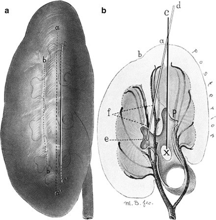
(a) Lateral view of left kidney, showing the location of the most advantageous incision through the parenchyma in kidneys which have a normal arterial arrangement. aa´: Lateral convex border of kidney. bb´: Position of lateral column of cortical substance containing the vessels. cc´: Best incision. (b) de: Incorrect direction of incision. cx: Correct direction of incision. From Max Brödel [206]. Original illustration 1901 Brödel Publication, reproduced with permission from the Collection of the Max Brödel Archives, in the Department of Art as Applied to Medicine, The Johns Hopkins University School of Medicine, Baltimore, Maryland, USA
- 7.
The percutaneous cystolithomy has been mentioned above (see section number 11.3.6).
- 8.
CLU (Club della litiasi Urinaria) is the Italian expert committee for urinary lithiasis.
- 9.
Citrus fruits: lemon, orange, mandarin, clementine, grapefruit, lime, sudachi, calamondin, etc.
- 10.
Non-citrus fruits: melon, grapes, apricots, pears, peaches, blackberries, raspberries, plums, etc.
- 11.
Febuxostat is FDA-approved since 2009.
- 12.
The chelating action of Penicillamine has been used for many other medical indications: Wilson’s disease (where it binds with copper and allows its elimination with urine), arsenic poisoning etc. Penicillamine has also been used as a disease-modifying agent in scleroderma and rheumatoid arthritis. However in all these indications its use was progressively confined to exceptional cases or banned because of the potentially dangerous side effects.

References
Türk C, Petřík A, Sarica K, et al. EAU guidelines on diagnosis and conservative management of urolithiasis. Eur Urol. 2016;69(3):468–74.
Dropkin BM, Moses RA, Sharma D, Pais VM Jr. The natural history of nonobstructing asymptomatic renal stones managed with active surveillance. J Urol. 2015;193(4):1265–9.
Assimos D, Krambeck A, Miller NL, et al. Surgical management of stones: American Urological Association/Endourological Society Guideline, PART I. J Urol. 2016;196(4):1153–60.
Afshar K, Jafari S, Marks AJ, et al. Nonsteroidal anti-inflammatory drugs (NSAIDs) and non-opioids for acute renal colic. Cochrane Database Syst Rev. 2015;(6). Art. No.: CD006027. doi:10.1002/14651858.CD006027.pub2.
Preminger GM, Tiselius HG, Assimos DG, et al. 2007 Guideline for the management of ureteral calculi. Eur Urol. 2007;52:1610–31. [PMID: 18074433].
Furyk JS, Chu K, Banks C, et al. Distal ureteric stones and tamsulosin: a double-blind, placebo-controlled, randomized, multicenter trial. Ann Emerg Med. 2016;67(1):86–95.e2.
Davenport K, Timoney AG, Keeley FX. A comparative in vitro study to determine the beneficial effect of calcium-channel and alpha(1)-adrenoceptor antagonism on human ureteric activity. BJU Int. 2006;98:651–5.
Liu C, Zeng G, Kang R, et al. Efficacy and safety of alfuzosin as medical expulsive therapy for ureteral stones: a systematic review and meta-analysis. PLoS One. 2015;10(8):e0134589.
Berger DA, Ross MA, Hollander JB, Ziadeh J, Chen C, Jackson RE, Swor RA. Tamsulosin does not increase 1-week passage rate of ureteral stones in ED patients. Am J Emerg Med. 2015;33(12):1721–4.
Pickard R, Starr K, MacLennan G, et al. Use of drug therapy in the management of symptomatic ureteric stones in hospitalised adults: a multicentre, placebo-controlled, randomised controlled trial and cost-effectiveness analysis of a calcium channel blocker (nifedipine) and an alpha-blocker (tamsulosin) (the SUSPEND trial). Health Technol Assess. 2015;19(63):vii–viii, 1–171.
Hollingsworth JM, Canales BK, Rogers MA, et al. Alpha blockers for treatment of ureteric stones: systematic review and meta-analysis. BMJ. 2016;355:i6112. doi:10.1136/bmj.i6112.
Yang D, Wu J, Yuan H, Cui Y. The efficacy and safety of silodosin for the treatment of ureteral stones: a systematic review and meta-analysis. BMC Urol. 2016;16(1):23.
Yuvanc E, Yilmaz E, Tuglu D, Batislam E. Medical and alternative therapies in urinary tract stone disease. World J Nephrol. 2015;4(5):492–9.
Ramsey S, Robertson A, Ablett MJ, et al. Evidence-based drainage of infected hydronephrosis secondary to ureteric calculi. J Endourol. 2010;24(2):185–9.
Türk C, Petřík A, Sarica K, et al. EAU guidelines on interventional treatment for urolithiasis. Eur Urol. 2016;69(3):475–82. doi:10.1016/j.eururo.2015.07.041. Epub 2015 Sep 4.
Assimos D, Krambeck A, Miller NL, et al. Surgical management of stones: American Urological Association/Endourological Society Guideline, PART II. J Urol. 2016;196(4):1161–9.
Doizi S, Raynal G, Traxer O. Evolution of urolithiasis treatment over 30 years in a French academic institution. Prog Urol. 2015;25(9):543–8.
Heers H, Turney BW. Trends in urological stone disease: a 5-year update of Hospital Episode statistics. BJU Int. 2016;118(5):785–9. doi:10.1111/bju.13520.
Chaussy C, Schmiedt E, Jocham D, et al. First clinical experience with extracorporeally induced destruction of kidney stones by shock waves. J Urol. 1982;127:417–20.
Chaussy C, Brendel W, Schmiedt E. Extracorporeally induced destruction of kidney stones by shock waves. Lancet. 1980;2:1265–8.
Gschwend JE, Paiss T, Gottfried HW, Hautmann RE. Extracorporeal shockwave lithotripsy in children. Complications and long-term results. Urologe A. 1995;34(4):324–8.
Zogović J. Extracorporeal shock wave lithotripsy in the urinary tract in patients with one kidney. Srp Arh Celok Lek. 2002;130(9–10):312–5.
Tailly G, Chaussy CG, Bohris C, et al. ESWL in a nutshell. 4th ed. Munich: Dornier MedTech Europe GmbH; 2014.
Pemberton J. Extra-corporeal shock wave lithotripsy. Postgrad Med J. 1987;63:1025–31.
Rassweiler JJ, Knoll T, Köhrmann KU, et al. Shock wave technology and application: an update. Eur Urol. 2011;59(5):784–96.
Sass W, Braunlich M, Dreyer H, Matura E. The mechanisms of stone disintegration by shock waves. Ultrasound Med Biol. 1991;17(3):239–43.
Duryea AP, Roberts WW, Cain CA, Hall TL. Controlled cavitation to augment SWL stone comminution: mechanistic insights in vitro. IEEE Trans Ultrason Ferroelectr Freq Control. 2013;60(2):301–9.
Semins MJ, Trock BJ, Matlaga BR. The effect of shock wave rate on the outcome of shock wave lithotripsy: a meta-analysis. J Urol. 2008;179:194–7.
Grasso M, Loisides P, Beaghler M, Bagley D. The case for primary endoscopic management of upper urinary tract calculi: I. A critical review of 121 extracorporeal shock-wave lithotripsy failures. Urology. 1995;45(3):363–71.
Mishra SK, Ganpule A, Manohar T, Desai MR. Surgical management of pediatric urolithiasis. Indian J Urol. 2007;23(4):428–34.
Telha KA, Alkohlany K, Alnono I. Extracorporeal shockwave lithotripsy monotherapy for treating patients with bladder stones. Arab J Urol. 2016;14(3):207–10.
Streem SB. Contemporary clinical practice of shock wave lithotripsy: a reevaluation of contraindications. J Urol. 1997;157(4):1197–203.
Lechevallier E, Traxer O, Saussine C. Extracorporeal shockwave lithotripsy for upper urinary tract stone. Prog Urol. 2008;18(12):878–85.
Vasavada SP, Streem SB, Kottke-Marchant K, Novick AC. Pathological effects of extracorporeally generated shock waves on calcified aortic aneurysm tissue. J Urol. 1994;152(1):45–8.
Atlee JL, Bernstein AD. Cardiac rhythm management devices (part II): perioperative management. Anesthesiology. 2001;95(6):1492–506.
Ector H, Janssens L, Baert L, De Geest H. Extracorporeal shock wave lithotripsy and cardiac arrhythmias. Pacing Clin Electrophysiol. 1989;12(12):1910–7.
Tonolini M, Villa F, Ippolito S, et al. Cross-sectional imaging of iatrogenic complications after extracorporeal and endourological treatment of urolithiasis. Insights Imaging. 2014;5(6):677–89.
Abdel-Khalek M, Sheir KZ, Mokhtar AA, et al. Prediction of success rate after extracorporeal shock-wave lithotripsy of renal stones--a multivariate analysis model. Scand J Urol Nephrol. 2004;38(2):161–7.
Madbouly K, Sheir KZ, Elsobky E, et al. Risk factors for the formation of a steinstrasse after extracorporeal shock wave lithotripsy: a statistical model. J Urol. 2002;167(3):1239–42.
El-Assmy A, El-Nahas AR, Elsaadany MM, et al. Risk factors for formation of steinstrasse after extracorporeal shock wave lithotripsy for pediatric renal calculi: a multivariate analysis model. Int Urol Nephrol. 2015;47(4):573–7.
Coptcoat MJ, Webb DR, Kellet MJ, et al. The steinstrasse: a legacy of extracorporeal lithotripsy? Eur Urol. 1988;14(2):93–5.
Resim S, Ekerbicer HC, Ciftci A. Role of tamsulosin in treatment of patients with steinstrasse developing after extracorporeal shock wave lithotripsy. Urology. 2005;66(5):945–8.
Puppo P. Steinstrasse 20 years later: still a problem after ESWL? Eur Urol. 2006;50(4):643–7.
Van Savage JG, Fried FA. Bilateral spontaneous steinstrasse and nephrocalcinosis associated with distal renal tubular acidosis. J Urol. 1993;150(2 Pt 1):467–8.
Kok HK, Donnellan JP, Torreggiani WC. Spontaneous steinstrasse from multiple ureteric calculi. Br J Hosp Med (Lond). 2012;73(8):474.
Biyani CS, Bhatia V, Baliga D. Urethral steinstrasse—clinical experience and radiographic findings. Clin Radiol. 1993;48(4):273–4.
Brahmbhatt YG, Schulsinger DA, Wadhwa NK. Urethral steinstrasse in renal transplantation. Kidney Int. 2009;75(3):344.
Kumar S, Sharma S, Ganesamoni R, Singh SK. Urethral steinstrasse with urethrocutaneous fistula. Urology. 2012;79(2):e1–2.
Vaddi SP, Devraj R, Reddy V, et al. Urethral steinstrasse causing acute urinary retention. Urology. 2011;77(3):594–5.
Kaynar M, Tekinarslan E, Keskin S, et al. Effective radiation exposure evaluation during a one year follow-up of urolithiasis patients after extracorporeal shock wave lithotripsy. Cent European J Urol. 2015;68(3):348–52.
Wrixon AD. New ICRP recommendations. J Radiol Prot. 2008;28(2):161–8.
Ackermann D, Merz V, Marth D, Zehntner C. Clinical experiences with extracorporeal shockwave lithotripsy. Schweiz Med Wochenschr. 1989;119(26):935–40.
Ghoneim IA, El-Ghoneimy MN, El-Naggar AE, et al. Extracorporeal shock wave lithotripsy in impacted upper ureteral stones: a prospective randomized comparison between stented and non-stented techniques. Urology. 2010;75(1):45–50.
Matsuoka Y, Ishizaka K, Machida T, et al. Treatment of 2019 cases with upper urinary tract calculi using a piezoelectric lithotriptor ESL-500A. Nihon Hinyokika Gakkai Zasshi. 2002;93(3):476–82.
Bon D, Doré B, Irani J, et al. Results of extracorporeal lithotripsy with ultrasonography-guided hydroelectric lithotriptor: study on 546 patients, prognosis factors. Prog Urol. 1995;5(5):671–8.
Park BH, Choi H, Kim JB, Chang YS. Analyzing the effect of distance from skin to stone by computed tomography scan on the extracorporeal shock wave lithotripsy stone-free rate of renal stones. Korean J Urol. 2012;53:40–3.
Müllhaupt G, Engeler DS, Schmid HP, Abt D. How do stone attenuation and skin-to-stone distance in computed tomography influence the performance of shock wave lithotripsy in ureteral stone disease? BMC Urol. 2015;15:72.
Yazici O, Tuncer M, Sahin C, et al. Shock wave lithotripsy in ureteral stones: evaluation of patient and stone related predictive factors. Int Braz J Urol. 2015;41(4):676–82.
Pareek G, Armenakas NA, Panagopoulos G, et al. Extracorporeal shock wave lithotripsy success based on body mass index and Hounsfield units. Urology. 2005;65(1):33–6.
Ouzaid I, Al-qahtani S, Dominique S, et al. A 970 Hounsfield units (HU) threshold of kidney stone density on non-contrast computed tomography (NCCT) improves patients’ selection for extracorporeal shockwave lithotripsy (ESWL): evidence from a prospective study. BJU Int. 2012;110:E438–42.
Bon D, Doré B, Irani J, et al. Radiographic prognostic criteria for extracorporeal shock-wave lithotripsy: a study of 485 patients. Urology. 1996;48:556–60. Discussion 560–1.
Bastian PJ, Bastian HP. Outpatient extracorporeal shock wave lithotripsy. Prospective evaluation of 2937 cases. Urologe A. 2004;43(7):829–35.
Kaczmarek K, Gołąb A, Słojewski M. Impact of ureteric stent on outcome of extracorporeal shockwave lithotripsy: a propensity score analysis. Cent European J Urol. 2016;69(2):184–9.
Argyropoulos AN, Tolley DA. Ureteric stents compromise stone clearance after shockwave lithotripsy for ureteric stones: results of a matched-pair analysis. BJU Int. 2009;103(1):76–80.
Sfoungaristos S, Gofrit ON, Pode D, et al. History of ureteral stenting negatively affects the outcomes of extracorporeal shockwave lithotripsy. Results of a matched-pair analysis. Prague Med Rep. 2015;116(3):225–32.
Singh V, Gupta A. Stenturia: a rare complication of indwelling ureteral stent. Urol J. 2009;6(3):226–7.
Heimbach D, Bäumler D, Schoeneich G, Hesse A. Percutaneous chemolysis: an important tool in the treatment of urolithiasis. Int Urol Nephrol. 1998;30(6):655–64.
Pfister RC, Dretler SP. Percutaneous chemolysis of renal calculi. Urol Radiol. 1984;6(2):138–43.
Kachrilas S, Papatsoris A, Bach C, et al. The current role of percutaneous chemolysis in the management of urolithiasis: review and results. Urolithiasis. 2013;41(4):323–6.
Zhang J, Wang S, Hong J, et al. New potential solutions for the chemolysis of urinary phosphate calculi determined by an in vitro study. Urolithiasis. 2015;43(2):147–53.
Young HH, McKay RW. Congenital valvular obstruction of the prostatic urethra. Surg Gynecol Obstet. 1929;48:509–12.
Rizkala ER, Monga M. Controversies in ureteroscopy: wire, basket, and sheath. Indian J Urol. 2013;29(3):244–8.
Goodman TM. Ureteroscopy with pediatric cystoscope in adults. Urology. 1977;9(4):394–7.
Lyon ES, Kyker JS, Schoenberg HW. Transurethral ureteroscopy in women: a ready addition to the urological armamentarium. J Urol. 1978;119(1):35–41.
Marshall VF. Fiber optics in urology. J Urol. 1964;91:110–4.
Bagley DH, Huffman JL, Lyon ES. Flexible ureteropyeloscopy: diagnosis and treatment in the upper urinary tract system. J Urol. 1987;138:280–5.
Kavoussi L, Clayman RV, Basler J. Flexible actively deflectable fiberoptic ureteronephroscopy. J Urol. 1989;142:949–54.
Gridley CM, Knudsen BE. Digital ureteroscopes: technology update. Res Rep Urol. 2017;9:19–25.
Jones P, Rai BP, Somani BK. Outcomes of ureteroscopy for patients with stones in a solitary kidney: evidence from a systematic review. Cent European J Urol. 2016;69(1):83–90.
Ishii H, Couzins M, Aboumarzouk O, et al. Outcomes of systematic review of ureteroscopy for stone disease in the obese and morbidly obese population. J Endourol. 2016;30(2):135–45.
Drake T, Ali A, Somani BK. Feasibility and safety of bilateral same-session flexible ureteroscopy (FURS) for renal and ureteral stone disease. Cent European J Urol. 2015;68(2):193–6.
Ishii H, Rai B, Traxer O, et al. Outcome of ureteroscopy for stone disease in patients with horseshoe kidney: Review of world literature. Urol Ann. 2015;7(4):470–4.
Rukin NJ, Somani BK, Patterson J, et al. Tips and tricks of ureteroscopy: consensus statement Part I. Basic ureteroscopy. Cent European J Urol. 2015;68(4):439–46.
Traxer O, Lechevallier E, Saussine C. Flexible ureteroscopy with Holmium laser: technical aspects. Progrès en urologie. 2008;18:929–37.
Noble MJ, Esac WE. Semirigid ureteroscopy: the Cleveland Clinic approach. In: Monga M, editor. Ureteroscopy: indications, instrumentation and technique. New York: Humana Press/Springer; 2013. p. 257–70.
Rabah DM, Fabrizio MD. Flexible fiberoptic ureteropyeloscopy. In: Smith AD, Badlani GH, Baglev DH, et al., editors. Smith’s textbook of endourology. 2nd ed. Hamilton, London: BC Decker Inc; 2007. p. 237–42.
Giusti G, Proietti S, Villa L, et al. Current standard technique for modern flexible ureteroscopy: tips and tricks. Eur Urol. 2016;70(1):188–94.
Georgescu D, Multescu R, Mirciulescu V, et al. Instruments. In: Geavlete PA, editor. Retrograde ureteroscopy. Handbook of endourology: Elsevier; 2016. p. 29–35.
Miller J, Stoller ML. Intracorporeal lithotripsy: electrohydraulic, pneumatic, and ultrasonic. In: Monga M, editor. Ureteroscopy: indications, instrumentation and technique. New York: Humana Press/Springer; 2013. p. 149–60.
Knudsen BE. Flexible ureteroscopy: holmium:YAG laser and optical fibers. In: Monga M, editor. Ureteroscopy: indications, instrumentation and technique. New York: Humana Press/Springer; 2013. p. 161–8.
Dretler SP, Watson G, Parrish JA, Murray S. Pulsed dye laser fragmentation of ureteral calculi: initial clinical experience. J Urol. 1987;137(3):386–9.
Marguet CG, Sung JC, Springhart WP, et al. In vitro comparison of stone retropulsion and fragmentation of the frequency doubled, double pulse Nd:YAG laser and the holmium:YAG laser. J Urol. 2005;173:1797–800.
Teichman JM, Chan KF, Cecconi PP, et al. Erbium : YAG versus holmium : YAG lithotripsy. J Urol. 2001;165(3):876–9.
Lee H, Kang HW, Teichman JM, et al. Urinary calculus fragmentation during Ho: YAG and Er:YAG lithotripsy. Lasers Surg Med. 2006;38(1):39–51.
Matlaga BR, Lingeman JE. Surgical management of stones: new technology. Adv Chronic Kidney Dis. 2009;16:60–4.
Keeley FX Jr, Pillai M, Smith G, Chrisofos M, Tolley DA. Electrokinetic lithotripsy: safety, efficacy and limitations of a new form of ballistic lithotripsy. BJU Int. 1999;84(3):261–3.
Ali AA, Ali ZA, Halstead JC, et al. A novel method to prevent retrograde displacement of ureteric calculi during intracorporeal lithotripsy. BJU Int. 2004;94(3):441–2.
Bastawisy M, Gameel T, Radwan M, et al. A comparison of stone cone versus lidocaine jelly in the prevention of ureteral stone migration during ureteroscopic lithotripsy. Ther Adv Urol. 2011;3(5):203–10.
Kreydin E, Eisner B. Stone migration devices. In: Monga M, editor. Ureteroscopy: indications, instrumentation and technique. New York: Humana Press/Springer; 2013. p. 169–78.
Hofbauer J, Hobarth K, Marberger M. Electrohydraulic versus pneumatic disintegration in the treatment of ureteral stones: a randomized, prospective trial. J Urol. 1995;153:623.
Garg S, Mandal AK, Singh SK, et al. Ureteroscopic laser lithotripsy versus ballistic lithotripsy for treatment of ureteric stones: a prospective comparative study. Urol Int. 2009;82(3):341–5.
Santa-Cruz RW, Leveillee RJ, Krongrad A. Ex vivo comparison of four lithotripters commonly used in the ureter: what does it take to perforate? J Endourol. 1998;12:417.
Hecht SL, Wolf JS. Techniques for holmium laser lithotripsy of intrarenal calculi. Urology. 2013;81:442–5.
Auge BK, Pietrow PK, Lallas CD, et al. Ureteral access sheath provides protection against elevated renal pressures during routine flexible ureteroscopic stone manipulation. J Endourol. 2004;18(1):33–6.
Geraghty RM, Ishii H, Somani BK. Outcomes of flexible ureteroscopy and laser fragmentation for treatment of large renal stones with and without the use of ureteral access sheaths: results from a university hospital with a review of literature. Scand J Urol. 2016;50(3):216–9.
Traxer O, Thomas A. Prospective evaluation and classification of ureteral wall injuries resulting from insertion of UAS during RIRS. J Urol. 2013;189:580–4.
Rukin NJ, Somani BK, Patterson J, et al. Tips and tricks of ureteroscopy: consensus statement. Part II. Advanced ureteroscopy. Cent European J Urol. 2016;69(1):98–104.
Kurahashi T, Miyake H, Oka N, et al. Clinical outcome of ureteroscopic lithotripsy for 2,129 patients with ureteral stones. Urol Res. 2007;35(3):149–53.
Takazawa R, Kitayama S, Kobayashi S, et al. Transurethral lithotripsy with rigid and flexible ureteroscopy for renal and ureteral stones: results of the first 100 procedures. Hinyokika Kiyo. 2011;57(8):411–6.
Tanriverdi O, Silay MS, Kadihasanoglu M, et al. Revisiting the predictive factors for intra-operative complications of rigid ureteroscopy: a 15-year experience. Urol J. 2012;9(2):457–64.
Alcaide JRC, Elbers JR, Perez DL, et al. Flexible ureterorenoscopy (URS): technique and results. Arch Esp Urol. 2010;63(10):862–70.
Grasso M, Ficazzola M. Retrograde ureteropyeloscopy for lower pole caliceal calculi. J Urol. 1999;162:1904–8.
Fuganti PE, Pires S, Branco R, Porto J. Predictive factors for intraoperative complications in semirigid ureteroscopy: analysis of 1235 ballistic ureterolithotripsies. Urology. 2008;72(4):770–4.
Baş O, Tuygun C, Dede O, et al. Factors affecting complication rates of retrograde flexible ureterorenoscopy: analysis of 1571 procedures-a single-center experience. World J Urol. 2016;35(5):819–26.
El Harrech Y, Abakka N, El Anzaoui J, et al. Ureteral stenting after uncomplicated ureteroscopy for distal ureteral stones: a randomized, controlled trial. Minim Invasive Surg. 2014;2014:892890.
Jung GH, Jung JH, Ahn TS, et al. Comparison of retrograde intrarenal surgery versus a single-session percutaneous nephrolithotomy for lower-pole stones with a diameter of 15 to 30 mm: a propensity score-matching study. Korean J Urol. 2015;56(7):525–32.
Klingler HC, et al. Stone treatment and coagulopathy. Eur Urol. 2003;43(1):75–9.
Al Busaidy SS, Kurukkal SN, Al Hooti QM, et al. Is RIRS emerging as the preferred option for the management of 2 cm-4 cm renal stones: our experience. Can J Urol. 2016;23(4):8364–7.
Liu K, Xiao CL, Liu YQ, et al. Management of calyceal diverticular calculi with stenotic infundibulum by flexible ureteroscopic holmium laser infundibulectomy and lithotripsy. Beijing Da Xue Xue Bao. 2015;47(4):618–21.
Cohen J, Cohen S, Grasso M. Ureteropyeloscopic treatment of large, complex intrarenal and proximal ureteral calculi. BJU Int. 2013;111(3 Pt B):E127–31.
Marguet CG, Springhart WP, Tan YH, et al. Simultaneous combined use of flexible ureteroscopy and percutaneous nephrolithotomy to reduce the number of access tracts in the management of complex renal calculi. BJU Int. 2005;96(7):1097–100.
Hamamoto S, Yasui T, Okada A, et al. Developments in the technique of endoscopic combined intrarenal surgery in the prone split-leg position. Urology. 2014;84(3):565–70.
Hamamoto S, Yasui T, Okada A, et al. Efficacy of endoscopic combined intrarenal surgery in the prone split-leg position for staghorn calculi. J Endourol. 2015;29(1):19–24.
Sternberg KM, Jacobs BL, King BJ, et al. The prone ureteroscopic technique for managing large stone burdens. Can J Urol. 2015;22(2):7758–62.
Bernardo N, Lopez-Silva M, Sanguinetti H, et al. Unplanned flexible ureteroscopy during percutaneous nephrolithotomy in the prone position. Actas Urol Esp. 2016;40(2):115–8.
Chew BH, Lange D. The future of ureteroscopy. Minerva Urol Nefrol. 2016;68(6):592–7.
Fernstrom I, Johansson B. Percutaneous pyelolithotomy, a new extraction technique. Scand J Urol Nephrol. 1976;10:257–9.
Alken P, Hutschenreiter G, Gunther R, Marberger M. Percutaneous stone manipulation. J Urol. 1981;125:463–6.
Thomas K, Smith NC, Hegarty N, Glass JM. The Guy’s stone score—grading the complexity of percutaneous nephrolithotomy procedures. Urology. 2011;78(2):277–81.
Vernez SL, Okhunov Z, Motamedinia P, et al. Nephrolithometric scoring systems to predict outcomes of percutaneous nephrolithotomy. Rev Urol. 2016;18(1):15–27.
Torricelli FC, Padovani GP, Marchini GS, et al. Percutaneous nephrolithotomy in patients with solitary kidney: a critical outcome analysis. Int Braz J Urol. 2015;41(3):496–502.
Ishibashi M, Morita S, Rabito CA, et al. Evaluation of the therapeutic effect of percutaneous nephroureterolithotomy by Tc-99m diethylenetiaminepentaacetic acid (DTPA) renal scintigraphy--alteration of the renal fraction of blood flow, split-GFR, and renal mean transit time. Kurume Med J. 1990;37(4):285–91.
Sharma GR, Maheshwari PN, Sharma AG, et al. Fluoroscopy guided percutaneous renal access in prone position. World J Clin Cases. 2015;3(3):245–64.
Valdivia Uria JG, Valle Gerhold J, Lopez Lopez JA, et al. Technique and complications of percutaneous nephroscopy: experience with 557 patients in the supine position. J Urol. 1998;160:1975–8.
Ibarluzea G, Scoffone CM, Cracco CM, et al. Supine Valdivia and modified lithotomy position for simultaneous anterograde and retrograde endourological access. BJU Int. 2007;100:233–6.
Serra S, Corona A, De Lisa A. Endoscopic combined intra renal surgery (ECIRS) in prone position. Urologia. 2012;79(Suppl 19):121–4.
Hoznek A, Rode J, Ouzaid I, et al. Modified supine percutaneous nephrolithotomy for large kidney and ureteral stones: technique and results. Eur Urol. 2012;61(1):164–70.
Kumar P, Bach C, Kachrilas S, et al. Supine percutaneous nephrolithotomy (PCNL): ‘in vogue’ but in which position? BJU Int. 2012;110(11 Pt C):E1018–21.
Kim SC, Lingeman JE. Percutaneous access to the urinary tract. In: Nakada SY, Pearle MS, editors. Advanced endourology: the complete clinical guide. Totowa, NJ: Humana Press Inc; 2006. p. 43–60.
Ko R, Soucy F, Denstedt JD, Razvi H. Percutaneous nephrolithotomy made easier: a practical guide, tips and tricks. BJU Int. 2008;101(5):535–9.
Munver R, Delvecchio FC, Newman GE, Preminger GM. Critical analysis of supracostal access for percutaneous renal surgery. J Urol. 2001;166:1242–6.
El-Karamany T. A supracostal approach for percutaneous nephrolithotomy of staghorn calculi: a prospective study and review of previous reports. Arab J Urol. 2012;10(4):358–66.
Li J, Xiao B, Hu W, et al. Complication and safety of ultrasound guided percutaneous nephrolithotomy in 8,025 cases in China. Chin Med J (Engl). 2014;127(24):4184–9.
Helal M, Black T, Lockhart J, Figueroa TE. The Hickman peel-away sheath: alternative for pediatric percutaneous nephrolithotomy. J Endourol. 1997;11:171–2.
Jackman SV, Hedican SP, Peters CA, Docimo SG. Percutaneous nephrolithotomy in infants and preschool age children: experience with a new technique. Urology. 1998;52:697–701.
Jackman SV, Docimo SG, Cadeddu JA, Bishoff JT, Kavoussi LR, Jarrett TW. The “mini-perc” technique: a less invasive alternative to percutaneous nephrolithotomy. World J Urol. 1998;16:371–4.
Ganpule AP, Desai MR. What's new in percutaneous nephrolithotomy. Arab J Urol. 2012;10(3):317–23.
Desai MR, Sharma R, Mishra S, et al. Single-step percutaneous nephrolithotomy (microperc): the initial clinical report. J Urol. 2011;186:140–5.
Sabnis RB, Ganesamoni R, Doshi A, et al. Micropercutaneous nephrolithotomy (microperc) vs retrograde intrarenal surgery for the management of small renal calculi: a randomized controlled trial. BJU Int. 2013;112(3):355–61.
Kaynar M, Sümer A, Şalvarcı A, et al. Micropercutaneous nephrolithotomy (Microperc®) in a two-year-old with the ‘all-seeing needle’. Urol Int. 2013;91(2):239–41.
Desai MR, Kukreja RA, Desai MM, et al. A prospective randomized comparison of type of nephrostomy drainage following percutaneous nephrostolithotomy: large bore versus small bore versus tubeless. J Urol. 2004;172(2):565–7.
Istanbulluoglu MO, Ozturk B, Gonen M, et al. Effectiveness of totally tubeless percutaneous nephrolithotomy in selected patients: a prospective randomized study. Int Urol Nephrol. 2009;41(3):541–5.
Chang CH, Wang CJ, Huang SW. Totally tubeless percutaneous nephrolithotomy: a prospective randomized controlled study. Urol Res. 2011;39(6):459–65.
Garofalo M, Pultrone CV, Schiavina R, et al. Tubeless procedure reduces hospitalization and pain after percutaneous nephrolithotomy: results of a multivariable analysis. Urolithiasis. 2013;41(4):347–53.
Javali T, Pathade A, Nagaraj HK. A Novel method of ensuring safe and accurate dilatation during percutaneous nephrolithotomy. Int Braz J Urol. 2015;41(5):1014–9.
Maheshwari PN, Sharma GR, Wagaskar VG. RE: A Novel method of ensuring safe and accurate dilalation during percutaneous nephrolithotomy. Int Braz J Urol. 2016;42(3):628–9.
Kumar S, Singh S, Singh P, Singh SK. Day care PNL using ‘Santosh-PGI hemostatic seal’ versus standard PNL: a randomized controlled study. Cent European J Urol. 2016;69(2):190–7.
Song Y, Ma Y, Song Y, Fei X. Evaluating the learning curve for percutaneous nephrolithotomy under total ultrasound guidance. PLoS One. 2015;10(8):e0132986.
Armitage JN, Withington J, van der Meulen J, et al. Percutaneous nephrolithotomy in England: practice and outcomes described in the Hospital Episode Statistics database. BJU Int. 2014;113(5):777–82.
Yamaguchi A, Skolarikos A, Buchholz NP, et al. Clinical Research Office Of The Endourological Society Percutaneous Nephrolithotomy Study Group. Operating times and bleeding complications in percutaneous nephrolithotomy: a comparison of tract dilation methods in 5,537 patients in the Clinical Research Office of the Endourological Society Percutaneous Nephrolithotomy Global Study. J Endourol. 2011;25(6):933–9.
Meng XJ, Mi QW, Hu T, Zhong WD. Value of CT angiography in reducing the risk of hemorrhage associated with mini-percutaneous nephrolithotomy. Int Braz J Urol. 2015;41(4):690–6.
Shokeir AA. Transurethral cystolitholapaxy in children. J Endourol. 1994;8(2):157–9. Discussion 159–60.
Comisarow RH, Barkin M. Electrohydraulic cystolitholapaxy. Can J Surg. 1979;22(6):525–6.
Teichman JM, Rogenes VJ, McIver BJ, Harris JM. Holmium:yttrium-aluminum-garnet laser cystolithotripsy of large bladder calculi. Urology. 1997;50(1):44–8.
Li A, Ji C, Wang H, et al. Transurethral cystolitholapaxy with the AH-1 stone removal system for the treatment of bladder stones of variable size. BMC Urol. 2015;15:9.
Gallego Vilar D, Beltran Persiva J, Pérez Mestre M, et al. Giant bladder lithiasis: case report and bibliographic review. Arch Esp Urol. 2011;64(4):383–7.
Cohen J, Giuliano K, Sopko N, et al. Cystolitholapaxy in Ileal Conduit. Urol Case Rep. 2015;3(6):185–7.
Floyd MS Jr, Stubington SR. Mitrofanoff cystolitholapaxy: an innovative method of stone clearance in a hostile abdomen with an inaccessible urethra. Urol J. 2015;12(2):2115–8.
Xiong Y, Yang S, Liao W, et al. Autonomic dysreflexia during cystolitholapaxy in patients with spinal cord injury. Minerva Urol Nefrol. 2015;67(2):85–90.
Al-Marhoon MS, Sarhan OM, Awad BA, et al. Comparison of endourological and open cystolithotomy in the management of bladder stones in children. J Urol. 2009;181(6):2684–7. Discussion 2687–8.
Gamal W, Eldahshoury M, Hussein M, Hammady A. Cystoscopically guided percutaneous suprapubic cystolitholapaxy in children. Int Urol Nephrol. 2013;45(4):933–7.
Metwally AH, Sherief MH, Elkoushy MA. Safety and efficacy of cystoscopically guided percutaneous suprapubic cystolitholapaxy without fluoroscopic guidance. Arab J Urol. 2016;14(3):211–5.
Meria P, Milcent S, Desgrandchamps F, et al. Management of pelvic stones larger than 20 mm: laparoscopic transperitoneal pyelolithotomy or percutaneous nephrolithotomy? Urol Int. 2005;75(4):322–6.
Basiri A, Tabibi A, Nouralizadeh A, et al. Comparison of safety and efficacy of laparoscopic pyelolithotomy versus percutaneous nephrolithotomy inpatients with renal pelvic stones: a randomized clinical trial. Urol J. 2014;11(6):1932–7.
Rui X, Hu H, Yu Y, et al. Comparison of safety and efficacy of laparoscopic pyelolithotomy versus percutaneous nephrolithotomy in patients with large renal pelvic stones: a meta-analysis. J Investig Med. 2016;64(6):1134–42.
Sasaki Y, Kohjimoto Y, Nishizawa S, et al. Laparoscopic pyelolithotomy in a horseshoe kidney. Hinyokika Kiyo. 2012;58(2):87–91.
Ölçücüoğlu E, Çamtosun A, Biçer S, Bayraktar AM. Laparoscopic pyelolithotomy in a horseshoe kidney. Turk J Urol. 2014;40(4):240–4.
Chipde SS, Agrawal S. Retroperitoneoscopic pyelolithotomy: a minimally invasive alternative for the management of large renal pelvic stone. Int Braz J Urol. 2014;40(1):123–4. Discussion 124
Agrawal S, Chipde SS, Kalathia J, Agrawal R. Renal stone in crossed fused renal ectopia and its laparoscopic management: case report and review of literature. Urol Ann. 2016;8(2):236–8.
Stein RJ, Turna B, Nguyen MM, et al. Laparoscopic pyeloplasty with concomitant pyelolithotomy: technique and outcomes. J Endourol. 2008;22:1251–5.
Gandhi HR, Thomas A, Nair B, Pooleri G. Laparoscopic pyelolithotomy: an emerging tool for complex staghorn nephrolithiasis in high-risk patients. Arab J Urol. 2015;13(2):139–45.
Agrawal V, Bajaj J, Acharya H, et al. Laparoscopic management of pediatric renal and ureteric stones. J Pediatr Urol. 2013;9(2):230–3.
Al-Hunayan A, Abdulhalim H, El-Bakry E, et al. Laparoscopic pyelolithotomy: is the retroperitoneal route a better approach? Int J Urol. 2009;16(2):181–6.
Pastore AL, Palleschi G, Silvestri L, et al. Combined laparoscopic pyelolithotomy and endoscopic pyelolithotripsy for staghorn calculi: long-term follow-up results from a case series. Ther Adv Urol. 2016;8(1):3–8.
Simforoosh N, Radfar MH, Nouralizadeh A, et al. Laparoscopic anatrophic nephrolithotomy for management of staghorn renal calculi. J Laparoendosc Adv Surg Tech A. 2013;23(4):306–10.
Ghani KR, Rogers CG, Sood A, et al. Robot-assisted anatrophic nephrolithotomy with renal hypothermia for managing staghorn calculi. J Endourol. 2013;27(11):1393–8.
Ghani KR, Trinh QD, Jeong W, et al. Robotic nephrolithotomy and pyelolithotomy with utilization of the robotic ultrasound probe. Int Braz J Urol. 2014;40(1):125–6. Discussion 126.
King SA, Klaassen Z, Madi R. Robot-assisted anatrophic nephrolithotomy: description of technique and early results. J Endourol. 2014;28(3):325–9.
Garg M, Singh V, Sinha RJ, et al. Prospective randomized comparison of open versus transperitoneal laparoscopic ureterolithotomy: experience of a single center from Northern India. Curr Urol. 2013;7(2):83–9.
Khalil M, Omar R, Abdel-Baky S, et al. Laparoscopic ureterolithotomy; which is better: Transperitoneal or retroperitoneal approach? Turk J Urol. 2015;41(4):185–90.
Torricelli FC, Monga M, Marchini GS, et al. Semi-rigid ureteroscopic lithotripsy versus laparoscopic ureterolithotomy for large upper ureteral stones: a meta-analysis of randomized controlled trials. Int Braz J Urol. 2016;42(4):645–54.
Şahin S, Aras B, Ekşi M, et al. Laparoscopic ureterolithotomy. JSLS. 2016;20(1):e2016.00004.
Ma L, Yu DM, Zhang ZG, et al. Transperitoneal laparoscopic ureterolithotomy for upper ureteral calculi: a report of 1171 cases. Zhonghua Yi Xue Za Zhi. 2013;93(20):1577–9.
Verit A, Savas M, Ciftci H, et al. Outcomes of urethral calculi patients in an endemic region and an undiagnosed primary fossa navicularis calculus. Urol Res. 2006;34(1):37–40.
Akhtar J, Ahmed S, Zamir N. Management of impacted urethral stones in children. J Coll Physicians Surg Pak. 2012;22(8):510–3.
Walker BR, Hamilton BD. Urethral calculi managed with transurethral Holmium laser ablation. J Pediatr Surg. 2001;36(9):E16.
Smith MJ, Boyce WH. Anatrophic nephrotomy and plastic calyrhaphy. J Urol. 1968;99(5):521–7.
Gil-Vernet JM, Caralps A. Human renal homotransplantation. New surgical technique. Urol Int. 1968;23(3):201–23.
Gil-Vernet JM, Carretero P, Caralps A, Ballesteros JJ. New surgical technique for balanitic hypospadias correction. J Urol. 1974;112(5):673.
Gil-Vernet JM. A new technique for surgical correction of vesicoureteral reflux. J Urol. 1984;131(3):456–8.
Gil-Vernet JM, Gutierrez del Pozo R, Carretero P, et al. Urogenital diaphragm raising maneuver. J Urol. 1988;140(3):555–8.
Gil-Vernet JM, Gil-Vernet A, Campos JA. New surgical approach for treatment of complex vesicovaginal fistula. J Urol. 1989;141(3):513–6.
Gil-Vernet JM, Gil-Vernet A, Caralps A, et al. Orthotopic renal transplant and results in 139 consecutive cases. J Urol. 1989;142(2 Pt 1):248–52.
Gil-Vernet S. Pathologia Urogenital: Biologia y pathologia de la prostata. T.1. Madird: Editorial Paz-Montalvo; 1953.
Crosby RW, Cody J. Max Brödel: the man who put art into medicine. New York: Springer; 1991.
Brödel M. The intrinsic blood-vessels of the kidney and their significance in nephrotomy. The Johns Hopkins Hospital Bulletin. XII/118; 1901.
Latarjet M, Ruiz Liard A. Systema urinario. Riñόn. In: Anatomía Humana, vol. 2. 4th ed. Buenos Aires: Editorial Medica Panamericana; 2008. p. 1509–26.
Lunardi P, Timsit MO, Roumiguie M, et al. Single procedure treatment of complex nephrolithiasis: about a modern series of anatrophic nephrolithotomy. Prog Urol. 2015;25(2):90–5.
Bove AM, Altobelli E, Buscarini M. Indication to open anatrophic nephrolithotomy in the twenty-first century: a case report. Case Rep Urol. 2012;2012:851020.
Aminsharifi A, Irani D, Masoumi M, et al. The management of large staghorn renal stones by percutaneous versus laparoscopic versus open nephrolithotomy: a comparative analysis of clinical efficacy and functional outcome. Urolithiasis. 2016;44(6):551–7.
Mahran MR, Dawaba MS. Cystolitholapaxy versus cystolithotomy in children. J Endourol. 2000;14(5):423–5.
Ma C, Lu B, Sun E. Giant bladder stone in a male patient: a case report. Medicine (Baltimore). 2016;95(30):e4323.
Randall A. Giant vesical calculus. J Urol. 1920;5(2):119–25.
Moran ME. The largest stone of all! In: Urolithiasis. A comprehensive history. Now York: Springer; 2014. p. 351–63.
Pearle MS, Goldfarb DS, Assimos DG, et al. Medical management of kidney stones: AUA guideline. J Urol. 2014;192(2):316–24.
Prezioso D, Strazzullo P, Lotti T, et al. Dietary treatment of urinary risk factors for renal stone formation. A review of CLU Working Group. Arch Ital Urol Androl. 2015;87(2):105–20.
Bates GP, Miller VS. Sweat rate and sodium loss during work in the heat. J Occup Med Toxicol. 2008;3:4.
Godek SF, Peduzzi C, Burkholder R, et al. Sweat rates, sweat sodium concentrations, and sodium losses in 3 groups of professional football players. J Athl Train. 2010;45(4):364–71.
Brake DJ, Bates GP. Fluid losses and hydration status of industrial workers under thermal stress working extended shifts. Occup Environ Med. 2003;60(2):90–6.
Pak CY, Sakhaee K, Fuller CJ. Physiological and physiochemical correction and prevention of calcium stone formation by potassium citrate therapy. Trans Assoc Am Physicians. 1983;96:294–305.
Sakhaee K, Nicar M, Hill K, Pak CY. Contrasting effects of potassium citrate and sodium citrate therapies on urinary chemistries and crystallization of stone-forming salts. Kidney Int. 1983;24(3):348–52.
Siener R. Can the manipulation of urinary pH by beverages assist with the prevention of stone recurrence? Urolithiasis. 2016;44(1):51–6.
Sorensen MD, Hsi RS, Chi T. Dietary intake of fiber, fruit and vegetables decreases the risk of incident kidney stones in women: a Women’s Health Initiative report. J Urol. 2014;192(6):1694–9.
Manfredini R, De Giorgi A, Storari A, Fabbian F. Pears and renal stones: possible weapon for prevention? A comprehensive narrative review. Eur Rev Med Pharmacol Sci. 2016;20(3):414–25.
Nijenhuis T, Vallon V, van der Kemp AW, et al. Enhanced passive Ca2+ reabsorption and reduced Mg2+ channel abundance explains thiazide-induced hypocalciuria and hypomagnesemia. J Clin Invest. 2005;115:1651–8.
Laerum E, Larsen S. Thiazide prophylaxis of urolithiasis. A double-blind study in general practice. Acta Med Scand. 1984;215:383–9. An early RCT to show the effect of HCTZ in the prevention of calcium stone recurrence.
Escribano J, Balaguer A, Roqué i Figuls M, et al. Dietary interventions for preventing complications in idiopathic hypercalciuria. Cochrane Database Syst Rev. 2014;(2):CD006022.
Escribano J, Balaguer A, Pagone F, et al. Pharmacological interventions for preventing complications in idiopathic hypercalciuria. Cochrane Database Syst Rev. 2009;(1):CD004754.
Nicar MJ, Hill K, Pak CY. Inhibition by citrate of spontaneous precipitation of calcium oxalate in vitro. J Bone Miner Res. 1987;2(3):215–20.
Barcelo P, Wuhl O, Servitge E, et al. Randomized double-blind study of potassium citrate in idiopathic hypocitraturic calcium nephrolithiasis. J Urol. 1993;150(6):1761–4.
Coe FL, Evan A, Worcester E. Pathophysiology-based treatment of idiopathic calcium kidney stones. Clin J Am Soc Nephrol. 2011;6(8):2083–92.
Xu H, Zisman AL, Coe FL, Worcester EM. Kidney stones: an update on current pharmacological management and future directions. Expert Opin Pharmacother. 2013;14(4):435–47.
Gul Z, Monga M. Medical and dietary therapy for kidney stone prevention. Korean J Urol. 2014;55(12):775–9.
Kok DJ. Metaphylaxis, diet and lifestyle in stone disease. Arab J Urol. 2012;10(3):240–9.
Carrasco A Jr, Granberg CF, Gettman MT, et al. Surgical management of stone disease in patients with primary hyperoxaluria. Urology. 2015;85(3):522–6.
Lorenzo V, Torres A, Salido E. Primary hyperoxaluria. Nefrologia. 2014;34(3):398–412.
Holmes RP, Goodman HO, Assimos DG. Contribution of dietary oxalate to urinary oxalate excretion. Kidney Int. 2001;59(1):270–6.
Ettinger B, Tang A, Citron JT, et al. Randomized trial of allopurinol in the prevention of calcium oxalate calculi. N Engl J Med. 1986;315:1386–9.
Beara-Lasic L, Pillinger MH, Goldfarb DS. Advances in the management of gout: critical appraisal of febuxostat in the control of hyperuricemia. Int J Nephrol Renovasc Dis. 2010;3:1–10.
Preminger GM, Sakhaee K, Skurla C, et al. Prevention of recurrent calcium stone formation with potassium citrate therapy in patients with distal renal tubular acidosis. J Urol. 1985;134:20–3.
Maalouf NM, Cameron MA, Moe OW, et al. Novel insights into the pathogenesis of uric acid nephrolithiasis. Curr Opin Nephrol Hypertens. 2004;13:181.
Biyani CS, Cartledge JJ. Cystinuria: diagnosis and management. EAU-EBU Update Series. 2006;4:175–83.
Preminger GM, Assimos DG, Lingeman JE, et al. AUA Nephrolithiasis Guideline Panel. Chapter 1: AUA guideline on management of staghorn calculi: diagnosis and treatment recommendations. J Urol. 2005;173(6):1991–2000.
Crawhall JC, Scowen EF, Watts RW. Effect of penicillamine on cystinuria. Br Med J. 1963;1:588–90.
Lotz M, Bartter FC. Stone dissolution with D-penicillamine in cystinuria. Br Med J. 1965;2:1408–9.
Goldfarb DS, Coe FL, Asplin JR. Urinary cystine excretion and capacity in patients with cystinuria. Kidney Int. 2006;69:1041–7.
Fattah H, Hambaroush Y, Goldfarb DS. Cystine nephrolithiasis. Transl Androl Urol. 2014;3(3):228–33.
Griffith DP, Gleeson MJ, Lee H, et al. Randomized, double-blind trial of Lithostat (acetohydroxamic acid) in the palliative treatment of infection-induced urinary calculi. Eur Urol. 1991;20:243.
Rodman JS, Williams JJ, Jones RL. Hypercoagulability produced by treatment with acetohydroxamic acid. Clin Pharmacol Ther. 1987;42:346.
Faridi P, Roozbeh J, Mohagheghzadeh A. Ibn-Sina’s life and contributions to medicinal therapies of kidney calculi. Iran J Kidney Dis. 2012;6(5):339–45.
Nasim MJ, Bin Asad MH, Durr-e-Sabih, et al. Gist of medicinal plants of Pakistan having ethnobotanical evidences to crush renal calculi (kidney stones). Acta Pol Pharm. 2014;71(1):3–10.
Bahmani M, Baharvand-Ahmadi B, Tajeddini P, et al. Identification of medicinal plants for the treatment of kidney and urinary stones. J Renal Inj Prev. 2016;5(3):129–33.
Boim MA, Heilberg IP, Schor N. Phyllanthus niruri as a promising alternative treatment for nephrolithiasis. Int Braz J Urol. 2010;36(6):657–64. Discussion 664.
Kieley S, Dwivedi R, Monga M. Ayurvedic medicine and renal calculi. J Endourol. 2008;22(8):1613–6.
Micali S, Sighinolfi MC, Celia A, et al. Can Phyllanthus niruri affect the efficacy of extracorporeal shock wave lithotripsy for renal stones? A randomized, prospective, long-term study. J Urol. 2006;176(3):1020–2.
Erickson SB, Vrtiska TJ, Canzanello VJ, Lieske JC. Cystone® for 1 year did not change urine chemistry or decrease stone burden in cystine stone formers. Urol Res. 2011;39(3):197–203.
Erickson SB, Vrtiska TJ, Lieske JC. Effect of Cystone® on urinary composition and stone formation over a one year period. Phytomedicine. 2011;18(10):863–7.
Atmani F, Slimani Y, Mimouni M, Hacht B. Prophylaxis of calcium oxalate stones by Herniaria hirsuta on experimentally induced nephrolithiasis in rats. BJU Int. 2003;92(1):137–40.
Rodgers AL, Webber D, Ramsout R, Gohel MD. Herbal preparations affect the kinetic factors of calcium oxalate crystallization in synthetic urine: implications for kidney stone therapy. Urolithiasis. 2014;42(3):221–5.
Monti E, Trinchieri A, Magri V, et al. Herbal medicines for urinary stone treatment. A systematic review. Arch Ital Urol Androl. 2016;88(1):38–46.
Iqbal MW, Shin RH, Youssef RF, et al. Should metabolic evaluation be performed in patients with struvite stones? Urolithiasis. 2017;45(2):185–92.
Alamuri P, Eaton KA, Himpsl SD, et al. Vaccination with proteus toxic agglutinin, a hemolysin-independent cytotoxin in vivo, protects against Proteusmirabilis urinary tract infection. Infect Immun. 2009;77(2):632–41.
Hoppe B, Groothoff JW, Hulton SA, et al. Efficacy and safety of Oxalobacter formigenes to reduce urinary oxalate in primary hyperoxaluria. Nephrol Dial Transplant. 2011;26(11):3609–15.
Jairath A, Parekh N, Otano N, et al. Oxalobacter formigenes: opening the door to probiotic therapy for the treatment of hyperoxaluria. Scand J Urol. 2015;49(4):334–7.
Rimer JD, An Z, Zhu Z, et al. Crystal growth inhibitors for the prevention of L-cystine kidney stones through molecular design. Science. 2010;330(6002):337–41.
Goldfarb DS. Potential pharmacological treatments for cystinuria and for calcium stones associated with hyperuricosuria. Clin J Am Soc Nephrol. 2011;6(8):2093–7.
Sahota A, Parihar JS, Yang M, et al. Novel cystine ester mimics for the treatment of cystinuria-induced urolithiasis in a knockout mouse model. Urology. 2014;84(5):1249.e9–15.
Lee MH, Sahota A, Ward MD, Goldfarb DS. Cystine growth inhibition through molecular mimicry: a new paradigm for the prevention of crystal diseases. Curr Rheumatol Rep. 2015;17(5):33.
Wendt-Nordahl G, Sagi S, Bolenz C, et al. Evaluation of cystine transport in cultured human kidney cells and establishment of cystinuria type I phenotype by antisense technology. Urol Res. 2008;36(1):25–9.
Author information
Authors and Affiliations
Rights and permissions
Copyright information
© 2017 Springer International Publishing AG
About this chapter
Cite this chapter
Al-Mamari, S.A. (2017). Treatment of Urolithiasis. In: Urolithiasis in Clinical Practice. In Clinical Practice. Springer, Cham. https://doi.org/10.1007/978-3-319-62437-2_11
Download citation
DOI: https://doi.org/10.1007/978-3-319-62437-2_11
Published:
Publisher Name: Springer, Cham
Print ISBN: 978-3-319-62436-5
Online ISBN: 978-3-319-62437-2
eBook Packages: MedicineMedicine (R0)