Differences in the imaging of Crohn’s disease patients between North America and Europe: are we ready to bridge the divide? Stuart A. TaylorFlorian RiederJoel G. Fletcher Perspective Open access 22 December 2018 Pages: 1637 - 1643
Ischiorectal fossa: benign and malignant neoplasms of this “ignored” radiological anatomical space S. C. FariaS. B. ElsherifP. R. Bhosale Review 06 April 2019 Pages: 1644 - 1674
Diagnostic performance of MRI- versus MDCT-categorized T3cd/T4 for identifying high-risk stage II or stage III colon cancers: a pilot study Soo Yeun ParkSeung Hyun ChoGyu-Seog Choi Hollow Organ GI 17 November 2018 Pages: 1675 - 1685
Mucinous appendiceal neoplasms: classification, imaging, and HIPEC David J. BartlettPaul G. Thacker Jr.Shannon P. Sheedy Review Paper 04 January 2019 Pages: 1686 - 1702
Diagnostic performance of F-18 FDG PET/CT for prediction of KRAS mutation in colorectal cancer patients: a systematic review and meta-analysis Seong-Jang KimKyoungjune PakKeunyoung Kim Hollow Organ GI 02 January 2019 Pages: 1703 - 1711
Morphological predictors for lymph node metastases on computed tomography in colon cancer Erik RollvénLennart BlomqvistMirna Abraham-Nordling Hollow Organ GI Open access 14 February 2019 Pages: 1712 - 1721
Analyzing the post-contrast attenuation of the esophageal wall on routine contrast-enhanced MDCT examination can improve the diagnostic accuracy in response evaluation of the squamous cell esophageal carcinoma to neoadjuvant chemoradiotherapy in comparison with the esophageal wall thickness Aleksandra Djuric-StefanovicAleksandra JankovicPredrag Pesko Hollow Organ GI 13 February 2019 Pages: 1722 - 1733
Acquired diverticular disease of the jejunum and ileum: imaging features and pitfalls P. LebertO. ErnstM. Zins Review 13 February 2019 Pages: 1734 - 1743
Perianal sepsis: surgical perspective and practical MRI reporting for radiologists Edwin HoMatthew J. F. X. RickardJessica Yang Perspective 15 February 2019 Pages: 1744 - 1755
Improving preoperative detection of synchronous liver metastases in pancreatic cancer with combined contrast-enhanced and diffusion-weighted MRI D. M. RiviereE. J. M. van GeenenJ. J. Hermans Pancreas Open access 18 January 2019 Pages: 1756 - 1765
Magnetic resonance cholangiopancreatography using optimized integrated combination with parallel imaging and compressed sensing technique Shoma NagataSatoshi GoshimaMasayuki Matsuo Hepatobiliary 18 January 2019 Pages: 1766 - 1772
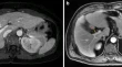
The utility of diffusion-weighted imaging in improving the sensitivity of LI-RADS classification of small hepatic observations suspected of malignancy Mohammad Abd Alkhalik BashaRania RefaatEman H. Abdelbary Hepatobiliary 02 January 2019 Pages: 1773 - 1784
Prediction of liver remnant regeneration after living donor liver transplantation using preoperative CT texture analysis Ji-Eun KimJung Hoon KimJoon Koo Han Hepatobiliary 05 January 2019 Pages: 1785 - 1794
Imaging post-stereotactic body radiation therapy responses for hepatocellular carcinoma: typical imaging patterns and pitfalls Katerina MastrocostasHyun-Jung JangTae Kyoung Kim Review 01 February 2019 Pages: 1795 - 1807
Intra-individual comparison of conventional and simultaneous multislice-accelerated diffusion-weighted imaging in upper abdominal solid organs: value of ADC normalization using the spleen as a reference organ Weon JangJi Soo SongMun Young Paek Hepatobiliary 08 February 2019 Pages: 1808 - 1815
Diagnostic value of MR-based texture analysis for the assessment of hepatic fibrosis in patients with nonalcoholic fatty liver disease (NAFLD) Roberto CannellaAmir A. BorhaniAlessandro Furlan Hepatobiliary 20 February 2019 Pages: 1816 - 1824
MR elastography of liver at 3 Tesla: comparison of gradient-recalled echo (GRE) and spin-echo (SE) echo-planar imaging (EPI) sequences and agreement across stiffness measurements Chenyang ZhanStephan KannengiesserKrishna Prasad Shanbhogue Hepatobiliary 22 February 2019 Pages: 1825 - 1833
3D MR elastography of the pancreas in children Suraj D. SeraiMaisam Abu-El-HaijaAndrew T. Trout Pancreas 25 January 2019 Pages: 1834 - 1840
Usefulness of rapid kV-switching dual energy CT in renal tumor characterization İlkay ÇamlıdağMehmet Selim NuralEnder Özden Kidneys, Ureters, Bladder, Retroperitoneum 14 January 2019 Pages: 1841 - 1849
Quantitative contrast-enhanced ultrasound of renal perfusion: a technology for the assessment of early diabetic nephropathy in cynomolgus macaques with type 2 diabetes mellitus Jieli LuoJianshe ChenPintong Huang Kidneys, Ureters, Bladder, Retroperitoneum 29 January 2019 Pages: 1850 - 1857
Ultrasound versus computed tomography for the detection of ureteral calculi in the pediatric population: a clinical effectiveness study Nathaniel P. RobersonJonathan R. DillmanAndrew T. Trout Kidneys, Ureters, Bladder, Retroperitoneum 11 February 2019 Pages: 1858 - 1866
Diffusion tensor imaging of the kidney in healthy controls and in children and young adults with autosomal recessive polycystic kidney disease Suraj D. SeraiHansel J. OteroErum A. Hartung Kidneys, Ureters, Bladder, Retroperitoneum 19 February 2019 Pages: 1867 - 1872
Prenatal planning of placenta previa: diagnostic accuracy of a novel MRI-based prediction model for placenta accreta spectrum (PAS) and clinical outcome Andrea Delli PizziAlessandra TavolettaRaffaella Basilico Pelvis 01 January 2019 Pages: 1873 - 1882
Prostate cancer detection with biparametric magnetic resonance imaging (bpMRI) by readers with different experience: performance and comparison with multiparametric (mpMRI) Marco GattiRiccardo FalettiPaolo Fonio Pelvis 20 February 2019 Pages: 1883 - 1893
Transnasal stent-assisted targeting technique for percutaneous jejunostomy placement in patients with hiatal hernias Jeffrey Forris Beecham ChickNeil JairathRavi N. Srinivasa Interventional Radiology 12 February 2019 Pages: 1894 - 1900
Complication rates of percutaneous biliary drainage in the presence of ascites Viren PatelShaun W. McLaughlinGregory Nadolski Interventional Radiology 06 February 2019 Pages: 1901 - 1906
Quantification of fat and skeletal muscle tissue at abdominal computed tomography: associations between single-slice measurements and total compartment volumes Anton FaronJulian A. LuetkensAlois M. Sprinkart Practice 29 January 2019 Pages: 1907 - 1916
Frequency and imaging features of abdominal immune-related adverse events in metastatic lung cancer patients treated with PD-1 inhibitor Francesco AlessandrinoSonia SahuMark M. Awad Practice 21 February 2019 Pages: 1917 - 1927
Effect of low tube voltage and low iodine concentration abdominal CT on image quality and radiation dose in children: preliminary study Sun Kyoung YouYoung Hun ChoiHyun-Hae Cho Hepatobiliary 25 January 2019 Pages: 1928 - 1935
The “enlarged hilar periportal space sign” in liver cirrhosis Giuseppe MamoneRoberto Miraglia Classics in Abdominal Radiology 01 January 2019 Pages: 1936 - 1937
Playboy bunny sign Subramaniyan RamanathanAdnan Sheikh Classics in Abdominal Radiology 01 January 2019 Pages: 1938 - 1939
The “maiden waist” sign of the ureters Janardhana PonnatapuraRaymond B. Dyer Classics in Abdominal Radiology 02 January 2019 Pages: 1940 - 1941
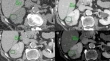
The “central stellate scar” sign in renal oncocytoma Dario GiambellucaSilvia PellegrinoGiuseppe Salvaggio Classics in Abdominal Radiology 29 January 2019 Pages: 1942 - 1943
The “sigmoid” esophagus Bradley UnruhJohn BillingsRaymond B. Dyer Classics in Abdominal Radiology 29 January 2019 Pages: 1944 - 1945
“Giant” hydronephrosis Bhavana BudigiRaymond B. Dyer Classics in Abdominal Radiology 16 April 2019 Pages: 1946 - 1948
The “spoke wheel” sign in mesenteric carcinoid Dario GiambellucaRoberto CannellaGiuseppe Salvaggio Classics in Abdominal Radiology 30 January 2019 Pages: 1949 - 1950
The “phantom” calyx Janardhana PonnatapuraRaymond B. Dyer Classics in Abdominal Radiology 06 February 2019 Pages: 1951 - 1952
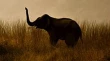
The “trumpeting elephant” sign Joseph WilsonRaymond B. Dyer Classics in Abdominal Radiology 06 February 2019 Pages: 1953 - 1954
Sentinel clot sign in hemoperitoneum Mohd IlyasMuiez BashirImran Hamid Classics in Abdominal Radiology 06 February 2019 Pages: 1955 - 1956
Response to Dr. Kamanahalli’s Letter to the Editor Mitchell A. Klein Letter to the Editor 29 April 2019 Pages: 1957 - 1958