Abstract
Background
Glioblastoma multiforme (GBM) is a highly aggressive primary malignant brain tumor characterized by rapid progression, poor prognosis, and high mortality rates. Understanding the relationship between cerebrospinal fluid (CSF) metabolites and GBM is crucial for identifying potential biomarkers and pathways involved in the pathogenesis of this devastating disease.
Methods
In this study, Mendelian randomization (MR) analysis was employed to investigate the causal relationship between 338 CSF metabolites and GBM. The data for metabolites were obtained from a genome-wide association study summary dataset based on 291 individuals, and the GBM data was derived from FinnGen included 91 cases and 174,006 controls of European descent. The Inverse Variance Weighted method was utilized to estimate the causal effects. Supplementary comprehensive assessments of causal effects between CSF metabolites and GBM were conducted using MR-Egger regression, Weighted Median, Simple Mode, and Weighted Mode methods. Additionally, tests for heterogeneity and pleiotropy were performed.
Results
Through MR analysis, a total of 12 identified metabolites and 2 with unknown chemical properties were found to have a causal relationship with GBM. 1-palmitoyl-2-stearoyl-gpc (16:0/18:0), 7-alpha-hydroxy-3-oxo-4-cholestenoate, Alpha-tocopherol, Behenoyl sphingomyelin (d18:1/22:0), Cysteinylglycine, Maleate, Uracil, Valine, X-12,101, X-12,104 and Butyrate (4:0) are associated with an increased risk of GBM. N1-methylinosine, Stachydrine and Succinylcarnitine (c4-dc) are associated with decreased GBM risk.
Conclusion
In conclusion, this study sheds light on the intricate interplay between CSF metabolites and GBM, offering novel perspectives on disease mechanisms and potential treatment avenues. By elucidating the role of CSF metabolites in GBM pathogenesis, this research contributes to the advancement of diagnostic capabilities and targeted therapeutic interventions for this aggressive brain tumor. Further exploration of these findings may lead to improved management strategies and better outcomes for patients with GBM.
Introduction
Glioblastoma multiforme (GBM) is one of the most aggressive primary malignant brain tumors, characterized by rapid progression, poor prognosis, and high mortality rates [1]. According to the 2016 WHO classification, gliomas are divided into four grades: low-grade gliomas (LGG grades I and II) and high-grade gliomas (HGG grades III and IV) [2]. GBM, the most malignant subtype, accounts for over 50% of all gliomas and more than 15% of primary brain tumors [3]. Due to its immunosuppressive microenvironment and tendency for recurrence, GBM stands out as one of the most challenging tumors, imposing a significant societal burden.
Cerebrospinal fluid (CSF) envelops the cerebral and spinal regions within the meningeal cavities, playing a crucial role in maintaining brain homeostasis, providing nutrients, and clearing waste. Thanks to the presence of the blood-brain barrier, CSF maintains its distinctive environment, safeguarding the normal functioning of neurons. Due to their direct exposure to GBM, CSF metabolites hold the potential to mirror metabolic alterations linked to tumor initiation, presence, and progression [4]. GBM, as a highly invasive brain tumor, can disrupt the normal flow and composition of CSF, leading to increased intracranial pressure and hydrocephalus. Additionally, GBM cells can infiltrate the soft meninges and spread along the CSF pathways [5]. Therefore, CSF analysis, including cytology and molecular spectrum analysis, is pivotal for diagnosing and monitoring GBM, as it may contain tumor cells and biomarkers reflecting the disease state. Understanding the dynamic interplay between GBM and CSF is crucial for enhancing diagnostic and therapeutic strategies against this devastating disease.
Studying CSF presents both challenges and valuable opportunities in understanding various neurological disorders. One of the primary hurdles lies in obtaining CSF samples, typically acquired through invasive procedures like lumbar punctures, posing risks and discomfort to patients. Consequently, most human metabolomics research has focused on more accessible sample types such as blood or urine. Furthermore, CSF composition is dynamic, influenced by factors like age, gender, and disease status, making standardization and interpretation complex [6]. Despite these challenges, researching CSF holds immense value. CSF serves as a direct window into the central nervous system, reflecting biochemical, cellular, and molecular changes associated with neurological disorders like Alzheimer’s disease, multiple sclerosis, and brain tumors [7,8,9]. Analysis of CSF biomarkers provides diagnostic, prognostic, and therapeutic insights, aiding in disease detection, monitoring, and treatment response assessment. Rogachev et al. found that there were partial differences in the correlation of plasma and CSF metabolites between HGG and healthy control groups [4]. Ji et al. demonstrated that CSF metabolites could be used to predict glioma grade and leptomeningeal metastasis [10]. So, by overcoming technical barriers and leveraging the rich information provided by CSF, researchers can significantly enhance our understanding and management of neurological diseases, ultimately improving patient outcomes.
In this study, we employed Mendelian randomization (MR) analysis to investigate the causal relationship between CSF metabolites and GBM. MR is a robust epidemiological method that utilizes single nucleotide polymorphisms (SNPs) as instrumental variables (IVs), less susceptible to unmeasured confounders and reverse causality [11]. This study aims to elucidate potential disease mechanisms, identify novel therapeutic avenues, and provide insights for the diagnosis, treatment, and prevention of GBM.
Materials and methods
Study design
Our study provides an overview of two sample MR surveys used to explore the causal relationship between CSF metabolites and GBM. In MR studies, adherence to three fundamental instrumental variable (IV) assumptions is crucial: (1) genetic variants must correlate with the exposure; (2) these variants should be independent of confounding factors; and (3) they should exclusively impact the outcome through the exposure [11]. Strict quality control measures, including tests for multicollinearity and heterogeneity, were conducted to enhance the reliability of causal results.
CSF metabolites data synopsis
The exposure factor we studied is 338 CSF metabolites, based on a metabolome-wide association study by Panyard et al. [12]. In this study, a total of 291 independent samples of individuals of European descent were retained after quality control and further data cleaning steps. All participants were in a state of cognitive health when CSF was extracted. Among the 338 metabolites, 38 are still chemically undefined, whereas 296 have undergone chemical validation and are categorized into eight primary metabolic groups, encompassing amino acid, carbohydrate, cofactor and vitamin, energy, lipid, nucleotide, peptide, and xenobiotic metabolism.
GBM data synopsis
The outcome variable under scrutiny is GBM, for which the genome-wide association study (GWAS) summary dataset was sourced from FinnGen included 91 cases and 174,006 controls of European descent and comprised 16,380,303 SNPs.
Selection of IVs
SNPs are commonly used genetic variations in MR Analysis, reflecting DNA sequence diversity resulting from a single nucleotide change at the genomic level. In our study, we meticulously constructed a unique set of IVs for each of the 338 CSF metabolites. This approach was taken to ensure that the genetic associations specific to each metabolite were accurately represented. Firstly, considering the limited number of SNPs available to achieve genome-wide significance for metabolites, we relaxed the threshold by testing with a p-value set to less than 1 × 10^−5 [13]. Then, linkage disequilibrium criteria were set at r2 < 0.001 and a genetic distance of 10,000 kb, with highly correlated SNPs excluded to ensure the independence of included SNPs. Furthermore, to mitigate bias resulting from weak instrumentality, the F-value of each SNP was computed, and SNPs with an F-value < 10 were identified as weak instruments [14]. F = R2(N − 2)/(1 − R2), in which R2 and N denotes the proportion of variance explained by the chosen SNPs and the sample size of the GWAS for the SNP respectively. What’s more, we utilized the IEU OpenGWAS database to meticulously identify and exclude any SNPs that showed significant associations with potential confounders. Finally, project the SNPs related to CSF metabolites onto the GWAS merged data of GBM and extract the corresponding statistical parameters.
Statistical analysis
In this study, we employed the Inverse Variance Weighted (IVW), MR-Egger, and Weighted Median, Weighted mode, and Simple mode methods to estimate dependent effects. Given the IVW method’s superior test efficacy compared to other MR methods, we selected it as the primary method. The IVW method assumes the validity of all genetic variants as instrumental variables. It calculates causal effect values for individual instrumental variables using the ratio method, then combines each estimate through weighted linear regression to derive the total effect value. Nonetheless, there might be unidentified confounding variables leading to genetic pleiotropy and bias in effect size estimation. Therefore, MR-Egger regression and weighted median methods were employed as supplementary approaches to corroborate the causal impact of exposure on the outcome. The “leave-one-out” method was employed to identify any instrumental variables that could influence the estimation of causal effects. Horizontal pleiotropy was assessed using the Egger intercept, and the Mendelian randomization pleiotropy residual sum and outlier (MR-PRESSO) test was performed to identify and exclude SNPs that may be influenced by pleiotropy. Heterogeneity tests were conducted using the Cochran Q test to evaluate the diversity of the SNPs. All analyses were performed using the TwoSampleMR R packages in R software 4.4.1.
Results
After investigating the quality control of IVs, we identified 14 CSF metabolites associated with GBM, including 21–196 SNPs (with Stachydrine genetically represented by 21 SNPs and Butyrate (4:0) having the highest representation with 196 SNPs). The F-statistic data range for CSF metabolites is from 19.37 to 78.49, all exceeding a threshold greater than 10, indicating they are less likely to be influenced by instrument biases (Supplementary Table S1). Among the 14 CSF metabolites associated with GBM, there are 12 identified metabolites and 2 with unknown chemical properties. The identified metabolites chemically classify into lipids, vitamins, amino acids, nucleotides, energy and xenobiotics (Supplementary Table S2).
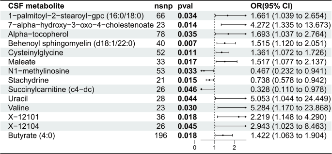
The causal relationship between the risk of GBM and 14 CSF metabolites is as follows (Fig. 1): 1-palmitoyl-2-stearoyl-gpc (16:0/18:0) (OR 1.661, 95% CI: 1.039–2.654, p = 0.034), 7-alpha-hydroxy-3-oxo-4-cholestenoate (7-HOCA) (OR 4.272, 95% CI: 1.335–13.673, p = 0.014), Alpha-tocopherol (12:0) (OR 1.693, 95% CI: 1.037–2.764, p = 0.035), Behenoyl sphingomyelin (d18:1/22:0) (OR 1.515, 95% CI: 1.120–2.051, p = 0.007), Cysteinylglycine (OR 1.361, 95% CI: 1.072–1.726, p = 0.011), Maleate (OR 1.517, 95% CI: 1.077–2.137, p = 0.017), N1-methylinosine (OR 0.467, 95% CI: 0.232–0.941, p = 0.033), Stachydrine (OR 0.738, 95% CI: 0.578–0.942, p = 0.015), Succinylcarnitine (c4-dc) (OR 0.328, 95% CI: 0.110–0.978, p = 0.046), Uracil (OR 5.053, 95% CI: 1.044–24.449, p = 0.044), Valine (OR 5.284, 95% CI: 1.170–23.868, p = 0.030), X-12,101 (OR 2.219, 95% CI: 1.148–4.290, p = 0.018), X-12,104 (OR 2.943, 95% CI: 1.023–8.463, p = 0.045), Butyrate (4:0) (OR 1.422, 95% CI: 1.063–1.904, p = 0.018).
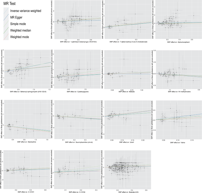
In brief, the IVW-derived estimates demonstrated significance (p < 0.05), with a consistent direction and magnitude observed across IVW, MR-Egger, Weighted mode, Weighted median and Simple mode estimates (Supplementary Table S3). The conclusions of the other four methods remain largely consistent with those of the IWV method. Both the MR-Egger intercept test and the Cochran Q test strongly support the absence of pleiotropy and heterogeneity, except the Egger intercept of Behenoyl sphingomyelin (d18:1/22:0) showing pleiotropy (Table 1). Scatter plots for 14 identified CSF metabolites across various tests are displayed in Fig. 2. The leave-one-out analysis confirmed that excluding any single SNP did not introduce bias into the MR estimation (Supplementary Figure S1). The funnel plots are presented in Supplementary Figure S2.
Discussion
Our study delved into the intricate relationship between CSF metabolites and GBM, shedding light on potential biomarkers and pathways implicated in the pathogenesis of this aggressive brain tumor. By investigating the relationship between CSF metabolites and GBM risk through MR analysis, we identified 14 CSF metabolites significantly associated with the risk of GBM. These metabolites encompass a variety of biochemical classes, including lipids, vitamins, amino acids, nucleotides, energy and xenobiotics, underscoring the multifactorial nature of GBM development and progression. Understanding the dynamic interplay between CSF metabolites and GBM not only enhances our diagnostic capabilities but also holds promise for the development of targeted therapeutic interventions aimed at disrupting key metabolic pathways implicated in GBM tumorigenesis and progression.
Lipids, including sterols, phospholipids, and diacyl/triacylglycerols, are components of biofilms, which emerge as pivotal players in the landscape of GBM research. Lipids serve not only as structural components of cell membranes but also as signaling molecules involved in various cellular processes, including proliferation, migration, and apoptosis. The dysregulation of lipid metabolism has been implicated in tumor initiation, progression, and therapy resistance, making lipid metabolites attractive candidates for biomarker discovery and therapeutic targeting in GBM [15]. So far, there have been no literature reports on the roles of 1-palmitoyl-2-steroyl-gpc (16:0/18:0) and Behenoyl sphingomyelin (d18:1/22:0) in cancer. We speculate that these two phospholipid metabolites may be associated with enhanced lipid synthesis and metabolism typically exhibited by cancer cells. Not only do they provide the structural support required for cancer cell proliferation, but they also participate in regulating cell signaling pathways and epigenetic events, promoting the survival and metastasis ability of cancer cells. Halama et al. have discovered that in colon and ovarian cancer, endothelial cells induce metabolic reprogramming by promoting the overexpression of glycerophospholipids and polyunsaturated fatty acids in cancer cells [16].
In our study, we are pioneering the association between 7-HOCA and GBM for the first time. 7-HOCA is the primary metabolite of oxysterol 27-hydroxycholesterol in the brain, and the increase of 7-HOCA in CSF has been shown to reflect blood-brain barrier damage [17, 18]. Feng et al. found that 7-HOCA is closely related to the occurrence of lung cancer, showing a significantly increased risk [19]. Combining our research findings, 7-HOCA may play a significant role in the development of cancer. Butyrate (4:0) is the salt form of butyric acid, which stands as a significant byproduct in fatty acid metabolism. In vivo, butyric acid can be generated through various pathways, including its production from the metabolism of fatty acids or its fermentation from dietary fiber by intestinal microbiota. Once produced, butyric acid actively participates in the regulation and modulation of lipid metabolism, influencing the synthesis, breakdown, and utilization of fats in the body. Tumor cells can obtain fatty acids through lipolysis to support their growth. During periods of adequate nutritional supply, fatty acids are stored in adipose tissue as triglycerides (TG), which are broken down to release fatty acids when energy levels are low. Recent studies have revealed the significant role of butyrate salts in the occurrence and progression of colorectal and lung cancers [20]. However, this is the first report on the relationship between butyrate salts and GBM. We are also presenting, for the first time, the causal relationship between maleate in CSF and GBM. Maleate is a salt form of malic acid, present in the human body as a metabolite or dietary source. Further research is needed to understand its involvement in the onset and progression of GBM.
The importance of amino acid metabolism in GBM lies in its role as the building blocks of proteins. Amino acids not only provide essential materials for cell growth and proliferation but also participate in regulating cellular signaling, gene expression, and metabolic pathways. GBM cells typically exhibit high metabolic activity, leading to an increased demand for amino acids. Therefore, abnormalities in amino acid supply and metabolism may influence the development and progression of glioblastoma. In our study, valine and cysteinylglycine was found to have a positive relationship with GBM. Similar to our results, Yao et al. found that the valine, leucine, and isoleucine biosynthesis metabolic pathway can be used for screening and diagnosis of ovarian cancer [21]. Valine, leucine and isoleucine constitute the group of branched-chain amino acids (BCAAs), which can be utilized and processed by astrocytes to perform various functions, including serving as fuel materials for brain energy metabolism [22]. The diagnostic role of valine metabolites in cancer has also been found in saliva. In another study, valine was found to play a dominant role in diagnosing multiple cancers through saliva [23]. Cysteinylglycine, produced during the breakdown of glutathione, has been proposed as a prooxidant, contributing to oxidative stress and lipid peroxidation, which are implicated in the development of human cancers. Consistent with our results, Lin et al. found that the increase of cysteinylglycine level was associated with the increased risk of breast cancer in the high oxidative stress group of middle-aged and elderly women [24]. However, there are also studies suggesting the opposite perspective. For instance, Miranti et al. found a negative correlation between serum cysteinylglycine and esophageal adenocarcinoma [25]. For understanding and intervening in the metabolic characteristics and therapeutic mechanisms of GBM, in-depth investigation into the role of amino acid metabolism is crucial.
As the fundamental substances that ensure the structure of DNA, the metabolism of nucleotides is closely intertwined with the initiation and progression of tumors. As shown in this study, uracil may act as a metabolite and participant in the onset and progression of tumors. UPP1, as a critical enzyme in uracil metabolism, catalyzes the dephosphorylation of uridine to uracil and ribose-1-phosphate. Its activation of the AKT signaling pathway can enhance tumorigenesis and resistance to anticancer drugs [26]. N1-methylinosine is a known modification of ribonucleosides, formed by adding a methyl group to the N1 position of the purine ring of a nucleotide. This methylation modification typically occurs within RNA molecules. In biological organisms, methylation modifications of ribonucleosides are common epigenetic regulatory mechanisms that can influence RNA stability, translation, and interactions. Studies by Li et al. detected N1-methylinosine in urine samples from cancer patients, suggesting its potential as a biomarker for cancer [27]. The presence of N1-methylinosine may indicate abnormalities in ribonucleoside metabolism within cells, which could be associated with the development and progression of cancer.
In our study, except N1-methylinosine, another two metabolites that showed a negative causal relationship with GBM were Stachydrine and Succinylcarnitine (c4-dc). Stachydrine is renowned for its antioxidant and anti-inflammatory properties [28]. Consistent with our results, some studies have demonstrated that Stachydrine can inhibit the proliferation of breast cancer and liver cancer cells, while inducing apoptosis, autophagy, and cellular senescence. Its mechanisms of action may include inhibiting survival signaling pathways such as Akt and ERK, as well as regulating the expression of cell cycle proteins [29, 30]. Liu et al. found that Stachydrine may prevent liver damage by attenuating inflammatory responses, inhibiting the ERK and AKT pathways, and suppressing the expression of macrophage stimulating protein [31]. Succinylcarnitine is an intermediate in the tricarboxylic acid (TCA) cycle and amino acid-based energy metabolism, responsible for fatty acid β-oxidation and mitochondrial function, serving as an energy-related metabolic product [32]. An MR study identified maternal succinylcarnitine during pregnancy as a significant factor contributing to the risk of congenital heart disease in offspring [33]. Kim et al. found that the decrease in plasma succinylcarnitine levels was associated with the inhibition of prostate tumor growth by a whole walnut diet, possibly achieved through influencing cellular energy metabolism status and regulating relevant gene expression [34]. However, further research is needed to elucidate the specific mechanisms of these two CSF metabolites.
Several limitations exist within our study. the Pleiotropy test result of Behenoyl sphingomyelin (d18:1/22:0) was less than 0.05, indicating the presence of pleiotropy. However, further SNP screening was performed using MR-PRESSO, but the results showed no SNPs that needed to be removed. There are many possible reasons for this result, such as sample size, insufficient statistical power, complexity of pleiotropy, and methodological limitations. Therefore, it is emphasized that readers need to be cautious when reviewing the results, which does not mean that there is no pleiotropy, but may be due to the influence of the above factors. Additionally, our study may have another important limitation, which is the lack of research on the potential correlation of metabolites at SNP and phenotype levels. This oversight may have implications for the interpretation of our findings, as unaccounted correlations could influence the results of the Mendelian Randomization analysis.
These findings from our study open several avenues for future research. It is essential to delve into the mechanistic roles of the identified CSF metabolites in GBM biology, exploring their interactions with cellular pathways and the tumor microenvironment. Additionally, longitudinal studies are needed to understand the dynamics of these metabolites over the course of the disease and in response to treatments. Investigating the generalizability of our findings across diverse populations and the integration of metabolomics data with other omics platforms will be critical for identifying robust biomarker panels. Furthermore, the potential of these metabolites as therapeutic targets should be rigorously tested in preclinical models to advance towards novel treatment strategies for GBM.
Conclusion
In conclusion, our study provides valuable insights into the intricate relationship between CSF metabolites and GBM, shedding light on potential biomarkers and pathways involved in the pathogenesis of this aggressive brain tumor. Through MR analysis, we identified 14 CSF metabolites significantly associated with GBM risk, spanning various biochemical classes such as lipids, vitamins, amino acids, nucleotides, energy, and xenobiotics. This underscores the multifactorial nature of GBM development and progression. Our findings highlight the importance of understanding the dynamic interplay between CSF metabolites and GBM, not only for enhancing diagnostic capabilities but also for potentially developing targeted therapeutic interventions aimed at disrupting key metabolic pathways implicated in GBM tumorigenesis and progression.
Data availability
The original contributions presented in the study are included in the article/supplementary material, further inquiries can be directed to the corresponding author.
Abbreviations
- 7-HOCA:
-
7-alpha-hydroxy-3-oxo-4-cholestenoate
- CSF:
-
Cerebrospinal fluid
- GBM:
-
Glioblastoma multiforme
- GWAS:
-
Genome-wide association study
- IVs:
-
Instrumental variables
- MR:
-
Mendelian randomization
- SNPs:
-
Single nucleotide polymorphisms
References
Ilkhanizadeh S, et al. Glial progenitors as targets for transformation in glioma. Adv Cancer Res. 2014;121:1–65.
Louis DN, et al. The 2016 World Health Organization Classification of Tumors of the Central Nervous System: a summary. Acta Neuropathol. 2016;131(6):803–20.
Topalian SL, et al. Safety, activity, and immune correlates of anti-PD-1 antibody in cancer. N Engl J Med. 2012;366(26):2443–54.
Rogachev AD et al. Correlation of metabolic profiles of plasma and Cerebrospinal Fluid of High-Grade Glioma patients. Metabolites, 2021. 11(3).
Lah TT, Novak M, Breznik B. Brain malignancies: Glioblastoma and brain metastases. Semin Cancer Biol. 2020;60:262–73.
Tashjian RS, Vinters HV, Yong WH. Biobanking of Cerebrospinal Fluid. Methods Mol Biol. 2019;1897:107–14.
Simrén J, et al. Fluid biomarkers in Alzheimer’s disease. Adv Clin Chem. 2023;112:249–81.
Deisenhammer F, et al. The cerebrospinal fluid in multiple sclerosis. Front Immunol. 2019;10:726.
Kopková A, et al. MicroRNAs in cerebrospinal fluid as biomarkers in Brain Tumor patients. Klin Onkol. 2019;32(3):181–6.
Im JH, et al. Comparative cerebrospinal fluid metabolites profiling in glioma patients to predict malignant transformation and leptomeningeal metastasis with a potential for preventive personalized medicine. Epma j. 2020;11(3):469–84.
Sekula P, et al. Mendelian randomization as an Approach to assess causality using Observational Data. J Am Soc Nephrol. 2016;27(11):3253–65.
Panyard DJ, et al. Cerebrospinal fluid metabolomics identifies 19 brain-related phenotype associations. Commun Biol. 2021;4(1):63.
Yang J, et al. Assessing the Causal effects of human serum metabolites on 5 Major Psychiatric disorders. Schizophr Bull. 2020;46(4):804–13.
Flatby HM, et al. Circulating levels of micronutrients and risk of infections: a mendelian randomization study. BMC Med. 2023;21(1):84.
Maan M, et al. Lipid metabolism and lipophagy in cancer. Biochem Biophys Res Commun. 2018;504(3):582–9.
Halama A, et al. Nesting of colon and ovarian cancer cells in the endothelial niche is associated with alterations in glycan and lipid metabolism. Sci Rep. 2017;7:39999.
Heverin M, et al. Crossing the barrier: net flux of 27-hydroxycholesterol into the human brain. J Lipid Res. 2005;46(5):1047–52.
Saeed A, et al. 7α-hydroxy-3-oxo-4-cholestenoic acid in cerebrospinal fluid reflects the integrity of the blood-brain barrier. J Lipid Res. 2014;55(2):313–8.
Feng Y, et al. Causal effects of genetically determined metabolites on cancers included lung, breast, ovarian cancer, and glioma: a mendelian randomization study. Transl Lung Cancer Res. 2022;11(7):1302–14.
Karim MR, et al. Butyrate’s (a short-chain fatty acid) microbial synthesis, absorption, and preventive roles against colorectal and lung cancer. Arch Microbiol. 2024;206(4):137.
Yao JZ et al. Diagnostics Ovarian cancer via Metabolite Anal Mach Learn Integr Biol (Camb), 2023. 15.
Murín R, et al. Glial metabolism of valine. Neurochem Res. 2009;34(7):1195–203.
Bel’skaya LV, Sarf EA, Loginova AI. Diagn Value Salivary Amino Acid Levels Cancer Metabolites, 2023. 13(8).
Lin J, et al. Plasma cysteinylglycine levels and breast cancer risk in women. Cancer Res. 2007;67(23):11123–7.
Miranti EH, et al. Prospective study of serum cysteine and cysteinylglycine and cancer of the head and neck, esophagus, and stomach in a cohort of male smokers. Am J Clin Nutr. 2016;104(3):686–93.
Du W, et al. UPP1 enhances bladder cancer progression and gemcitabine resistance through AKT. Int J Biol Sci. 2024;20(4):1389–409.
Li HY, et al. Separation and identification of purine nucleosides in the urine of patients with malignant cancer by reverse phase liquid chromatography/electrospray tandem mass spectrometry. J Mass Spectrom. 2009;44(5):641–51.
Zhao L, et al. Stachydrine ameliorates isoproterenol-induced cardiac hypertrophy and fibrosis by suppressing inflammation and oxidative stress through inhibiting NF-κB and JAK/STAT signaling pathways in rats. Int Immunopharmacol. 2017;48:102–9.
Bao X, et al. Stachydrine hydrochloride inhibits hepatocellular carcinoma progression via LIF/AMPK axis. Phytomedicine. 2022;100:154066.
Wang M, et al. Stachydrine hydrochloride inhibits proliferation and induces apoptosis of breast cancer cells via inhibition of akt and ERK pathways. Am J Transl Res. 2017;9(4):1834–44.
Liu FC et al. The modulation of Phospho-Extracellular Signal-regulated kinase and phospho-protein kinase B Signaling pathways plus Activity of Macrophage-stimulating protein contribute to the Protective Effect of Stachydrine on Acetaminophen-Induced Liver Injury. Int J Mol Sci, 2024. 25(3).
Mai M, et al. Serum levels of acylcarnitines are altered in prediabetic conditions. PLoS ONE. 2013;8(12):e82459.
Taylor K et al. The relationship of maternal gestational Mass spectrometry-derived metabolites with offspring congenital heart disease: results from multivariable and mendelian randomization analyses. J Cardiovasc Dev Dis, 2022. 9(8).
Kim H, Yokoyama W, Davis PA. TRAMP prostate tumor growth is slowed by walnut diets through altered IGF-1 levels, energy pathways, and cholesterol metabolism. J Med Food. 2014;17(12):1281–6.
Acknowledgements
The authors thank all the participants and researchers who contributed and collected data.
Funding
This research was supported by Research Foundation for Talented Scholars of Xuzhou Medical University (D2021011).
Author information
Authors and Affiliations
Contributions
HjB: Conceptualization, Data curation, Formal analysis, Investigation, Methodology, Projection administration; Software, Validation, Writing – original draft. YyC: Formal analysis, Investigation, Writing – original draft. ZjM: Conceptualization, Data curation, Investigation, Writing – review & editing. ZC: Conceptualization, Data curation, Formal analysis, Funding acquisition, Investigation, Methodology, Projection administration; Software, Validation, Writing – review & editing.
Corresponding author
Ethics declarations
Ethical approval
All data analyzed in this study were sourced from publicly available databases. Ethical approval was secured for each cohort, and informed consent was obtained from all participants prior to their involvement. Written informed consent was provided by the patients/participants for their participation in this study.
Consent for publication
Not Applicable.
Conflict of interest
The authors declare no competing interests.
Additional information
Publisher’s note
Springer Nature remains neutral with regard to jurisdictional claims in published maps and institutional affiliations.
Electronic supplementary material
Below is the link to the electronic supplementary material.
Rights and permissions
Open Access This article is licensed under a Creative Commons Attribution-NonCommercial-NoDerivatives 4.0 International License, which permits any non-commercial use, sharing, distribution and reproduction in any medium or format, as long as you give appropriate credit to the original author(s) and the source, provide a link to the Creative Commons licence, and indicate if you modified the licensed material. You do not have permission under this licence to share adapted material derived from this article or parts of it. The images or other third party material in this article are included in the article’s Creative Commons licence, unless indicated otherwise in a credit line to the material. If material is not included in the article’s Creative Commons licence and your intended use is not permitted by statutory regulation or exceeds the permitted use, you will need to obtain permission directly from the copyright holder. To view a copy of this licence, visit http://creativecommons.org/licenses/by-nc-nd/4.0/.
About this article
Cite this article
Bao, H., Chen, Y., Meng, Z. et al. The causal relationship between CSF metabolites and GBM: a two-sample mendelian randomization analysis. BMC Cancer 24, 1119 (2024). https://doi.org/10.1186/s12885-024-12901-7
Received:
Accepted:
Published:
DOI: https://doi.org/10.1186/s12885-024-12901-7


