
Avoid common mistakes on your manuscript.
Correction to: Cellular and Molecular Life Sciences https://doi.org/10.1007/s00018-019-03236-9
In the published article, few errors were noticed and this has been corrected with this erratum publication.
-
1.
In page 3, the GRP57 in lines 1 and 4 (right column) should be GRP75.
-
2.
In page 7, line 13 (right column), the text under section heading C-terminal of EI24 is essential for autophagy flux currently is read as follows:
Considering the possibility that phagophore formation begins at the MAM site, we studied the correlation of MAM number with autophagy flux. Results showed that overexpression of EI24, but not EI24-ΔC, decreased the level of p62 and LC3II (Fig. 7a, b). The average number of p62 and LC3 punctate per cells by immunostaining was also significantly decreased in the EI24 overexpression group, but not in the EI24-ΔC group (Fig. 7c, d), further indicating that the C-terminal of EI24 is essential for autophagy flux.
It should be read as
Considering the possibility that phagophore formation begins at the MAM site, we studied the correlation of MAM number with autophagy flux. We first examined whether artificially formed MAM by tethering ER and the mitochondrial could rescue autophagy flux in EI24 KO group. Since SpyCatcher can form an irreversible covalent bond to SpyTag, SpyCatcher-mApple-Sec61β and Tom20-EGFP-Spytag were co-overexpressed in EI24 KO group. We found that the ER and mitochondrial were colocalized well indicating the MAM were formed (Fig. 7a). Compared to EI24 KO group, a decreased level of p62 and an increased level of LC3II were observed in co-overexpressed EI24 KO group (Fig. 7b, upper panel), which might indicate that the autophagy flux was rescued. However, considering that this may be due to the plasmid overexpression, the SpyCatcher-mApple-Sec61β and Tom20-EGFP-Spytag were transfected individually as control groups or co-overexpressed as experiment group in EI24 KO cells, respectively. Results showed that the levels of p62 and LC3II were comparable between the control groups and the co-overexpression group (Fig. 7b, lower panel). These results indicated that artificially tethering ER and the mitochondrial cannot induce and rescue the autophagy flux in EI24 KO cells, which further demonstrated that EI24 took essential roles in the autophagy flux. Next, we examined which domain of EI24 is essential for autophagy flux. Results showed that overexpression of EI24, but not EI24-ΔC, decreased the level of p62 and LC3II (Fig. 7c, d). The average number of p62 and LC3 punctate per cell by immunostaining was also significantly decreased in the EI24 overexpression group, but not in the EI24-ΔC group (Fig. 7e, f), further indicating that the C-terminal of EI24 is essential for autophagy flux.
-
3.
Figure 7 and its legend have been updated.
The new Fig. 7 is as follows:
Fig. 7 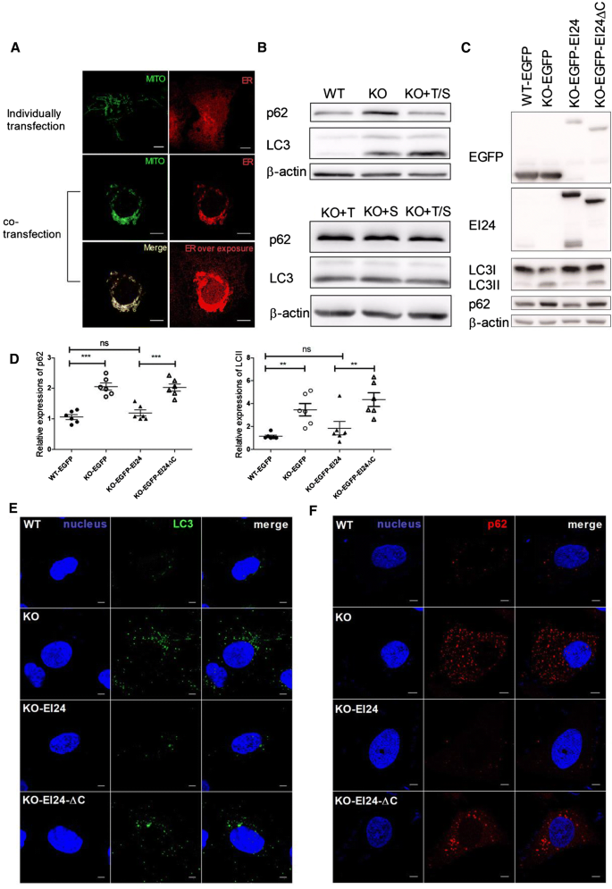
C-terminal of EI24 is essential for autophagy flux. a Images of U-2 OS cells transfected with SpyCatcher-mApple-Sec61β or Tom20-EGFP-Spytag individually or co-overexpression, respectively. Green: Tom20, red: Sec61β, yellow: merged image. b Expression levels of p62 and LC3 in WT and EI24-KO U-2 OS cells transfected with Tom20 (T), Sec61β (S), or co-overexpressed Tom20/Sec61β (T/S). c U-2 OS cells, stable transfected with EGFP, EGFP-EI24 or EGFP-EI24ΔC, were applied using Western blot analysis with the indicated antibodies. d Quantification analysis of p62 and LC3II, normalized to β-actin. e Immunostaining of nucleus (blue) and LC3 (green) in U-2 OS cells of WT, EI24-KO, EI24-KO stably expressing mApple-EI24 (KO-EI24) or mApple-EI24ΔC (KO-EI24-ΔC). Bar = 50 μm. f Immunostaining of nucleus (blue) and p62 (red) in U-2 OS cells of WT, EI24-KO, EI24-KO stably expressing EGFP-EI24 (KO-EI24) or EGFP-EI24ΔC (KO-EI24-ΔC). Bar = 50 μm

Author information
Authors and Affiliations
Corresponding author
Rights and permissions
About this article

Cite this article
Yuan, L., Liu, Q., Wang, Z. et al. Correction to: EI24 tethers endoplasmic reticulum and mitochondria to regulate autophagy flux. Cell. Mol. Life Sci. 77, 2255–2256 (2020). https://doi.org/10.1007/s00018-019-03355-3
Published:
Issue Date:
DOI: https://doi.org/10.1007/s00018-019-03355-3