Abstract
The use of antisense oligonucleotides (AONs) is a promising therapeutic strategy for central nervous system disorders. However, the delivery of AONs to the central nervous system is challenging because their size does not allow them to diffuse over the blood–brain barrier (BBB) when injected systemically. The BBB can be bypassed by administering directly into the brain. Here we describe a method to perform single and repeated intracerebroventricular injections into the lateral ventricle of the mouse brain.
You have full access to this open access chapter, Download protocol PDF
Key words
1 Introduction
There has been a recent revival of interest in the use of antisense oligonucleotides (AONs) to treat neurodegenerative disorders with one approved central nervous system AON therapy and several in clinical trials [1]. This is largely due to the remarkably widespread distribution and cellular uptake of AONs once delivered into the brain . However, for drugs to reach the nervous system, they first have to cross the blood–brain barrier (BBB). Since the molecular weight of AONs is approximately 6000–10,000 Da, they are too large to cross the BBB by simple diffusion when delivered systemically. During the study of therapeutic efficacy of AONs in mouse models, AONs are often infused intracerebroventricularly (ICV). The BBB is bypassed by injecting the AON directly into the lateral ventricle, after which the AONs pass the ependymal cell layer that lines the ventricular system and enters the brain parenchyma.
One group of disorders for which these AON therapies are being studied in mouse models are the polyglutamine disorders. In these disorders, the disease is caused by CAG triplet repeat expansions in the coding region of a gene that is subsequently translated into an expanded stretch of glutamine amino acids in the protein. The disease-causing proteins for each of these polyglutamine (polyQ) disorders are different, but in each case, the expanded stretch of glutamines results in a toxic gain of function of the protein leading to neurodegeneration. To date, a total of nine polyQ disorders have been described: dentatorubral-pallidoluysian atrophy (DRPLA), Huntington’s disease (HD), spinal bulbar muscular atrophy (SBMA), and spinocerebellar ataxias (SCA1, 2, 3, 6, 7, and 17) [2, 3]. There is an inverse correlation of disease onset and polyQ length in the protein; the longer the CAG repeat, the earlier the age of onset of the disease [2]. Protein aggregates are found in the nucleus and cytoplasm of cells, indicating that protein misfolding is a common feature of these disorders. Using RNA breakdown [4] or RNA splice modulating AONs [5, 6], it is possible to reduce the toxicity of the mutant proteins, which can hopefully halt the disease progression in patients.
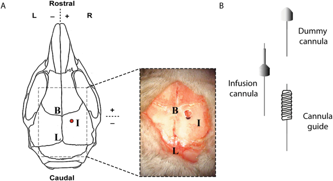
Because of the limited volume that can be injected into the mouse brain , multiple injections can be required to reach the desired dose. Repeated injections of AONs lead to a widespread distribution throughout the entire mouse brain , and the protein modifying effect can be detected in the cortex, cerebellum, and brainstem [6]. In this chapter, we describe a method for ICV delivery of AONs through a single injection or repeated injections using a cannula (Fig. 1).
Location and tools for intracerebroventricular injection. (a) Schematic and photographic outline of the injection site (I), bregma (B), and lambda (L) as points of reference. (b) Cannula types required for intracerebroventricular injection. The infusion cannula is connected to the syringe tubing and guided through the cannula guide to inject into the lateral ventricle. The cannula guide is attached to the skull and can be closed using a dummy cannula
2 Materials
2.1 Mice
(See Note 1).
2.2 Disposables
-
1.
Infusion cannula (PlasticsOne C315IS-5/SPC).
-
2.
Tubing (PlasticsOne C313CT).
-
3.
Cannula guide (PlasticsOne C315GS-5-SP).
-
4.
Dummy cannula (PlasticsOne C315DCS-5-SPC).
-
5.
MRI-compatible cannula guide (PlasticsOne C315GS-5-Pk/SPC) (see Note 2).
-
6.
MRI-compatible dummy cannula (PlasticsOne C315DCNS-5/SPC) (see Note 2).
-
7.
Dental cement (DiaFil Flow 1928–5005-01 DiaDent).
-
8.
Primer (OptiBond® All-In-One 33381-E; Kerr Dental, Bioggio, Switzerland).
-
9.
Analgesia (Carprofen 50 mg/mL).
-
10.
Ocular lubricant (Added Pharma 220201).
-
11.
Petroleum jelly.
-
12.
Small piece of paper.
-
13.
Anesthesia (oxygen/isoflurane system with box and mouthpiece).
-
14.
70% ethanol.
-
15.
Isopropanol.
-
16.
MilliQ water.
-
17.
25 G disposable needle (for subcutaneous injection of analgesia).
-
18.
23 G disposable needle (for flushing cannula tubing).
-
19.
1 mL disposable syringe.
-
20.
Tape (to remove dust from skull after drilling).
-
21.
Cotton cloths.
2.3 Fixed Equipment
-
1.
Stereotactic setup.
-
(a)
Noninvasive ear bars.
-
(b)
Stereotactic arm that fits the cannula guide.
-
(a)
-
2.
Drill (Meisinger 310104001001005).
-
3.
Macroscope + light.
-
4.
UV light source.
-
5.
Hamilton syringe (gastight Hamilton #1701, including blunt needle 32 G).
-
6.
Heating pad.
-
7.
Heating lamp.
-
8.
Animal shaver.
-
9.
Waterproof marker.
-
10.
Forceps (2×).
-
11.
Scissors (skin).
2.4 Solutions
-
1.
Reconstituted AON in DPBS (Gibco 14190144).
3 Methods
The limit of a bolus injection in the adult mouse brain is about 10 μL. In order to reach a sufficient dose of AON in the mouse brain , multiple injections over a longer period of time can be used (see Note 3). To make consecutive injections more easy and more animal-friendly, a cannula guide is placed on the mouse skull (see Note 4). The following injections can be given without surgery (see Note 5). The animal only has to be anesthetized and injected, which will not take longer than 10 min.
3.1 Preparation
-
1.
Prepare the stereotactic setup.
-
(a)
Stereotactic apparatus with arm with holder for cannula.
-
(b)
Isoflurane setup.
-
(c)
Heating pad.
-
(a)
-
2.
Prepare the AON at the desired concentration.
-
3.
Clean the infusion cannula, tubing, cannula guide, and dummy cannula with 70% ethanol.
-
4.
Attach about 30 cm of tubing to an infusion cannula (see Note 6).
-
5.
Flush the tubing and infusion cannula using a 23 G needle and a 1-mL syringe filled with MilliQ water.
-
6.
Withdraw the syringe slightly, creating an air bubble in the tubing (see Note 7).
-
7.
Attach the tubing to the 10-μL Hamilton syringe.
-
8.
Withdraw 10 μL of the AON into the tubing using the Hamilton syringe and make sure the AON is at room temperature when injecting.
-
9.
Prepare carprofen in NaCl at 0.5 mg/mL from 50 mg/mL stock.
3.2 Surgical Procedure
-
1.
Weigh the mouse .
-
2.
Induce anesthesia by placing the mouse in a box with 4% isoflurane (0.8 L/min) to induce anesthesia.
-
3.
Prepare carprofen analgesia (5 mg/kg) in a 1-mL syringe with 25 G needle at room temperature.
-
4.
When the mouse is fully sedated, shave the head of the mouse from the ears until the eyes.
-
5.
Lower the isoflurane to 2% (0.4 L/min), switch the isoflurane from the box to the stereotactic apparatus, place the mouse in the stereotactic setup and make sure the mouthpiece is over the entire snout.
-
6.
Make sure the mouse is placed on the heating pad, place the temperature probe in the anus of the mouse using lubricant gel and set the heating pad to 37 °C.
-
7.
Cover the eyes of the mouse with ocular lubricant in order to prevent corneal dehydration.
-
8.
Place a small piece of paper over the eyes to protect against UV light.
-
9.
Apply carprofen subcutaneously using a 25 G needle.
-
10.
Mount the mouse with noninvasive ear bars.
-
11.
Turn on macroscope light.
-
12.
Use 70% ethanol to clean the shaved area.
-
13.
Make an incision in the skin from the ears until the eyes using a pair of scissors and a pair of forceps.
-
14.
Use forceps and, under the macroscope, remove meninges and apply isopropanol to the skull where the cement will be located to make sure that the skull surface is dry.
-
15.
The head of the mice should be placed parallel to the stereotactic apparatus, so that bregma and lambda are at the same Z coordinate in accordance with the flat-skull principle used in stereotactic rodent surgery.
-
16.
Set the isoflurane to 1.5% (see Note 8).
-
17.
Secure the cannula guide in the arm of the stereotactic device.
-
18.
Assess bregma and mark it with a marker.
-
19.
Bring the cannula guide onto the marked point on bregma and note the coordinates of bregma.
-
20.
Apply primer to the whole area of the skull and also apply primer on the edge of the skin (see Note 9). Harden the primer with UV light 2× 10 s. Ensure the primer has hardened properly and is dry.
-
21.
Set the coordinates of the injection site in the stereotactic device: 0.2 mm posterior and 1.0 mm lateral to bregma (see Note 1).
-
22.
Apply a mark in the area where the cannula guide is expected. Bring the cannula guide down to the surface of the skull, which will leave a mark in the ink of the marker.
-
23.
Drill a hole in the skull on the place where the cannula guide made a mark in the ink using a dental drill and remove any dust by applying and removing tape. Remove any blood or liquid with a cotton cloth.
-
24.
Lower the cannula guide to a depth of 2.2 mm from the skull surface.
-
25.
Mount the infusion cannula into the cannula guide (click-on).
-
26.
Inject by hand at 1 μL/s and let the syringe in place to prevent outflow.
-
27.
Apply dental cement in layers, starting between the cannula guide and the skull working up to the skin (see Note 10). Use 2× 10 s of UV light on each layer to harden the cement.
-
28.
Detach the cannula guide from the stereotactic arm and carefully move the stereotactic arm upwards while leaving the cannula guide in place.
-
29.
Add a last layer of dental cement and harden with UV light.
-
30.
Screw the dummy cannula onto the cannula guide (see Note 11).
-
31.
Unmount the mouse from the stereotactic device and place it under a heating lamp for 15 min to recover (see Note 12).
3.3 Consecutive Injection
-
1.
Weigh the mouse .
-
2.
Induce anesthesia by placing the mouse in a box with 4% isoflurane (0.8 L/min) to induce anesthesia until it is fully sedated.
-
3.
Lower the isoflurane to 2% (0.4 L/min), switch the isoflurane from the box to the stereotactic apparatus and place the mouse in the stereotactic setup (or other isoflurane mask).
-
4.
Make sure the mouse is placed on the heating pad, place the temperature probe in the anus of the mouse using lubricant gel, and set the heating pad to 37 °C.
-
5.
Cover the eyes of the mouse with ocular lubricant in order to prevent corneal dehydration.
-
6.
Gently remove the dummy cannula from the cannula guide (see Note 11).
-
7.
Mount the infusion cannula into the cannula guide (click-on).
-
8.
Inject by hand at 1 μL/s and let the syringe in place to prevent outflow.
-
9.
Screw the dummy cannula onto the cannula guide.
-
10.
Unmount the mouse from the stereotactic device and place it under a heating lamp for 15 min to recover (see Note 13).
4 Notes
-
1.
For our experiments, we have used different mouse models with a C57BL/6 background in the age range of 2–6 months. The coordinates of the injection site may vary among mouse strains and ages at injection.
-
2.
When MRI is part of the experiment, use MRI-compatible cannula guide and dummy cannula (Fig. 2a).
-
3.
The technique described here has been applied to do three AON injections over a period of 4 weeks. There should be at least 1 week between injections to let the mice recover. Sometimes the cannula guide comes off the skull of the mouse ; however, in our hands, the cannula guide stays attached to the skull for a period of 6 months. After 6 months, we observed indentation of the skull in around 20% of the animals (Fig. 2b). Animals do not seem to suffer from this, but it could potentially influence the experiment depending on outcome measurements. For example, mice can have such a deformation that their MRI scans cannot be used.
-
4.
When a single injection is sufficient to reach the desired dose, a fixed cannula guide is not needed. The cannula guide is not attached to the skull after injection and the skin is sutured.
-
5.
Because of the protrusion of the cannula from the skull, the environment of the mouse might need adjustments. The cannula can get stuck in cage enrichment and experimental setups might need adjusting (MRI-coil, behavioral assays).
-
6.
Infusion cannula and tubing can be used multiple times, if cleaned with MilliQ water and 70% ethanol.
-
7.
The air bubble in the tubing creates a barrier between the water and the AON. By filling up the dead volume with water, less AON is wasted.
-
8.
Keep an eye on the temperature and breathing of the mouse . When the breathing pattern becomes too low, isoflurane should be decreased. If the mouse starts waking up (you can check by pinching its paw), increase the isoflurane.
-
9.
Make sure that the primer is not past its expiration date, the cement will detach if the primer is out of date. The primer should be kept at 4 °C, but it should be room temperature before applying. Make sure the skull is very dry before applying primer, if necessary use a cloth.
-
10.
It is important to add cement one layer at the time and let it set before adding a next layer. Build the cement a few millimeters up the cannula guide, but you still need to be able to screw the dummy onto the cannula guide.
-
11.
It happens that the dummy cannula is unscrewed from the cannula guide. This can be prevented by tightening the dummy onto the cannula guide using forceps.
-
12.
We found that mice can be placed back into group housing after cannula placement.
-
13.
Be careful with postmortem removal of the cannula guide, the inner part of the cannula guide can cause damage to the underlying brain tissue if not removed properly.
MRI of mouse brains holding an ICV cannula. (a) Axial image of mouse brain depicting the lateral ventricles (La) showing the protrusion of the cannula (C, white dashed line) into the right lateral ventricle. (b) Axial image of mouse brain showing skull malformation by intrusion of cement (Ce, white dashed line) oppressing the brain
References
Aartsma-Rus A (2017) FDA approval of Nusinersen for spinal muscular atrophy makes 2016 the year of splice modulating oligonucleotides. Nucl Acid Ther 27(2):67–69. https://doi.org/10.1089/nat.2017.0665
Cummings CJ, Zoghbi HY (2000) Fourteen and counting: unraveling trinucleotide repeat diseases. Hum Mol Genet 9(6):909–916. https://doi.org/10.1093/hmg/9.6.909
Nakamura K, Jeong SY, Uchihara T, Anno M, Nagashima K, Nagashima T, Ikeda S, Tsuji S, Kanazawa I (2001) SCA17, a novel autosomal dominant cerebellar ataxia caused by an expanded polyglutamine in TATA-binding protein. Hum Mol Genet 10(14):1441–1448. https://doi.org/10.1093/hmg/10.14.1441
Tabrizi SJ, Leavitt BR, Landwehrmeyer GB, Wild EJ, Saft C, Barker RA, Blair NF, Craufurd D, Priller J, Rickards H, Rosser A, Kordasiewicz HB, Czech C, Swayze EE, Norris DA, Baumann T, Gerlach I, Schobel SA, Paz E, Smith AV, Bennett CF, Lane RM (2019) Targeting huntingtin expression in patients with Huntington’s disease. N Engl J Med 380(24):2307–2316. https://doi.org/10.1056/NEJMoa1900907
Evers MM, Tran HD, Zalachoras I, Meijer OC, den Dunnen JT, van Ommen GJ, Aartsma-Rus A, van Roon-Mom WM (2014) Preventing formation of toxic N-terminal huntingtin fragments through antisense oligonucleotide-mediated protein modification. Nucl Acid Ther 24(1):4–12. https://doi.org/10.1089/nat.2013.0452
Toonen LJA, Rigo F, van Attikum H, van Roon-Mom WMC (2017) Antisense oligonucleotide-mediated removal of the polyglutamine repeat in spinocerebellar ataxia type 3 mice. Mol Ther Nucl Acids 8:232–242. https://doi.org/10.1016/j.omtn.2017.06.019
Acknowledgments
The research of the group is supported by: Campagne Team Huntington. “Antisense oligonucleotide disease modifying treatment for Huntington’s disease”; AFM Telethon. “Final proof of concept for the advancement of antisense oligonucleotide treatment for SCA3 towards the clinic” (Project number 20577); and ZonMW Memorabel. “RNA modulating therapy for Alzheimer’s disease” (Project number 733050818).
Author information
Authors and Affiliations
Corresponding author
Editor information
Editors and Affiliations
Rights and permissions
Open Access This chapter is licensed under the terms of the Creative Commons Attribution 4.0 International License (http://creativecommons.org/licenses/by/4.0/), which permits use, sharing, adaptation, distribution and reproduction in any medium or format, as long as you give appropriate credit to the original author(s) and the source, provide a link to the Creative Commons license and indicate if changes were made.
The images or other third party material in this chapter are included in the chapter's Creative Commons license, unless indicated otherwise in a credit line to the material. If material is not included in the chapter's Creative Commons license and your intended use is not permitted by statutory regulation or exceeds the permitted use, you will need to obtain permission directly from the copyright holder.
Copyright information
© 2022 The Author(s)
About this protocol
Cite this protocol
Metz, T., Kuijper, E.C., van Roon-Mom, W.M.C. (2022). Delivery of Antisense Oligonucleotides to the Mouse Brain by Intracerebroventricular Injections. In: Arechavala-Gomeza, V., Garanto, A. (eds) Antisense RNA Design, Delivery, and Analysis. Methods in Molecular Biology, vol 2434. Humana, New York, NY. https://doi.org/10.1007/978-1-0716-2010-6_23
Download citation
DOI: https://doi.org/10.1007/978-1-0716-2010-6_23
Published:
Publisher Name: Humana, New York, NY
Print ISBN: 978-1-0716-2009-0
Online ISBN: 978-1-0716-2010-6
eBook Packages: Springer Protocols


