Gastrointestinal stromal tumor risk classification: spectral CT quantitative parameters Xueling ZhangLiangcai BaiJunlin Zhou Hollow Organ GI 12 April 2019 Pages: 2329 - 2336
Tumors of the jejunum and ileum: a pattern-based imaging approach on CT Sang Won KimHyun Cheol KimDal Mo Yang Review 14 March 2019 Pages: 2337 - 2345
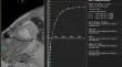
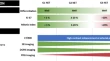
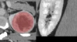
Feasibility of using computed tomography texture analysis parameters as imaging biomarkers for predicting risk grade of gastrointestinal stromal tumors: comparison with visual inspection In Young ChoiSuk Keu YeomJungwoo Choi Hollow Organ GI 28 March 2019 Pages: 2346 - 2356
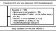
Single institutional experience with initial ultrasound followed by computed tomography or magnetic resonance imaging for acute appendicitis in adults Priyanka JhaNora EspinozaTara Morgan Practice 04 April 2019 Pages: 2357 - 2365
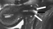
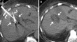
Diagnosis of recurrent HCC: intraindividual comparison of gadoxetic acid MRI and extracellular contrast-enhanced MRI Jae Hyun YimYoung Kon KimSoon Jin Lee Hepatobiliary 08 March 2019 Pages: 2366 - 2376
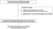
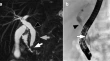
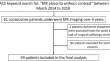
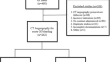
Diagnostic performance of magnetic resonance cholangiopancreatography (MRCP) versus endoscopic retrograde cholangiopancreatography (ERCP) in the pediatric population: a clinical effectiveness study Jonathan R. DillmanRakesh M. PatelAndrew T. Trout Hepatobiliary 14 March 2019 Pages: 2377 - 2383
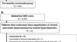
Abdominal manifestations of hereditary hemorrhagic telangiectasia: a series of 333 patients over 15 years Christopher L. WelleBrian T. WelchChristopher P. Wood Hepatobiliary 19 March 2019 Pages: 2384 - 2391
Performance of ultrasound for detection of transjugular intrahepatic portosystemic shunt dysfunction: a meta-analysis Wuttiporn ManatsathitHrishikesh SamantThammasin Ingviya Review 23 March 2019 Pages: 2392 - 2402
Assessment of liver T1 mapping in fontan patients and its correlation with magnetic resonance elastography-derived liver stiffness Preeti RamachandranSuraj D. SeraiRyan A. Moore Hepatobiliary 22 March 2019 Pages: 2403 - 2408
Clinicopathological findings and imaging features of intraductal papillary neoplasm of the bile duct: comparison between contrast-enhanced ultrasound and contrast-enhanced computed tomography Qiao ZhengSi-Min RuanWei Wang Hepatobiliary 15 May 2019 Pages: 2409 - 2417
Comparison of CT methods for determining graft steatosis in living donor liver transplantation Mehmet ŞekerCengiz ErolAfak Durur Karakaya Hepatobiliary 01 April 2019 Pages: 2418 - 2429
Value of 18F-FDG PET/CT in the diagnosis of portal vein tumor thrombus in patients with hepatocellular carcinoma Bing WuYiqiu ZhangHongcheng Shi Hepatobiliary 03 April 2019 Pages: 2430 - 2435
Hepatic disorders associated with exogenous sex steroids: MR imaging findings Cathryn L. HuiZhen Jiang Lee Review 06 April 2019 Pages: 2436 - 2447
Differentiation of aggressive from non-aggressive pancreatic solid pseudopapillary neoplasms using computed tomography Jianhua WangXiao ChenZhongqiu Wang Pancreas 08 March 2019 Pages: 2448 - 2458
Interobserver agreement of computed tomography reporting standards for chronic pancreatitis Ahmed Abdel Khalek Abdel RazekElsayed ElfarShefeek Abubacker Pancreas 06 April 2019 Pages: 2459 - 2465
110 Patients with adenosquamous carcinomas of the pancreas (PASC): imaging differentiation of small (≤ 3 cm) versus large (> 3 cm) tumors Yun-Feng FengJie-Yu ChenRi-Sheng Yu Pancreas 01 April 2019 Pages: 2466 - 2473
The role of multimodal imaging in guiding resectability and cytoreduction in pancreatic neuroendocrine tumors: focus on PET and MRI Laura RozenblumFatima-Zohra MokraneLaurent Dercle Review 12 April 2019 Pages: 2474 - 2493
Association between chronic asymptomatic pancreatic hyperenzymemia and pancreatic ductal anomalies: a magnetic resonance cholangiopancreatography study Wataru GonoiTakana Yamakawa HayashiOsamu Abe Pancreas 03 April 2019 Pages: 2494 - 2500
A review of the principles of texture analysis and its role in imaging of genitourinary neoplasms Richard ThomasLei QinAtul Shinagare Special section: Radiogenomics 17 November 2018 Pages: 2501 - 2510
The inferior vena cava: a pictorial review of embryology, anatomy, pathology, and interventions David S. ShinClaire K. SandstromGuy E. Johnson Review 01 April 2019 Pages: 2511 - 2527
CT-based machine learning model to predict the Fuhrman nuclear grade of clear cell renal cell carcinoma Fan LinEn-Ming CuiLiang-ping Luo Kidneys, Ureters, Bladder, Retroperitoneum 27 March 2019 Pages: 2528 - 2534
Precision and accuracy of magnetic resonance imaging for lobar classification of benign prostatic hyperplasia Neil F. WassermanEric NiendorfBenjamin Spilseth Pelvis 30 March 2019 Pages: 2535 - 2544
68Ga-PSMA-11 PET/CT in newly diagnosed prostate cancer: diagnostic sensitivity and interobserver agreement Mohammad Abd Alkhalik BashaMaged Abdel Galil HamedOla Harb Pelvis 08 April 2019 Pages: 2545 - 2556
MRI of cervical cancer with a surgical perspective: staging, prognostic implications and pitfalls Patricia BalcacerArvind ShergillBabak Litkouhi Review 22 March 2019 Pages: 2557 - 2571
Placenta accreta spectrum: value of placental bulge as a sign of myometrial invasion on MR imaging Priyanka JhaJoseph RabbanLiina Poder Pelvis 09 April 2019 Pages: 2572 - 2581
Transrectal and transvaginal catheter drainages and aspirations for management of pelvic fluid collections: technique, technical success rates, and outcomes in 150 patients David H. BallardMichael C. GatesHoracio B. D’Agostino Interventional Radiology 13 March 2019 Pages: 2582 - 2593
Using principal component analysis for the prediction of tumor response to transarterial chemoembolization Jessica P. MillerRaja RamaswamyOlaguoke Akinwande Interventional Radiology 19 April 2019 Pages: 2594 - 2601
Imaging findings during and after percutaneous cryoablation of hepatic tumors Lisa RatanaprasatpornNisha SainaniPaul B. Shyn Review 19 April 2019 Pages: 2602 - 2626
Body composition changes after left gastric artery embolization in overweight and obese individuals Edwin A. TakahashiNaoki TakahashiSanjay Misra Interventional Radiology 04 April 2019 Pages: 2627 - 2631
Recent use of NSAID and NOAC medications are associated with a positive CT arteriogram Muhammad A. ShafqetAlexander TonthatFrank K. Friedenberg Interventional Radiology 04 April 2019 Pages: 2632 - 2638
Diffusion-weighted imaging as a part of PET/MR for small lesion detection in patients with primary abdominal and pelvic cancer, with or without TOF reconstruction technique Tianbin SongBixiao CuiJie Lu Practice 12 March 2019 Pages: 2639 - 2647
A comparison of segmented abdominopelvic fluid volumes with conventional CT signs of abdominal compartment syndrome in a trauma population Thomas W. K. BatteyDavid DreizinWilliam Chiu Peritoneum 05 April 2019 Pages: 2648 - 2655
In comparison with other abdominal imaging modalities, which radiologists interpret abdominal MRI? Andrew B. RosenkrantzKrishna P. ShanbhogueRichard Duszak Jr. Practice 09 April 2019 Pages: 2656 - 2662
The “hot air balloon” sign Johnny LingRaymond B. Dyer Classics in Abdominal Radiology 08 March 2019 Pages: 2663 - 2664
Fadeout sign of liver Venkatraman IndiranJagannathan Kokilavani Classics in Abdominal Radiology 15 March 2019 Pages: 2665 - 2666
The stalk-of-corn sign of ureter Minghao YangChunni HeJun Li Classics in Abdominal Radiology 18 March 2019 Pages: 2667 - 2668
Thimble bladder Kalaichezhian MariappanVenkatraman Indiran Classics in Abdominal Radiology 29 March 2019 Pages: 2669 - 2670
“Nodule-in-nodule” architecture of hepatocellular carcinoma Dario GiambellucaRoberto CannellaGiuseppe Brancatelli Classics in Abdominal Radiology 02 April 2019 Pages: 2671 - 2673
Bare area sign Venkatraman IndiranJagannathan Kokilavani Classics in Abdominal Radiology 09 April 2019 Pages: 2674 - 2675
Clinical evaluation of ureteral pseudodiverticulosis Kenji Yorita Letter to the Editor 30 April 2019 Pages: 2676 - 2676
Reply to letter to the editor: “clinical evaluation of ureteral pseudodiverticulosis” Matthew A. MorganHanna M. ZafarParvati Ramchandani Letter to the Editor 27 April 2019 Pages: 2677 - 2677