The role of MR–PET in abdominal and pelvic oncology Julia R. Fielding Editorial 28 July 2015 Pages: 1351 - 1351
How does PET/MR work? Basic physics for physicians Gaspar DelsoEdwin ter VoertPatrick Veit-Haibach Review Article 24 April 2015 Pages: 1352 - 1357
Hybrid PET/MR imaging: physics and technical considerations Shetal N. ShahSteve S. Huang Review Article 19 May 2015 Pages: 1358 - 1365
Practical guide for implementing hybrid PET/MR clinical service: lessons learned from our experience Nainesh ParikhKent P. FriedmanHersh Chandarana OriginalPaper 19 May 2015 Pages: 1366 - 1373
Whole-body FDG PET-MR oncologic imaging: pitfalls in clinical interpretation related to inaccurate MR-based attenuation correction Ulrike AttenbergerCiprian CatanaAlexander R. Guimaraes Review Article 30 May 2015 Pages: 1374 - 1386
PET/MRI for the body imager: abdominal and pelvic oncologic applications Tyler J. FraumKathryn J. FowlerBarry A. Siegel Pictorial Essay 25 March 2015 Pages: 1387 - 1404
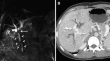
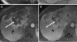
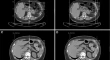
MR–PET evaluation of 1-month post-ablation therapy for hepatocellular carcinoma: preliminary observations Miguel RamalhoMamdoh AlObaidyRichard C. Semelka OriginalPaper 24 April 2015 Pages: 1405 - 1414
Comparison of hybrid FDG PET/MRI compared with PET/CT in colorectal cancer staging and restaging: a pilot study Raj Mohan PaspulatiSasan PartoviNghi C. Nguyen OriginalPaper 26 June 2015 Pages: 1415 - 1425
MR–PET co-registration in upper abdominal imaging: quantitative comparison of two different T1-weighted gradient echo sequences: initial observations Miguel RamalhoMamdoh AlObaidyRichard C. Semelka OriginalPaper 21 May 2015 Pages: 1426 - 1431
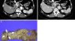
Simultaneous 68Ga-DOTA-TOC PET/MRI with gadoxetate disodium in patients with neuroendocrine tumor Thomas A. HopeMiguel Hernandez PampaloniEmily Bergsland OriginalPaper 28 March 2015 Pages: 1432 - 1440
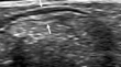
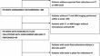
Use of transabdominal ultrasonography to preoperatively determine T-stage of proven colon cancers Susumu ShibasakiNorihiko TakahashiAkinobu Taketomi OriginalPaper 16 November 2014 Pages: 1441 - 1450
Optimal section thickness for detection of polyps at MR: resolution phantom study Courtney C. MorenoPardeep K. MittalW. Thomas Dixon OriginalPaper 14 December 2014 Pages: 1451 - 1456
18F-FDG PET/CT as a potential predictor of survival in patient with oesophageal cancer: a preliminary result A. S. Fathinul FikriR. DharmendranA. J. Nordin OriginalPaper 10 January 2015 Pages: 1457 - 1464
Prediction of esophageal varices in cirrhotic patients with apparent diffusion coefficient of the spleen Ahmed Abdel Khalek Abdel RazekSamia Mohammed Ali MassoudEnas Mohamed Motawea OriginalPaper 03 March 2015 Pages: 1465 - 1469
Apparent diffusion coefficient quantification as an early imaging biomarker of response for unresectable infiltrative hepatocellular carcinoma Ali Kemal SivriogluKemal KaraHakan Mutlu Letter 16 November 2014 Pages: 1470 - 1470
Can volumetric ADC measurement help predict response to Y90 radioembolization in HCC? Michael VoucheRiad SalemFrank H. Miller OriginalPaper 21 November 2014 Pages: 1471 - 1480
Retrospective cohort study of portacaval lymphadenopathy identified on multidetector CT and implications for follow-up Veena R. IyerSandeep S. HedgireMukesh G. Harisinghani OriginalPaper 17 December 2014 Pages: 1481 - 1486
A case of β-catenin-positive hepatocellular adenoma with MR imaging sign of diffuse intratumoral fat deposition Masaya IshiiJunichi KazaokaMasaaki Akahane OriginalPaper 21 January 2015 Pages: 1487 - 1491
Intratumoral artery on contrast-enhanced computed tomography imaging: differentiating intrahepatic cholangiocarcinoma from poorly differentiated hepatocellular carcinoma Seiji TsunematsuMakoto ChumaNaoya Sakamoto OriginalPaper 13 January 2015 Pages: 1492 - 1499
Budd–Chiari syndrome: a prospective analysis of hepatic vein obstruction on ultrasonography, multidetector-row computed tomography and MR imaging Sid Ahmed FaraounMohamed El Amine BoudjellaSalah Eddine Bendib OriginalPaper 17 February 2015 Pages: 1500 - 1509
Not to forget portal cavernoma cholangiopathy: the great mimicker! Ankur AroraKalpana BansalYashwant Patidar Letter 22 January 2015 Pages: 1510 - 1511
Quantification of liver, pancreas, kidney, and vertebral body MRI-PDFF in non-alcoholic fatty liver disease Ilkay S. IdilmanAli TuzunMusturay Karcaaltincaba OriginalPaper 26 February 2015 Pages: 1512 - 1519
Primary biliary tract malignancies: MRI spectrum and mimics with histopathological correlation Pardeep K. MittalCourtney Coursey MorenoWilliam C. Small OriginalPaper 22 November 2014 Pages: 1520 - 1557
Dynamic enhancement pattern of intrahepatic cholangiocarcinoma on contrast-enhanced ultrasound: the correlation with cirrhosis and tumor size Qing LuLi-Yun XueCui-Xian Li OriginalPaper 01 March 2015 Pages: 1558 - 1566
Imaging of choledochal cysts Vanessa A. LewisSharon Z. AdamFrank H. Miller Pictorial Essay 15 February 2015 Pages: 1567 - 1580
CT evaluation of common duct dilation after cholecystectomy and with advancing age Tatum A. McArthurVirginia PlanzMark E. Lockhart OriginalPaper 25 November 2014 Pages: 1581 - 1586
Clinical value of spectral CT in diagnosis of negative gallstones and common bile duct stones Huanguo LiDong HeLongxia Chen OriginalPaper 01 March 2015 Pages: 1587 - 1594
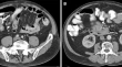
MDCT findings of pancreatic metastases according to primary tumors Tae Won ChoiSe Hyung KimByung Ihn Choi OriginalPaper 27 November 2014 Pages: 1595 - 1607
CT and MRI assessment of symptomatic organized pancreatic fluid collections and pancreatic duct disruption: an interreader variability study using the revised Atlanta classification 2012 Ayesha KamalVikesh K. SinghAtif Zaheer OriginalPaper 26 November 2014 Pages: 1608 - 1616
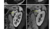
Lymphatic pathway around the pancreatic head and extrahepatic bile duct: evaluation using MR imaging at 3.0-T Yasunari YamadaHiromu MoriRika Tanoue OriginalPaper 07 January 2015 Pages: 1617 - 1628
Uncommon presentations of common pancreatic neoplasms: a pictorial essay Mirko D’OnofrioRiccardo De RobertisPaolo Pederzoli Pictorial Essay 15 March 2015 Pages: 1629 - 1644
Non-oncologic applications of diffusion-weighted imaging (DWI) in the genitourinary system Dell P. DunnNathan R. KelseyKoenraad J. Mortele Review Article 25 June 2015 Pages: 1645 - 1654
Diagnostic performances of FDG-PET/CT and diffusion-weighted imaging indices for differentiating benign pheochromocytoma from other benign adrenal tumors Masatoyo NakajoMasayuki NakajoTakashi Yoshiura OriginalPaper 11 November 2014 Pages: 1655 - 1665
Histogram analysis for characterization of indeterminate adrenal nodules on noncontrast CT Michael F. LinLauren Q. Chang-SenKyongtae T. Bae ReviewPaper 09 December 2014 Pages: 1666 - 1674
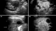
Histotype differentiation of hypo-echoic renal tumors on CEUS: usefulness of enhancement homogeneity and intensity Qing LuLi-yun XueCui-xian Li OriginalPaper 31 December 2014 Pages: 1675 - 1683
Radiogenomics of clear cell renal cell carcinoma: preliminary findings of The Cancer Genome Atlas–Renal Cell Carcinoma (TCGA–RCC) Imaging Research Group Atul B. ShinagareRaghu VikramStuart G. Silverman OriginalPaper 10 March 2015 Pages: 1684 - 1692
MDCT of extranodal mantle cell lymphoma: a single institute experience Akshay D. BahetiSree Harsha TirumaniNikhil H. Ramaiya OriginalPaper 28 February 2015 Pages: 1693 - 1699
Quantitative T2* magnetic resonance imaging for renal iron overload assessment: normal values by age and sex Emanuele GrassedonioAntonella MeloniAlessia Pepe OriginalPaper 12 March 2015 Pages: 1700 - 1704
Response assessment to neoadjuvant therapy in soft tissue sarcomas: using CT texture analysis in comparison to tumor size, density, and perfusion Fang TianKoichi HayanoDushyant V. Sahani OriginalPaper 21 December 2014 Pages: 1705 - 1712
Retroperitoneal low-flow vascular malformations: characteristic MRI findings correlated with histopathological findings Hirotaka AkitaYoshitake YamadaMasahiro Jinzaki OriginalPaper 10 December 2014 Pages: 1713 - 1720
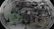
Hysterosalpingography: an imaging Atlas with cross-sectional correlation Karyn A. LedbetterMonisha ShettyDaniel T. Myers Pictorial Essay 12 November 2014 Pages: 1721 - 1732
Use of enhanced T2 star-weighted angiography (ESWAN) and R2* values to distinguish ovarian cysts due to endometriosis from other causes Ye LiQing-Wei SongAi-Lian Liu OriginalPaper 11 December 2014 Pages: 1733 - 1741
Differential diagnosis of uterine smooth muscle tumors using diffusion-weighted imaging: correlations with the apparent diffusion coefficient and cell density Akiko TasakiMina O. AsataniHidefumi Aoyama OriginalPaper 21 December 2014 Pages: 1742 - 1752
Magnetic resonance imaging findings of mucinous borderline ovarian tumors: comparison of intestinal and endocervical subtypes Sungmin WooSeung Hyup KimJeong Yeon Cho OriginalPaper 12 December 2014 Pages: 1753 - 1760
Preoperative CT-based nomogram for predicting overall survival in women with non-endometrioid carcinomas of the uterine corpus Yulia LakhmanDerya YakarEvis Sala OriginalPaper 31 December 2014 Pages: 1761 - 1768
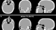
68Ga-PSMA PET/MR with multimodality image analysis for primary prostate cancer Matthias EiberStephan G. NekollaMarkus Schwaiger Case Report 21 November 2014 Pages: 1769 - 1771
Value of bimodal 18F-choline-PET/MRI and trimodal 18F-choline-PET/MRI/TRUS for the assessment of prostate cancer recurrence after radiation therapy and radical prostatectomy Francesco PaparoArnoldo PiccardoGian Andrea Rollandi Pictorial Essay 13 January 2015 Pages: 1772 - 1787
Recent advances in image-guided targeted prostate biopsy Anna M. BrownOsama ElbulukBaris Turkbey Pictorial Essay 18 January 2015 Pages: 1788 - 1799
Celiomesenteric and hepatosplenomesenteric trunks: characterization of two rare vascular anomalies with CT Pierre D. MaldjianMichael A. Chorney OriginalPaper 29 November 2014 Pages: 1800 - 1807
Digital subtraction angiography during transjugular intrahepatic portosystemic shunt creation or revision: data on radiation exposure and image quality obtained using a standard and a low-dose acquisition protocol in a flat-panel detector-based system Roberto MiragliaLuigi MaruzzelliAngelo Luca OriginalPaper 02 December 2014 Pages: 1808 - 1812