Guideline from Japanese Society of Echocardiography: 2018 focused update incorporated into Guidance for the Management and Maintenance of Echocardiography Equipment Masao DaimonMakoto AkaishiCommittee for Guideline Writing, the Japanese Society of Echocardiography Review Article Open access 23 January 2018 Pages: 1 - 5
Echocardiographic evaluation of cardiac function after cancer chemotherapy Tomoko NegishiKazuaki Negishi Review Article 11 July 2017 Pages: 20 - 27
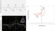
Increased apical rotation in patients with severe aortic stenosis assessed by three-dimensional speckle tracking imaging Maidar TumenbayarKazuto YamaguchiKazuaki Tanabe Original Investigation 11 August 2017 Pages: 28 - 33
Causes of an increased pressure gradient through the left ventricular outflow tract: a West Coast experience Sayuki KobayashiYoshihiko SakaiTakahiro Shiota Original Investigation Open access 18 September 2017 Pages: 34 - 41
Perforated mitral valve aneurysm diagnosed 3 years after etiology-unknown iliopsoas muscle abscess: illustrative case of ‘self-attack’ endocarditis of the mitral valve Shun NishinoNozomi WatanabeYoshisato Shibata Case image in cardiovascular ultrasound 29 July 2017 Pages: 42 - 44
Difficult diagnosis of ruptured mitral valve aneurysm because of severe mitral annular calcification Takahiro SakamotoHiroyuki YoshitomiKazuaki Tanabe Case image in cardiovascular ultrasound 10 August 2017 Pages: 45 - 46
Prominent v wave as a result of left atrial stiffening Katsuji InoueChiharuko IioShuntaro Ikeda Case image in cardiovascular ultrasound 08 August 2017 Pages: 47 - 48
Platypnea–orthodeoxia syndrome: additive value of three-dimensional echocardiography Andrea Rueda LiñaresJose Alberto de AgustinLeopoldo Pérez de Isla Case image in cardiovascular ultrasound 22 August 2017 Pages: 49 - 51
Paravalvular leakages between sewing cuffs after the second surgery for double valve replacement: evaluation by three-dimensional color-Doppler transesophageal echocardiography Hideta TakushiAkihiro HayashidaKiyoshi Yoshida Case image in cardiovascular ultrasound 20 September 2017 Pages: 52 - 53