
Abstract
The evaluation of skeletal health has progressed from diagnostic labeling (normal-low bone mass-osteoporosis) to quantifying future fracture risk. Thus, the recognition of prevalent and incident fractures has increased in importance because they influence risk assessment. Non-spinal osteoporotic fractures rarely pose diagnostic problems, although those of the proximal femur and pelvis may require computed tomography (CT) and/or magnetic resonance imaging for diagnosis. Spinal fractures remain a paradox. Whereas radiological diagnosis in general benefits increasingly from powerful diagnostic tools to examine disease, many osteoporotic spinal fractures are asymptomatic, and their recognition is often incidental to chest radiography or body CT. Moreover, there are no uniformly agreed criteria by which to decide if a vertebra is fractured. Lastly, it has become apparent that some osteoporosis treatments may themselves contribute to so-called atypical femoral fractures.
Introduction
Post-menopausal osteoporosis (PMO) is silent and only becomes manifest when bones fracture [1]. Bone strength is a function of bone mineral density (BMD) and bone quality—implying micro-architecture and macro-architecture [2•]. BMD is measurable [3], but the quality element of in vivo bone strength can, in day-to-day practice, only be inferred when and if a fragility fracture occurs. While the clinical challenge is to diagnose and, if necessary, treat the disease before such fractures happen, recognition of a low-trauma fracture has three important implications:
-
It provides an unequivocal statement about bone fragility and trumps BMD in indicating that the patient has osteoporosis.
-
It impacts fracture risk assessment, for example, using the FRAX model [4•], otherwise based on BMD and/or other clinical factors, since a low-trauma fracture is associated with an enhanced future fracture risk [5].
-
It affords an opportunity to investigate and treat a patient and thus try to prevent future fractures [6].
Fractures, Guidelines, and Risk Estimation
The management of patients with PMO has evolved from applying a diagnostic label (normal-osteopenia–osteoporosis) based on BMD [7] to a model based on fracture risk assessment. Thus, most guidelines now incorporate [8–11] a low-trauma fracture as an important factor in estimating an individual’s future fracture risk, along with other factors, as does the FRAX tool—calibrated and validated for use in a number of countries [4•]. Thus, the correct diagnosis of low-trauma fractures and their explicit attribution to osteoporosis are often key to management decisions. Secondary osteoporosis, for example, from glucocorticoid administration, is also included in the FRAX model as a weighting factor.
Clinical Aspects
Any systematic approach to understanding fragility fractures has to avoid a narrow osteocentric view of low-trauma fractures. While these are often the result of trauma acting upon weak bones, other factors are very important—ranging from a person’s muscle strength, balance, and agility to her or his consumption of sedatives, and to such physical hazards as scatter rugs, etc., which may cause falls [12].
Epidemiology
PMO is the most prevalent metabolic bone disease in the developed world [13–20]. It is a multifactorial disorder in which age, sex, and genetic factors are important. Older women suffer osteoporotic fractures both earlier and in greater number than men at the same age, by a factor of about two, but fracture types are themselves age-dependent (14–19: wrist fractures increase in the 50s, vertebral fractures in the 60s, and hip fractures thereafter [21]. Furthermore, there are substantial differences in the rates of hip fracture in different countries [22], while white populations suffer a greater incidence of hip fractures irrespective of abode [22]. PMO itself can be associated with dietary factors including low vitamin D levels, the latter usually without radiological evidence of osteomalacia.
Definition
Any given fracture is the result of a patient’s exposure to injury, the magnitude of that injury, and the ability of the skeleton to withstand damage caused by the forces acting upon it. Thus, a universally applicable definition of an osteoporotic fragility fracture is always going to be subject to interpretation. In a given patient, his or her balance, muscle strength in resisting a fall, the site of the trauma, and the mechanical strength of the bones bearing the brunt of an injury all play a part in the outcome. For practical purposes, a low-trauma or fragility fracture, as complicating PMO, is often defined as a fracture resulting from a fall from a standing height or less [23]. Thus, any fracture that results from a fall down stairs is likely to be predominantly due to the trauma involved, while the same fracture resulting from a fall on a sidewalk is likely to be osteoporotic in provenance. Patients too may point to the diagnosis when asked if they might have expected the injury in question to have caused a fracture had it occurred when they were a couple of decades younger.
What is apparent in population studies is that fractures of the cranium, facial bones, hands, and feet (carpus, tarsus, and bones of the fingers and toes) are not usually associated with osteoporosis and are usually disregarded in this context, bearing in mind that fractures of the distal radius are indeed often osteoporotic [24]. Rib fractures (for example, “cough fractures”) are also commonly due to osteoporosis [24].
An important distinction that is made in describing fractures due to PMO is the difference between prevalent (pre-existing) and incident (newly observed) fractures.
Diagnosis
In considering osteoporotic fracturing, it is convenient to consider fractures in general and vertebral fractures as a special case.
Non-spine Fractures
Non-spine fractures usually occur either spontaneously or as a result of trauma, and do so acutely and painfully. The common sites for traumatic extra-spinal fragility fractures are the ribs, distal forearm, proximal humerus, and proximal femur. Radiography is used to confirm the diagnosis, assess the position of the bone fragments, and determine need for surgery. Some patients with an undisplaced proximal femoral fracture may have normal radiographs, and, although both computed tomography (CT) and magnetic resonance imaging (MRI) have been used to confirm a fracture in this context [25, 26] (Fig. 1), MRI seems to be the superior modality [25, 26] and has been shown to be cost-effective in at least one setting [27]. Otherwise, supplementary examinations with CT or MRI are rarely required unless, for example, there is a suspicion of pathological fracturing (Fig. 2).

A CT scan section. There is an impacted fracture of the femoral neck (arrow). Plain radiographs had been inconclusive

Lumbar spine sagittal reconstruction: pathological fractures with a soft-tissue mass (arrows)
An exception to the above is that pelvic fractures are becoming more common in developed countries and require CT for assessment [28•].
Serial radiography is used to record fracture reduction and healing.
Spinal Fractures
Spinal fractures differ from fractures elsewhere in that:
-
There is no consensus about the definition of a vertebral fracture [13, 29, 30••].
-
About 60 % occur with few or no symptoms [31] and may only be detected by radiography, perhaps coincidentally on a chest radiograph or body CT [32–35].
-
They may occur progressively and not entirely instantaneously, so that the vertebral configuration changes over time.
-
Other than in children who are still growing, while some callus formation may occur, there is no healing in the sense that long bones heal. Growing children have the distinct potential to completely reshape and restore previously fractured vertebral bodies if the underlying cause of the fracture is treated [36•].
-
Unlike other fractures, excepting those of the proximal femur, spinal fractures are associated with an increased mortality, which at 5 years is nearly identical to that from fractures of the proximal femur [37]. It is not clear whether this is cause and effect or if the fracture is a marker for frailty. Multiple spinal fractures also result in morbidity resulting from changes in posture—a stoop, protuberant abdomen, and compromised chest volume, not to mention distress to the patient as her wardrobe is made obsolete by changes in her body habitus [31, 37–40].
-
Rarely, acute spinal fractures may only be seen on standing (“stress”) images [41].
Thus, the radiological diagnosis of spinal fractures in PMO represents a paradox. While the evolution of radiology has resulted in powerful diagnostic tools to examine specific localizing symptoms, because spinal fractures are often asymptomatic, the diagnosis tends to rely on plain film radiography used in either fracture risk assessment or, if the spine is seen incidentally, on chest radiography or CT [35, 36•]. Indeed, to an increasing extent, dual X-ray absorptiometry (DXA) scans are used to provide images (vertebral fracture assessments, VFA) that are useful for grade 2 and 3 vertebral fracture identification [42, 43] (Fig. 3).

A VFA lateral spine image of L3. A superior end-plate fracture of L3 is apparent
Osteoporotic spinal fractures are rarely of the “burst” type often seen with high-energy trauma. While there may be a small degree of retropulsion in osteoporotic vertebral fracturing, neurological compromise from pressure on the spinal cord is very rare.
Spinal Fracture Diagnosis
One recurrent theme in the diagnosis of spinal fractures, first identified by Gellbach et al. [32] but since repeated [33, 34], is the way in which they may be ignored. Radiologists, for example, may fail to report them as incidental findings on a chest radiograph. Moreover, if noted, clinicians may ignore the observation and, in a hospital setting, even the diagnosis. Thus, a diagnosis of osteoporosis may not make its way into the discharge summary. An opportunity to treat the underlying disease may thus be missed—a care gap. More recently, this same care gap has also been observed in respect to fractures not recognized on body CT images [35, 36•].
Recognition of this issue [44] has resulted in pedagogical initiatives, for example, by the International Skeletal Society (ISS) [45], to improve the recognition and diagnosis of vertebral fractures by radiologists [46].
At the same time, uncertainty about the precise characterization of vertebral fractures may lead to equivocation in describing fractures and in linking them to a potential diagnosis of osteoporosis [47, 48].
Indeed, spinal fracture recognition is far from a simple issue, particularly in respect of minor degrees of fracturing [49]. Recent reviews have documented this uncertainty in respect to spinal fractures and described a number of methods being used for diagnosing spinal fractures without being able to endorse any one [29, 30••]. The methods used have included:
-
Simple observational diagnosis comparing the self-similarity of adjacent vertebrae complemented by the conventional signs of fracturing (end plate breaks and disruption, loss of end-plate parallelism, end-plate attenuation, and anterior cortical buckling).
-
Absolute measurements of vertebral dimensions compared with either a data base [50] or between the anterior and posterior heights of a vertebral body [51••].
-
Relative morphometric measures comparing the vertical heights of vertebral bodies [52].
-
The Genant semiquantitative variation classifying fractures by degree as well as type: “wedge,” “bi-concave,” or “crush’ [53].
-
An algorithm for recognizing end-plate fractures [54, 55], which, the proponents argue, are the underlying and unifying feature of osteoporotic vertebral fractures—the algorithm-based qualitative (ABQ) tool. This algorithm is designed to exclude false-positive diagnoses that might result from a Schmorl node, for example (Fig. 4).
Fig. 4 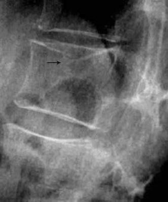
A superior end-plate fracture of a vertebra (arrow) meeting the ABQ criteria

Unfortunately, while these techniques have been compared, correlated with BMD, and examined for observer error, there have been no long-term outcome studies published to validate any one particular method.
The semiquantitative tool of Genant is the most widely used [53] and has been validated to the extent that it facilitated the pivotal trials of drugs currently used in osteoporosis. However, experience has tended to suggest that Genant grade 1 fractures in the thoracic spine have a low predictive power in terms of future fracturing. Moreover, using the Genant paradigm, there is a discrepancy between the distribution of fractures in the spine such that prevalent fractures are much more common in the mid- and upper-thoracic region than incident fractures [54–56], suggesting that at least some of the these apparent lesions are not true fractures. The Genant tool remains in wide use. However, the fact of proposals of alternative diagnostic criteria as well as experience of over-interpretation of the normal wedge shape of thoracic vertebrae (on account of the normal kyphosis) suggests caution. Indeed, at least one recent report used the Genant tool for classification with the rider that to be diagnosed as a fracture a vertebral body must provide evidence of end-plate damage [57•].
Imaging Tools
Conventional Radiographs
Conventional radiography is crucial to the diagnosis and follow-up of fractures.
DXA and Vertebral Fracture Assessment (VFA)
DXA examinations provide an image of the lumbar spine from which fractures may sometimes be identified, although such images cannot be used to exclude a fracture. Later generations of DXA machines provide for imaging the spine from T4 to L4. Although these images are photon-poor, they involve smaller radiation doses than conventional radiography, and, because the technique is a scanning technique, the X-ray beam is orthogonal to the spine long-axis along its length. Thus, the image provides good visualization of the vertebral end plates at least below the mid-thorax [42, 43] (Fig. 3).
Radionuclide Bone Scintigraphy
Radionuclide scintigraphy may complement radiography in two contexts:
-
Providing evidence to suggest that spinal fractures are recent or healed.
-
Demonstrating evidence of radiographically occult injuries, such as sacral fractures [58] and atypical femoral fractures (AFF) as described below.
Computed Tomography (CT)
CT is not necessary in the diagnosis of most peripheral fractures, and in the absence of symptoms in most patients, it is not used as a primary tool for the detection of osteoporotic spinal fractures. However, as noted above, scans of the chest or abdomen may enable the fortuitous detection of occult spinal fractures, particularly if the images are reformatted in sagittal projection [35, 36•]. Patients with DXA-detected osteoporosis have been shown to have low CT-attenuation values in the lumbar spine, and it has been suggested that abdominal CT scans may be used for the opportunistic diagnosis of osteoporosis [59].
Magnetic Resonance Imaging (MRI)
Similarly, MRI is not used for the routine detection of osteoporotic fractures. Vertebral MR performed for specific conditions such as spinal cord compression or assessment of the para-spinal soft tissues allows fortuitously detection of osteoporotic spinal fractures [30••]. The demonstration of bone marrow edema, thought to represent hemorrhage associated with microfracturing, helps to identify acute or sub-acute fractures [60].
Some patients with the clinical diagnosis of a femoral neck or intertrochanteric fracture may have normal radiographs, and although both CT and MRI have been used to confirm a fracture in this context, MRI seems to be the superior modality [26]. Otherwise, supplementary examinations with CT or MRI are rarely required unless, for example, there is a suspicion of pathological fracturing.
CT and/or MRI can usually be used to decide whether a vertebral fracture is osteoporotic [51].
Particular Considerations
Atypical Femoral Fractures (AFF)
From the earliest use of agents that inhibit bone resorption to treat osteoporosis, there has been concern that excessive inhibition of bone turnover might ultimately compromise bone strength. No such issues were observed in the pivotal drug trials of alendronate, risedronate, and other bisphosphonates. However, with expanded use of such drugs, an unusual pattern of femoral fractures has been observed. AFFs are very uncommon, but a relationship with the administration of agents such as bisphosphonates appears likely, although the relationship is unlikely to be simply one of cause and effect.
From a number of patients with such fractures a probable sequence of radiographic events can be inferred. Changes in the femoral cortex, potentially bilateral, occur, leading, if the condition progresses, to stress fractures. These are thought to be precursors of full-blown fractures, which are “atypical” in the sense that, unlike more usual femoral fractures, they are most often short, transverse fractures that have been likened to chalk stick fractures. There may be symptoms in the prodromal phase—chiefly thigh discomfort. At this stage, a radionuclide bone scan may reveal a focal abnormality or abnormalities in the femur. Also focal new bone may be formed on the outer or inner cortical surfaces, sometimes with a transverse cortical stress fracture when it is called “beaking” and, of interest, since some of these early signs may be detected on DXA examinations (Fig. 5) [61•]. The American Society of Bone and Mineral Research has recently published a revised set of clinical and radiological criteria for the diagnosis of AFF [62••].

Cortical stress lesion in the proximal femur thought to be a precursor of an atypical femoral fracture
The fractures, if and when they occur, may be spontaneous and not trauma related. Of note, all of these features are also present in the upper femoral fractures that occur spontaneously in hypophosphatasia [63]. To put AFF in perspective, it has been estimated that perhaps 100 typical hip fractures may be prevented for every atypical hip fracture that may be caused by bisphosphonate use.
Glucocorticoid-Induced Vertebral Fractures
Spinal fractures of this type are associated with exuberant callus formation near the end plate, which has been described as marginal condensation [64] so that there may be a region of increased density adjacent to the vertebral end plate (Fig. 6). As a sign, this is of low sensitivity but high specificity.

Lumbar spine with a glucocorticoid-induced fracture and the characteristic excess callus (upper arrow). The lower arrow points to similar changes presumably due to occult fracturing
Fracture-Related Necrosis
Excavation of the nucleus pulposus at any given intervertebral space may result in a void that fills with gas—the so-called vacuum phenomenon. Less commonly, gas may accumulate in the vertebral body (an intraosseous vacuum phenomenon) [65] (Fig. 7). This finding, also called a vertebral cleft, has been shown to be due to tissue necrosis [66]. It is not specific to osteoporosis but may occur in aseptic necrosis of the vertebra because of any cause, and such necrosis has also been called Kümmel disease.

Vertebral aseptic necrosis and an intraosseous vacuum phenomenon (arrow), sometimes described as Kümmel disease or a vertebral cleft, and a potential cause of pain
Summary
The prevention and treatment of PMO are directed to avoiding low-trauma fractures. However, recognizing these fractures when they occur is as important, as is explicitly attributing them to osteoporosis, since such recognition affords an opportunity, sometimes missed, to initiate care not only of the fracture but also the underlying disease [67].
References
Papers of particular interest, published recently, have been highlighted as: • Of importance. •• Of significant importance
Consensus Development Conference. Diagnosis, prophylaxis and treatment of osteoporosis. Am J Med. 1991;90:170–210.
• Link TM. Osteoporosis imaging: state of the art and advanced imaging. Radiology. 2012;263:3–17. This article describes the radiological diagnosis of osteoporosis and analyzes imaging methods being developed for examining bone quality.
Marshall D, Johnell O, Wedel H. Meta-analysis of how well measures of bone density predict occurrence of osteoporotic fractures. BMJ. 1996;312:1254–9.
• World Health Organization. WHO facture risk assessment tool. Available at: http://www.shef.ac.uk/FRAX. Accessed 17 Nov 2013. A widely used web-based tool to estimate both all and femoral fracture risk on the basis of risk factors and radiological findings.
Klotzbuecher CM, Ross PD, Landsman PB, Abbott TA 3rd, Berger M. Patients with prior fractures have an increased risk of future fractures: a summary of the literature and statistical synthesis. J Bone Miner Res. 2000;15:721–39.
Port L, Center J, Briffa NK, Nguyen T, Cumming R, Eisman J. Osteoporotic fracture: missed opportunity for intervention. Osteoporos Int. 2003;9:780–4.
World Health Organization. Assessment of osteoporotic fracture risk and its role in screening for postmenopausal osteoporosis. Geneva: WHO Technical Report Series, No 843; 1994. p. 5.
www.aace.com/pub/pdf/guidelines/osteoporosis2001Revised.pdf. Accessed 25 Aug 2013.
The North American Menopause Society. Management of osteoporosis in postmenopausal women: 2006 position statement of The North American Menopause Society. Menopause. 2006;13:340–67.
Papaioannou A, Morin S, Cheung AM, et al. 2010 clinical practice guidelines for the diagnosis and management of osteoporosis in Canada: summary. CMAJ. 2010;182:1864–73.
Kanis JA, Delmas P, Burckhardt P, Cooper C, Torgerson D. Guidelines for diagnosis and management of osteoporosis. Osteoporos Int. 1997;7:390–406.
O’Loughlin JL, Robitaille Y, Boivin JF, Suissa S. Incidence of and risk factors for falls and injurious falls among the community dwelling elderly. Am J Epidemiol. 1993;137:32–354.
Wasnich RD. Epidemiology of osteoporosis. In: Favus MJ, editor. Primer on the metabolic bone diseases and disorders of mineral metabolism. Philadelphia: Lipincott-Raven; 1996. p. 249–51.
Johnell O, Gullberg B, Allander E, Kanis JA. The apparent incidence of hip fracture in Europe: a study of national register sources. MEDOS study group. Osteoporos Int. 1992;2:298–302.
Bacon WE, Maggi S, Looker A, Harris T, Nair CR, Giaconi J, et al. International comparison of hip fracture rates in 1988–1989. Osteoporos Int. 1996;6:69–75.
Landin LA. Fracture patterns in children. Analysis of 8,682 fractures with special reference to incidence, etiology and secular changes in a Swedish urban population 1950–1979. Acta Orthop Scand Suppl. 1983;202:1–109.
Tiderius CJ, Landin L, Duppe H. Decreasing incidence of fractures in children: an epidemiological analysis of 1,673 fractures in Malmo, Sweden, 1993–1994. Acta Orthop Scand. 1999;70:622–6.
Cooper C, Dennison EM, Leufkens HG, Bishop N, van Staa TP. Epidemiology of childhood fractures in Britain: a study using the general practice research database. J Bone Miner Res. 2004;19:1976–81.
Rennie L, Court-Brown CM, Mok JY, Beattie TF. The epidemiology of fractures in children. Injury. 2007;38:913–22.
Mayranpaa MK, Makitie O, Kallio PE. Decreasing incidence and changing pattern of childhood fractures: a population-based study. J Bone Miner Res. 2010;25:2752–9.
Brudvik C, Hove LM. Childhood fractures in Bergen, Norway: identifying high-risk groups and activities. J Pediatr Orthop. 2003;23:629–34.
Hagino H, Yamamoto K, Ohshiro H, Nose T. Increasing incidence of distal radius fractures in Japanese children and adolescents. J Orthop Sci. 2000;5:356–60.
Kleerekoper M. The evaluation of patients with osteoporosis. In: Marcus R, Feldman D, Kelsey J, editors. Osteoporosis. San Diego, CA: Academic Press; 1966. p. 101–8.
Warriner AH, Patkar NM, Curtis JR, Delzell E, Gary L, Kilgore M, Saag K. Which fractures are most attributable to osteoporosis? J Clin Epidemiol. 2011;64:46–53.
Lubovsky O, Liebergall M, Mattan Y, Weil Y, Moshieff R. Early diagnosis of occult hip fractures MRI versus CT scan. Injury. 2005;36:788–92.
Sankey RA, Turner J, Lee J, Healy J, Gibbons CER. The use of MRI to detect occult fractures of the proximal femur: a study of 102 consecutive cases over a ten-year period. J Bone Joint Surg Br. 2009;91(8):1064–8.
Pool FJ, Crabbe JP. Occult femoral neck fractures in the elderly: optimisation of investigation. N Z Med J. 1996;109:235–7.
• Henry PDG, Kreder HJ, Jenkinson RJ. The osteoporotic acetabular fracture. Orthop Clin N Am. 2013;44:201–205. One other fracture that may only be recognized on CT/MRI and missed on plain images.
Grados F, Fechtenbaum J, Flipon E, Kolta S, Roux C, Fardellone P. Radiographic methods for evaluating osteoporotic vertebral fractures. Joint Bone Spine. 2009;76:241–7.
•• Oei L, Rivandeira F, Ly F, Breda SJ, Zillikens MC, Hofman A, Uitterlinden AG, Krestin GP, Oei EHG. Review of radiological scoring methods of osteoporotic vertebral fractures for clinical and research settings. Eur Radiol. 2013;23:476–486. A review of the methods by which vertebral fractures may be recognized while concluding that only circumstantial evidence favors the use of the Genant tool.
Cooper C, Atkinson EJ, Jacobsen SJ, O’Fallon WM, Melton LJ III. Population-based study of survival after osteoporotic fractures. Am J Epidemiol. 1993;13:1001–5.
Gehlbach SH, Bigelow C, Heimisdottir M, et al. Recognition of vertebral fractures in a clinical setting. Osteoporos Int. 2000;11:577–82.
Kim N, Rowe BH, Raymond G, Jen H, Colman I, Jackson SA, Siminoski KG, Chahal AM, Folk D, Majumdar SR. Underreporting of vertebral fractures on routine chest radiography. AJR Am J Roentgenol. 2004;182:297–300.
Majumdar SR, Kim N, Colman I, Chahal AM, Raymond G, Jen H, Siminoski KG, Hanley DA, Rowe BH. Incidental vertebral fractures discovered with chest radiography in the emergency department: prevalence, recognition, and osteoporosis management in a cohort of elderly patients. Arch Intern Med. 2005;165:905–9.
Woo EK, Mansoubi H, Alyas F. Incidental vertebral fractures on multidetector CT images of the chest: prevalence and recognition. Clin Radiol. 2008;63:160–4.
• Carberry Chan PL, Reddy T, Milne D, Bolland MJ. Incident vertebral fractures on computed tomography. N Z Med J. 2012;125:45–50. The importance of recognizing osteoporotic fractures as incidental findings.
Pandya NA, Meller ST, MacVicar D, Atra AA, Pinkerton CR. Vertebral compression fractures in acute lymphoblastic leukaemia and remodelling after treatment. Arch Dis Child. 2001;85:492–3.
Pluijm SM, Tromp AM, Smit JH, Deeg DJ, Lips P. Consequences of vertebral deformities in older men and women. J Bone Miner Res. 2000;15:1564–72.
Hall SE, Criddle RA, Comito TL, Prince RL. A case control study of quality of life and functional impairment in women with long-standing vertebral osteoporotic fracture. Osteoporos Int. 1999;9:508–15.
Fink HA, Ensrud KE, Nelson DB, Kerani RP, Schreiner PJ, Zhao Y, et al. Disability after clinical fracture in postmenopausal women with low bone density: the fracture intervention trial (FIT). Osteoporos Int. 2003;14:69–76.
McKiernan F, Jensen R, Faciszewski T. The dynamic mobility of vertebral compression fractures. J Bone Miner Res. 2003;18:24–9.
McCloskey EV, Vasireddy S, Threlkeld J, Eastaugh J, Parry A, Bonnet N, et al. Vertebral fracture assessment (VFA) with a densitometer predicts future fractures in elderly women unselected for osteoporosis. J Bone Miner Res. 2008;23:1561–8.
Schousboe JT, Debold CR. Reliability and accuracy of vertebral fracture assessment with densitometry compared to radiography in clinical practice. Osteoporos Int. 2006;17:281–9.
Delmas PD, van de Langerijt L, Watts NB, Eastell R, Genant H, Grauer A, Cahall DL, IMPACT Study Group. Underdiagnosis of vertebral fractures is a worldwide problem: the IMPACT study. J Bone Miner Res. 2005;20:557–63.
International Osteoporosis Foundation and European Society of Musculoskeletal Radiology. Vertebral fracture initiative. http://www.iofbonehealth.org/vertebral-fracture-teaching-program. Accessed 17 Nov 2013.
Lenchik L, Rogers LF, Delmas PD, Genant HK. Diagnosis of vertebral fractures: importance of recognition and description by radiologists. AJR. 2004;183:949–58.
Clark EM, Hutchinson AP, McCloskey EV, Stone MD, Martin JC, Bhalla AK, Tobias JH. Lateral back pain identifies prevalent vertebral fractures in postmenopausal women: cross-sectional analysis of a primary-care based cohort. Rheumatology. 2010;49:505–12.
http://courses.washington.edu/bonephys/fxdef.html. Accessed 10 Aug 2013.
McKiernan FE. The broadening spectrum of osteoporotic vertebral fracture. Skeletal Radiol. 2009;38:303–8.
Davies KM, Recker RR, Heaney RP. Normal vertebral dimensions and normal variation in serial measurements of vertebrae. J Bone Miner Res. 1989;4:341–9.
•• Guglielmi G, Muscarella S, Bazzocchi A. Integrated imaging approach to osteoporosis: state-of-the-art review and update. Radiographics. 2011;31:1343–1364. A pedagogical overview of the radiology of osteoporosis. The authors use a 4-mm difference between the anterior and posterior heights of a thoracic vertebra as indicative of fracturing but absolute measurements may be difficult to reconcile with computed radiography without inclusion of a reference measure in the field of view.
Guglielmi G, Diacinti D. Vertebral morphometry. In: Grampp S, editor. Radiology of Osteoporosis. 2nd ed. Berlin: Springer; 2008. p. 125–36.
Genant HK, Wu CY, Van Kuijk C, Nevitt MC. Vertebral fracture assessment using a semiquantitative technique. J Bone Miner Res. 1993;8:1137–48.
Jiang G, Eastell R, Barrington NA, Ferrar L. Comparison of methods for the visual identification of vertebral fractures. Osteoporos Int. 2004;15:887–96.
Ferrar L, Jiang G, Adams J, Eastell R. Identification of vertebral fractures: an update. Osteoporos Int. 2005;16:717–28.
http://www.fracturedefinition.uk.com/pdfs/Diagnosis%20of%20vertebral%20fracture%20using%20an%20ABQ%20method.pdf. Accessed 8 Aug 2013.
• Fabrequet I, Fechtenbaum J, Briot K, Patternotte S, Roux C. Lumbar disc degeneration in osteoporotic men: prevalence and assessment of the relation with presence of vertebral fracture. J Rheumatol. 2013;40(7):1183–90. A research study that is notable in reconciling the semi-quantitative and qualitative methods of vertebral fracture diagnosis by using the Genant formalism while requiring that for diagnosis there has to be evidence of end-plate fracture (such a combined approach having been been proposed earlier (52)).
Balseiro J, Brower AC, Ziessman HA. Scintigraphic diagnosis of sacral fractures. Am J Roentgenol. 1987;148(1):111–3.
Pickhardt PJ, Pooler BD, Lauder T, Del Rio AM, Bruce RJ, Binkley N. Opportunistic screening for osteoporosis using abdominal computed tomography scans obtained for other indications. Ann Intern Med. 2013;158:588–95.
Voormolen MHJ, van Rooij WJ, van der Graaf Y, Lohle PNM, Lampmann LEH, Juttman JR, Sluzewski M. Bone marrow edema in osteoporotic vertebral compression fractures after percutaneous vertebroplasty and relation with clinical outcome. Am J Neurorad. 2006;27:983–8.
• Prater G, Jankowski L, Peace F, Nunnally N, Burroughs L, Morgan SL. The effect of extending femur scan length on bone mineral density results on the Hologic Discovery-W scanner. J Clin Densitom. 2013;16:271–272. An example of a simple method to enhance detection of AFF on DXA examinations.
•• Shane E, Burr D, Abrahamsen B, Adler RA, Brown TD, Cheung AM, et al. Atypical subtrochanteric and diaphyseal femoral fractures: second report of a task force of the American Society of Bone and Mineral Research. J Bone Miner Res. 2013. doi:10.1002/jbmr.1998. [Epub ahead of print] This article provides a review of the pathophysiology and presentation of atypical femoral fractures. It also sets out an update on the ASBMR-approved criteria for diagnosis.
Sutton RA, Mumm S, Coburn SP, Ericson KL, Whyte MP. “Atypical femoral fractures” during bisphosphonate exposure in adult hypophosphatasia. JBMR. 2012;27:987–94.
Tan TCF, Sartoris DJ, Resnick D. Differential diagnosis of osteoporotic vertebral fractures: pathology and radiology. In: Genant HK, Jergas M, van Kujik C, editors. Vertebral fracture in osteoporosis. San Francisco: Osteoporosis Research Group, University of California; 1995. p. 71–94.
McKiernan F, Faciszewski T. Intravertebral clefts in osteoporotic vertebral compression fractures. Arthritis Rheum. 2003;48:1414–9.
Libicher M, Appelt A, Berger I, Meeder P-J, Grafe I, DaFonseca K, et al. The intravertebral vacuum phenomenon as a specific sign of osteonecrosis in vertebral compression fractures: results from a radiological and histological study. Eur Radiol. 2007;17:225–2248.
Link TM, Guglielmi G, van Kuijk C, Adams JE. Radiologic assessment of osteoporotic fractures: diagnostic and prognostic implications. Eur Radiol. 2005;15:125–1521.
Compliance with Ethics Guidelines
Conflicts of Interest
Brian C. Lentle, Ian Hammond, Gregory B. Firth, and Roger A.L. Sutton declare that they have no conflicts of interest.
Human and Animal Rights and Informed Consent
This article does not contain any studies with human or animal subjects performed by the authors.
Author information
Authors and Affiliations
Corresponding author
Additional information
This article is part of the Topical Collection on Osteoporosis Imaging.
Rights and permissions
About this article
Cite this article
Lentle, B.C., Hammond, I., Firth, G.B. et al. Imaging of Osteoporotic Fractures on XR, CT, and MR. Curr Radiol Rep 2, 32 (2014). https://doi.org/10.1007/s40134-013-0032-x
Published:
DOI: https://doi.org/10.1007/s40134-013-0032-x