

Abstract
Water pollution is a significant issue due to industrialization and population growth, and one of the main sources of wastewater is synthetic dyes. The textile sector is particularly affected by dyes like azo and anthraquinone dyes, which are difficult to degrade and produce toxic organic waste. Currently, synthetic dyes are processed through physical and chemical methods, which have financial and methodological disadvantages. Horseradish peroxidase (HRP) is a widely studied enzyme for purifying pollutants like dyes and phenols in wastewater. However, their high cost makes them a costly option. Recombinant protein production is suitable for the mass production of stable and resistant enzymes. In this study, the decolorization potential of recombinant HRP A2A (rHRP A2A) isoenzyme secreted by Komagataella phaffii and purified by affinity technique in a single step on Acid blue 113, Alizarin red, and Remazol brilliant blue R was presented for the first time, and the optimal conditions for the highest decolorization rate were determined. Fe2+ and Mn2+ metal ions increased enzyme activity by 158.62% and 79.54%, respectively. Color removal with 0.006 EU/mL rHRP A2A for Acid blue 113, Alizarin red, and Remazol brilliant blue R was observed at 71.27, 62.26, and 31.22%, respectively. ABTS served as a redox mediator, significantly increasing the rate of dye decolorization in a shorter period at the specified concentration.
Introduction
Increasing water pollution due to industrialization and rapid population growth has been a major concern in the last century. The discharge of large quantities of industrial waste contaminated with synthetic dyes into natural water reservoirs is one of the leading causes of water pollution [1]. Synthetic dyes are commonly utilized across various industrial sectors such as chemical, biomedical, pharmaceutical, food, textile, leather, printing, and plastics [1]. It is estimated that their production can reach 7 × 10⁵ tons per year, and the industries use 10,000 types of pigments and dyes annually [2].
Over the past 20 years, environmental problems related to the textile sector have drawn much attention. For the past few decades, the textile sector has been a major source of highly polluted wastewater due to its extensive use of dyes of various kinds. Up to 80% of synthetic azo dyes used in dyeing applications are thought to be produced annually by the industry. During dyeing, 10–15% of the colors are disposed in wastewater because they do not bond to fibers. Therefore, wastewater from processing plants is estimated to increase geometrically due to industrial growth and expansion [3, 4]. The primary issue arising from the release of this wastewater is water pollution. Wastewater discharge impacts the aquatic ecology and, therefore, the health of the flora and fauna [5]. These colorants threaten marine life, the environment, and human health when they contaminate water. The chemical nature of these dyes, which tends to linger in nature, has negative effects. Moreover, these dyes harm aquatic organisms’ ability to survive and alter the makeup of aquatic ecosystems [6,7,8,9].
Based on chemical structure, synthetic dyes are classified into 20–30 groups, with the majority of these dye groups being composed of azo (60–70%) and anthraquinone (15%) [10]. Azo dyes are the most widely used class of synthetic dyes in the textile industry and the most commonly found in industrial wastewater [11]. Around 90% of the azo dyes are assumed to be released into the environment without adequate treatment [12]. Because of their stability and ease of synthesis, these dyes are favored. However, the toxicity, carcinogenicity, and mutagenicity of synthetic azo dyes and/or their metabolites (aromatic amines) are well described [13]. Acid blue 113 (AB 113), an azo dye used in textile production, accounts for about 60% of the total market [14]. The anionic azo dye Acid blue 113 has aromatic sulfonic groups that cause ecological toxicity [15].
The second most significant family of dyes used extensively in the textile industry, anthraquinone dyes, are simply applied and offer a wide range of colors and shades [2]. Remazol brilliant blue R (RBBR, disodium 1-amino-9,10-dioxo-4-{3-[2-(sulfonatooxy)ethane-1-sulfonyl]anilino}−9,10-dihydroanthracene-2-sulfonate) is an anthraquinone dye mostly used in textile production. This dye is difficult to degrade and produces toxic organic waste [16]. Another anthraquinone dye, Alizarin red (AR; 3,4-dihydroxy-9,10-dioxo-9,10-dihydroanthracene-2-sulfonic acid), is a widely used dye and acid–base indicator. Approximately 10 to 15% of the dye used in the textile industry ends up in the dye effluent. AR can double the efficiency of oxidative damage by inserting between base pairs of the DNA double helix [17].
Many approaches have been used to remove dye from textile wastewater and lower overall process costs, including chemical oxidation, physicochemical techniques (adsorption, coagulation/flocculation, and reverse osmosis), microbial or electrochemical discoloration, and, most recently, the use of various enzymes [18]. In contrast to physical/chemical procedures, biological techniques have been developed to clean up many toxic pollutants in a more cost-effective, efficient, and ecologically friendly manner [19,20,21]. The capacity of pollutants to function in a wide range of concentrations, increased specificity, ease of use, improved standardization, and storage have all contributed to the growing interest in the enzyme degradation of dyes [22, 23]. An array of oxidoreductive enzymes, including horseradish peroxidase (HRP), several fungal lignin peroxidases (LiP), cytochrome C peroxidase, manganese peroxidase (MnP), and chloroperoxidase, are implicated in the process of dye decolorization. Together with hydrogen peroxide, these enzymes are excellent oxidant agents for degrading dyes [24,25,26,27,28].
Horseradish peroxidase (HRP; EC 1.11.1.7) is an oxidoreductase enzyme that contains heme and is mostly isolated from the root of the horseradish plant (Armoracia rusticana) [29]. It breaks down hydrogen peroxide (H2O2) to accelerate the oxidation process of various substrates. Hazardous substances such as dyes, pharmaceuticals, phenols, and xenobiotics have recently been decontaminated using HRP [30]. Numerous investigations revealed that HRP effectively cleaved aromatic azo compounds in the presence of H2O2, degraded and precipitated the azo dyes [31,32,33,34], and promoted the breakdown of anthraquinone dyes [35,36,37].
This study aimed to investigate the decolorization potential of recombinant HRP A2A (rHRP A2A) isoenzyme produced extracellularly by Komagataella phaffii (previously described as Pichia pastoris) and purified by affinity technique in a single step from the culture medium on Acid blue 113, Alizarin red, and Reactive blue 19 (Remazol brilliant blue R, RBBR) and to determine the optimum conditions for the highest rate of decolorization.
Material and Methods
Materials
Acid blue 113, an azo dye, Remazol brillant blue R and Alizarin red, anthraquinone dyes (Fig. 1), o-dianisidine, 2,2′-azino-bis (3-ethylbenzothiazoline-6-sulfonic acid (ABTS), and H2O2, (hydrogen peroxide) were purchased from Sigma-Aldrich. 3-Amino 4-kloro benzohydrazide affinity column was synthesized as in our previous study [38]. Sodium hydroxide (NaOH), yeast extract, and potassium dihydrogen phosphate (KH2PO4) were obtained from Merck. Yeast extract peptone dextrose (YPD) (Sigma-Aldrich), bacteriological peptone (Neogen), yeast nitrogen base (YNB), glycerol (ChemCruz), casamino acid (USBiological), and agar (Neogen) were also used.
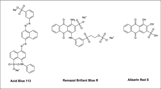
Dyes used for decolorization experiments
Extracellular Production of Recombinant HRP A2A (rHRP A2A)
In our previous study, we successfully achieved heterologous protein expression of HRP A2A by Komagataella phaffii (Pichia pastoris) [38]. The recombinant yeast cells were grown on YPD agar at 30 °C for 48–60 h. YPD agar was prepared by dissolving 5 g of YPD and 2 g of agar in 100 mL of purified water and sterilized by autoclaving at 121 °C for 15 min. After incubation, the cells were inoculated in 12.5 mL of pre-culture (BMGY, Buffered Minimal Glycerol-Complex Medium) medium containing 1% KH2PO4 (100 mM), 1% yeast extract, 1.34% YNB, 2% peptone, 1% glycerol, and 25 μL of 500 × B (0.02% Biotin) and then incubated at 30 °C at 225 rpm until the culture reached an OD600 value of approximately 2. After incubation, cells were harvested from culture medium by centrifugation and resuspended in 25 mL of the production (BMMY, Buffered Minimal Methanol-Complex Medium) medium containing 1% KH2PO4 (100 mM), 1% yeast extract, 1% casamino acid, 2% peptone, 1% sorbitol, 50 μL 500X B, and 1.34% YNB to induce protein expression. Methanol was added to the production medium every 24 h to a final concentration of 0.5% for induction maintenance. The obtained culture liquid at the end of 72 h of incubation was used to load onto the affinity column.
Purification of rHRP A2A and Enzyme Activity Assay
As obtained in our previous study, the rHRP A2A enzyme was purified in a single step using a 3-amino-4-chloro benzohydrazide affinity column [38]. Sodium dodecyl sulfate–polyacrylamide gel electrophoresis (SDS-PAGE) was performed to visualize the purified enzyme [39]. By measuring the absorbance increase brought on by the colored derivative compound (maximum: 420 nm) created by the oxidation of the o-dianisidine substrate in the presence of H2O2, the rHRP A2A enzyme activity was determined. The final reaction medium consisted of 0.8 mM o-dianisidine (200 μL), 60 mM phosphate buffer (pH 6.0) (600 μL), 2.25 mM H2O2 (100 μL), and 100 μL of enzyme. The activity (EU) was determined as the amount of enzyme catalyzing the formation of one µmol of derivative product per minute.
Effects of Metal Ions on Enzyme Activity
To determine the effects of different metal ions on the activity of the pure rHRP A2A isoenzyme, the initial activity of the enzyme was determined using a fixed volume of enzyme (100 µL) [40, 41]. Compounds: CaCI2. 2H2O, FeCI2. 4H2O, FeCI3. 6H2O, MnSO4. H2O, and Cu(NO3)2. 5H2O were used in the studies. Solutions of the compounds were prepared with distilled water. Before examining the effect of metal ions on the enzyme, the absorbance of the solutions containing metal and substrate in the enzyme-free medium was checked against the blank in the spectrophotometer. This control was performed before all studies. Prior to studying each metal ion, the enzyme’s activity was determined using the provided method. Then, various metal solution concentrations (0–1 mM) were added to the reaction medium (0.8 mM o-dianisidine (200 µL), 60 mM phosphate buffer (pH 6.0) (600 µL), 2.25 mM H2O2 (100 µL), and 100 µL of enzyme) while maintaining the same amount of enzyme (100 µL), and by measuring the absorbances with a spectrophotometer, the enzyme activity was recalculated. The results were evaluated together with the initial enzyme activity. The absorbance of the metals was checked against the blank in a spectrophotometer, and no absorbance was detected. This check was done before all studies.
Decolorization of Dyes with rHRP A2A
The parameters affecting the degree of biodegradation of peroxidase-catalyzed dye are the pH of the reaction mixture, dye concentration, hydrogen peroxide concentration, enzyme concentration, and temperature. The optimal pH value was determined by monitoring the biodegradation rate between pH 4–7.5. For this purpose, 50 mM buffers at pH 4–5 (acetate buffer) and pH 6–7 (phosphate buffer) were prepared and used. To determine the effect of enzyme concentration on dye removal, biodegradation was varied between 0.004 and 0.04 EU/mL and monitored for 24 h for rHRP A2A. Absorbance was recorded at different time intervals. Then, under optimal pH and enzyme concentration, the effect of dye and hydrogen peroxide concentration on biodegradation was examined. Dyes were prepared at concentrations ranging from 10 to 100 mg/L, and their absorbance was recorded at the wavelength of each dye. Enzyme and H2O2 were added to the dye solution prepared at the determined concentrations, and the solution was incubated. Absorbances were recorded at different times. Thus, the dye concentrations with the highest removal were determined. Then, while holding the other variables constant, the effects of pH, H2O2 concentration, enzyme quantity, and temperature on dye removal were assessed for each parameter under study. Also, color removal studies were performed with a redox mediator on the dye with the highest removal according to the % decolorization results. For this, various amounts of 1 mM stock ABTS solution were added to the reaction, and the resulting concentrations were calculated and graphed. The ABTS concentration providing the highest color removal was determined. It was compared with the dye solution without ABTS.
The decolorization of dyes was calculated as % by the following equation.
Ai: initial absorbance value of the dye;
At: absorbance value at the end of the specified time.
Result and Discussion
Production and Purification of rHRP A2A
Eukaryotic systems are utilized to produce recombinant HRP because native HRP has disulfide bridges and glycosylation. In 1992, hyperglycosylated HRP was produced using Saccharomyces cerevisiae [42]. Additionally, 19 different HRP isoenzymes and HRP mutants with higher activity and stability were produced in methylotrophic K. phaffii (P. pastoris) [43, 44]. In this study, the selected clone was grown in a BMMY medium with methanol added every 24 h for rHRP A2A isoenzyme expression in K. phaffii [45]. Methanol acts as an inducer and carbon source, while glycerol offers higher biomass yield but has repressive effects. Co-substrate glycerol increases biomass yield but inhibits the AOX1 promoter, reducing the amount of recombinant proteins produced [46,47,48,49]. The medium was supplemented with 1% (w/v) sorbitol as a carbon source and 1% (w/v) casamino acid, a hydrolysate of proteins, to reduce protease production [50].
The horseradish plant’s root contains a combination of distinct isoenzymes, including the HRP enzyme, and it is exceedingly challenging to isolate these enzymes [51]. Therefore, there is a need for alternative methods that can be faster and cheaper, where the enzyme can be obtained and purified more easily. Recombinant production is very advantageous since it makes it possible to produce the desired isoenzyme. As reported in the previous study, rHRP A2A isoenzyme was produced extracellularly in K. phaffii cells and purified by affinity chromatography in a single step [38]. In this study, the recombinant enzyme produced and purified using the same method was demonstrated by silver staining after being run on SDS-PAGE (Fig. 2).

SDS-PAGE analysis of purified rHRP A2A (M: protein marker, 1: purified enzyme)
The Effect of Metal Ions on the Activity of rHRP A2A
Peroxidases require different metal ions as their prosthetic groups or activators to perform their oxidative agent function [52,53,54]. In general, metal ions can coordinate with active site residues and lead to activation. Conversely, such coordination can also cause inhibition by blocking substrate interaction [55]. Recently, the effects of some metals on the functional stability of peroxidases have been reported [56]. The activation, inhibition, and denaturation effects of transition metals (Mn2+, Co2+, Ni2+, and Cu2+) on HRP in the presence of guaiacol as an aromatic donor were investigated. Depending on the applied concentration, they have been shown to affect horseradish peroxidase (HRP) activity. Such metal ions are strong inducers of the conformational and functional stability of HRP [57].
The effect of various metal ions (final concentration 0.2 mM) such as Cu2+, Fe2+, Ni2+, Hg2+, Ca2+, Mg2+, Zn2+, Mn2+, and K+ on Citrus medica leaf peroxidase enzyme activity was investigated. The enzyme activity was observed to rise in the presence of Cu2+, Co2+, Mg2+, and K+, but to drop to 84.33, 39.26, 62.33, and 63% of the initial activity in the presence of Hg2+, Fe2+, Mn2+, and Zn2+ [58]. In light of the literature, the effect of metal ions on pure rHRP A2A isoenzyme activity was investigated in this study. The activities of purified rHRP A2A isoenzyme were determined by adding salts containing Mn2+, Fe2+, Fe3+, and Ca2+ metal ions in the range of 0.03–0.6 mM. The activity of the control group (without addition of metal ions) was compared (Fig. 3). Peroxidases perform oxidation–reduction reactions with iron ions in their active center [59]. Iron is considered essential for the activities of plant peroxidases because it plays a role in H2O2 binding and the formation of compound I [60]. The effects of chloride salts of metal ions Ca2+, Mn2+, and Fe3+ at different concentrations on the enzyme were investigated by Goyal and Chugh. It was stated that the addition of Fe3+ increased the activity of peroxidase isolated from pearl millet grains, but Mn2+ moderately inhibited the enzyme activity [61]. There is also evidence for the inhibitory effects of Mn2+ (MnCI2) on peroxidase isolated from cotton cell suspension [62] and black lentil peels [63].

Effect of metal ions on rHRP A2A activity. Effect of A Fe2+, B Fe3+, C Mn2+, and D Ca2+. Control: without addition of metal ions
The data showed that the activity of pure rHRP A2A isoenzyme increased by 158.62 and 79.54%, respectively, when 0.150 mM Fe2+ and 0.473 mM Mn2+ were added to the activity measurement medium. The results revealed that the Fe2+ ion is an important factor in the activity of the rHRP A2A isoenzyme.
Ca2+ is a cofactor that helps to maintain the conformational integrity of the active site of the peroxidase enzyme [64]. Avocado, cereal, and wheat germ peroxidase enzyme activities have been reported to be activated by Ca2+ [65, 66]. Onsa et al. reported that the activities of mPOD isoenzymes isolated from Metroxylon sagu were variously affected by the presence of metal ions. It was determined that 1 mM Fe3+ significantly increased the activity of both mPOD-I (339%) and mPOD-II (328%) [67]. However, while it was observed that Fe3 + ion caused inhibition, the enzyme activity remained stable in a certain concentration range of Ca2+ ions. This could imply that metals do not have the same effect on the activities of various source peroxidases and isoenzymes. It was observed that the Cu2+-containing compound interacted with the substrate and formed a precipitate, and it was determined that the formed particles caused an increase in absorbance. The impact of Cu2+ ions on enzyme activity could not therefore be investigated.
Decolorization of Dyes with rHRP A2A
Isolated enzymes are highly efficient and considered environmentally friendly, as they are highly specific catalysts that produce by-products with lower toxicity and volume. The enzymes responsible for dye degradation mainly belong to the family of oxidoreductases, including peroxidases, reductases, and laccases [68, 69]. Horseradish peroxidase (HRP), the most important representative of peroxidases, is the most studied enzyme derived from plant material, used as a very effective biocatalyst for the treatment of various recalcitrant pollutants (i.e., dyes, phenols) in wastewater [70,71,72,73,74].
Optimization conditions vary in enzymatic dye degradation studies. Souza et al. studied the decolorization of different dyes and observed that the reaction time is directly dependent on the different structures of the dyes, resulting in variation in the reaction time [75]. In the present study, the decolorization potential of pure rHRP A2A isoenzyme on one azo dye (Acid blue 113) and two anthraquinone dyes (Alizarin red and Remazol brillant blue), which are widely used in the textile field, was investigated, and optimization studies were carried out. The % decolorization rates of Acid blue 113, Remazol brilliant blue, and Alizarin red dyes with a concentration of 10 mg/L at the end of 1, 3, and 5 h were calculated and graphed. The graphs show that for all three dyes, the greatest amount of decolorization occurred after the 5 th hour. The dye with the best color decolorization was Acid blue 113 (71.27%), followed by Alizarin red (62.26%) and Remazol brilliant blue (31.22%) (Fig. 4A). According to the % decolorization values obtained with the buffers used in the pH 4–7 range to determine the effect of pH on the color decolorization of the dyes studied, for Acid blue 113, the maximum decolorization was attained at pH 6.0, whereas Remazol brilliant blue and Alizarin red dyes accomplished this at pH 4.5 (Fig. 4B). Enzymes isolated from different sources have also different optimum pHs. Trichosanthes dioica peroxidase functioned better in acidic conditions with a pH range of 3–5, whereas its decolorizing/degrading activity was negatively affected in alkaline conditions [76]. Degradation of industrially important dyes by enzymes such as horseradish peroxidase, polyphenol oxidase, bitter melon peroxidase, and laccase is maximal in acidic pH buffers [77]. In decolorization experiments with HRP, a decrease in the activity of HRP was observed at pH values above 6.0 [78]. The optimum pH for rHRP A2A enzyme activity was determined as 6.0 in our previous study [38]. In the literature, to determine the HRP-catalyzed degradation of Remazol brilliant blue, the absorbance decrease of 15 mg/L dye was determined at various pH values (3.0–8.0) with plant-derived HRP enzyme (0.14 EU/mL) [78]. For Remazol brilliant blue, the HRP enzyme showed much better dye decolorization at acidic pH than at neutral pH [79].

A The change of dye decolorization with time. B Effect of pH. C. Effect of temperature. D. Effect of H2O2 concentration
To determine the effect of temperature on dye removal, dye absorbance changes between 20 and 55 °C were determined, and % decolorization values were calculated. The reaction temperature is an important parameter affecting the color removal of dyes. In the literature, it was determined that the optimal temperature varies according to the structure of the dye in the decolorization studies carried out with HRP [36]. It is believed that the decrease in dye removal at high temperatures may be linked to the loss of enzymatic activity [80,81,82]. According to the obtained data, the maximum % decolorization of Acid blue 113 was reached at 45 °C. The best decolorization for Remazol brilliant blue was determined at 25 °C. No effect of temperature on decolorization for Alizarin red dye was observed (Fig. 4C). As seen in the graph, besides the different effects of temperature on each dye, the decrease in dye removal at increasing temperatures suggests that it may be related to the deactivation of the enzyme.
To determine the effect of H2O2 on dye decolorization, H2O2 was added to the reaction medium in the range of 0.0075–0.75 mM. Absorbances were determined spectrophotometrically, and % decolorization was calculated. As seen in the graph, maximum decolorization of Acid blue 113 was observed in the presence of 0.375 mM H2O2. As for Remazol brilliant blue and Alizarin red, the highest % decolorization was observed in the presence of 0.09 mM and 0.0375 mM H2O2, respectively (Fig. 4D). The sensitivity of peroxidases to high concentrations of H2O2 has also been reported in previous studies [30, 31, 82,83,84,85]. It has also been observed that low concentrations of H2O2 inhibit enzyme action, and excess of this reagent causes enzyme inactivation [86]. According to the results of this study, the concentration of H2O2 required for maximum oxidation of each dye has shown changes. The difference in the level of oxidation between dyes with various structures explains this. In addition, decolorization percentages decreased as the H2O2 concentration increased. This indicates that high H2O2 inhibits the enzyme as stated in the literature.
In the decolorization studies performed at different dye concentrations (10–100 mg/L), the maximum % decolorization of Acid blue 113, Alizarin red, and Remazol brilliant blue for 10 mg/L dye was 71.27, 62.26, and 31.22%, respectively. No removal was observed at dye concentrations above 25 mg/L (Fig. 5).

Graph of % decolorization at different dye concentrations
It was also aimed to increase the color removal by increasing the enzyme activity for Acid blue 113 dye, which was best removed with 71.27% decolorization for 10 mg/L dye concentration. For this purpose, dye decolorization was studied using rHRP A2A with increasing activity in the range of 0.004–0.04 EU/mL given in Fig. 6A. While 71.27% decolorization was obtained in 5 h with 0.006 EU/mL activity, 74.83% dye % decolorization was achieved when the activity was increased to 0.04 EU/mL. Consequently, it was determined that dye removal increased as the enzyme activity increased. The enzyme activity also increased with the addition of metal ions. Thus, it enabled dye removal studies to be carried out using less amount (mL) of the enzyme. Sadhanandam et al. used 0.08 EU/mL plant-derived HRP enzyme and degraded 30 mg/L acid blue 113 dye up to 75% in 45 min at pH 6.6 and 30 °C [87]. Compared to this study, three times more dye can be removed by using 13 times more active enzymes with about 75%, while 71.27% decolorization of Acid blue 113 dye was achieved with rHRP A2A with an activity of 0.006 EU/mL. This indicates that even at low enzyme activity, high decolorization rates can be attained.

A Enzyme activity (EU/mL)-%decolorization graph. B ABTS (mM)-% decolorization graph
Additionally, for the Acid blue 113 dye with the best removal, % decolorization values were calculated and graphed by adding different concentrations of ABTS to the medium. According to Husain (2006), enzymes alone cannot degrade some recalcitrant dyes, and therefore, they need some low molecular weight redox mediators to increase the dye degradation efficiency. Redox mediators speed up the degradation process by moving electrons from biological electron donors to electron acceptor dye compounds [88]. A redox mediator such as ABTS is known to increase substrate diversity and accelerate the decolorization efficiency of resistant textile dyes [89]. In this context, the decolorization of Acid blue 113 dye was examined by adding ABTS at different concentrations. According to the results, in the non-ABTS % decolorization study, 12.74% removal was detected after 3 h, while 33.123% decolorization was achieved after 2 h with the addition of 0.003 mM ABTS (Fig. 6B). Thus, it was demonstrated that the presence of a redox mediator at the appropriate concentration provides dye decolorization in a shorter time and at a higher rate.
Conclusion
This study highlights the potential of the rHRP A2A enzyme as an effective biological agent for the decolorization of toxic dyes Acid blue 113, Alizarin red, and Remazol brilliant blue R. While numerous physical and chemical methods have been proposed for the decolorization of synthetic dyes, these approaches often have drawbacks, including high costs, low efficiency, and energy-intensive processes. In contrast, biodegradation of dyes using enzymes is recognized as a cost-effective and environmentally friendly option. In this study, the azo and anthraquinone dye removal potential of the rHRP A2A enzyme, which was produced extracellularly by recombinant K. phaffii cells and purified in a single step by the affinity chromatography technique using the 3-amino-4-chloro benzohydrazide ligand, was demonstrated. The optimum conditions for maximum dye degradation, including time, pH, temperature, dye concentration, and substrate concentration, were determined. Under these optimal conditions, the highest percentage of decolorization was achieved for Acid blue 113 dye, with a removal rate of 71.27%. This was followed by Alizarin red at 62.26% and Remazol brilliant blue R at 31.22%. These results indicate that rHRP A2A has significant decolorization capabilities, particularly for Acid blue 113 and Alizarin red dyes, highlighting its potential for treating textile wastewater.
Data Availability
Data will be made available on reasonable request.
References
Othman, A. M., Elsayed, M. A., Elshafei, A. M., & Hassan, M. M. (2018). Purification and biochemical characterization of two isolated laccase isoforms from Agaricus bisporus CU13 and their potency in dye decolorization. International Journal of Biological Macromolecules, 1(113), 1142–1148. https://doi.org/10.1016/j.ijbiomac.2018.03.043
Vikrant, K., Giri, B. S., Raza, N., Roy, K., Kim, K. H., Rai, B. N., & Singh, R. S. (2018). Recent advancements in bioremediation of dye: Current status and challenges. Bioresource Technology. https://doi.org/10.1016/j.biortech.2018.01.029
P Shah, M., KA, P., & SS, N. (2013). Bioremoval of azo dye reactive red by Bacillus spp. ETL-1982. Journal of Bioremediation & Biodegradation, 4(3), 1000186. https://doi.org/10.4172/2155-6199.1000186
Pattnaik, P., Dangayach, G. S., & Bhardwaj, A. K. (2018). A review on the sustainability of textile industries wastewater with and without treatment methodologies. Reviews on Environmental Health. https://doi.org/10.1515/reveh-2018-0013
Meng, X., Liu, G., Zhou, J., Shiang, F. Q., & Wang, G. (2012). Azo dye decolorization by Shewanella aquimarina under saline conditions. Bioresource Technology, 114, 95–101. https://doi.org/10.1016/j.biortech.2012.03.003
Fernandes, F. H., Bustos-Obregon, E., & Salvadori, D. M. F. (2015). Disperse Red 1 (textile dye) induces cytotoxic and genotoxic effects in mouse germ cells. Reproductive Toxicology, 53, 75–81. https://doi.org/10.1016/j.reprotox.2015.04.002
Rápó, E., & Tonk, S. (2021). Factors affecting synthetic dye adsorption; desorption studies: A review of results from the last five years (2017–2021). Molecules. https://doi.org/10.3390/molecules26175419
Khan, N. A., Saeed, K., Khan, I., Gul, T., Sadiq, M., Uddin, A., & Zekker, I. (2022). Efficient photodegradation of orange II dye by nickel oxide nanoparticles and nanoclay supported nickel oxide nanocomposite. Applied Water Science, 12(132). https://doi.org/10.1007/s13201-022-01647-x
Ngo, A. C. R., & Tischler, D. (2022). Microbial degradation of azo dyes: Approaches and prospects for a hazard-free conversion by microorganisms. International Journal of Environmental Research and Public Health. https://doi.org/10.3390/ijerph19084740
Routoula, E., & Patwardhan, S. V. (2020). Degradation of anthraquinone dyes from effluents: A review focusing on enzymatic dye degradation with industrial potential. Environmental Science and Technology. https://doi.org/10.1021/acs.est.9b03737
Rawat, D., Mishra, V., & Sharma, R. S. (2016). Detoxification of azo dyes in the context of environmental processes. Chemosphere. https://doi.org/10.1016/j.chemosphere.2016.04.068
Jin, X., Li, S., Long, N., & Zhang, R. (2018). A robust and stable nano-biocatalyst by co-immobilization of chloroperoxidase and horseradish peroxidase for the decolorization of azo dyes. Journal of Chemical Technology & Biotechnology, 93(2), 489–497. https://doi.org/10.1002/jctb.5379
Forootanfar, H., Rezaei, S., Zeinvand-Lorestani, H., Tahmasbi, H., Mogharabi, M., Ameri, A., & Faramarzi, M. A. (2016). Studies on the laccase-mediated decolorization, kinetic, and microtoxicity of some synthetic azo dyes. Journal of Environmental Health Science and Engineering, 14, 7–9. https://doi.org/10.1186/s40201-016-0248-9
Asghar, A., Bello, M. M., Raman, A. A. A., Daud, W. M. A. W., Ramalingam, A., & Zain, S. B. M. (2019). Predicting the degradation potential of Acid blue 113 by different oxidants using quantum chemical analysis. Heliyon, 5, 1–11. https://doi.org/10.1016/j.heliyon.2019.e02396
Pai, S., Kini, M. S., Rangasamy, G., & Selvaraj, R. (2023). Mesoporous calcium hydroxide nanoparticle synthesis from waste bivalve clamshells and evaluation of its adsorptive potential for the removal of Acid Blue 113 dye. Chemosphere, 313, 137476. https://doi.org/10.1016/j.chemosphere.2022.137476
Hadibarata, T., Yusoff, A. R. M., & Kristanti, R. A. (2012). Decolorization and metabolism of anthraquionone-type dye by laccase of white-rot fungi polyporus sp. S133. Water, Air, and Soil Pollution, 223, 933–941. https://doi.org/10.1007/s11270-011-0914-6
Kwok, W. Y., Xin, J. H., & Sin, K. M. (2002). Quantitative prediction of the degree of pollution of effluent from reactive dye mixtures. Coloration Technology, 118(4), 174–180. https://doi.org/10.1111/j.1478-4408.2002.tb00096.x
Robinson, T., McMullan, G., Marchant, R., & Nigam, P. (2001). Remediation of dyes in textile effluent: A critical review on current treatment technologies with a proposed alternative. Bioresource Technology, 77(3), 247–255. https://doi.org/10.1016/S0960-8524(00)00080-8
McMullan, G., Meehan, C., Conneely, A., Kirby, N., Robinson, T., Nigam, P., … Smyth, W. F. (2001). Microbial decolourisation and degradation of textile dyes. Applied Microbiology and Biotechnology, 50(1–2), 81–87. https://doi.org/10.1007/s002530000587
Arroyo-Figueroa, G., Ruiz-Aguilar, G. M. L., López-Martínez, L., González-Sánchez, G., Cuevas-Rodríguez, G., & Rodríguez-Vázquez, R. (2011). Treatment of a textile effluent from dyeing with cochineal extracts using Trametes versicolor fungus. TheScientificWorldJOURNAL, 11, 1005–1016. https://doi.org/10.1100/tsw.2011.99
Andleeb, S., Atiq, N., Robson, G. D., & Ahmed, S. (2012). An investigation of anthraquinone dye biodegradation by immobilized Aspergillus flavus in fluidized bed bioreactor. Environmental Science and Pollution Research, 19(5), 1728–1737. https://doi.org/10.1007/s11356-011-0687-x
Husain, Q. (2010). Peroxidase mediated decolorization and remediation of wastewater containing industrial dyes: A review. Reviews in Environmental Science and Biotechnology, 9(2), 117–140. https://doi.org/10.1007/s11157-009-9184-9
Arabaci, G., & Usluoglu, A. (2014). The enzymatic decolorization of textile dyes by the immobilized polyphenol oxidase from quince leaves. The Scientific World Journal, 2014, 1–5. https://doi.org/10.1155/2014/685975
Ollikka, P., Harjunpää, T., Palmu, K., Mäntsälä, P., & Suominen, I. (1998). Oxidation of crocein orange G by lignin peroxidase isoenzymes kinetics and effect of H2O2. Applied Biochemistry and Biotechnology - Part A Enzyme Engineering and Biotechnology, 75(2–3), 307–321. https://doi.org/10.1007/BF02787783
Ghasemi, F., Tabandeh, F., Bambai, B., & Sambasiva Rao, K. R. S. (2010). Decolorization of different azo dyes by phanerochaete chrysosporium RP78 under optimal condition. International Journal of Environmental Science and Technology, 7(3), 457–464. https://doi.org/10.1007/BF03326155
Bansal, N., & Kanwar, S. S. (2013). Peroxidase(s) in environment protection. The Scientific World Journal. https://doi.org/10.1155/2013/714639
Zhang, A., Fang, L., Wang, J., & Liu, W. (2013). Enzymatic decolorization of Orange II: Optimization by response surface methodology and pathway. Environmental Progress and Sustainable Energy, 32(2), 294–301. https://doi.org/10.1002/ep.11628
Liu, L., Zhang, J., Tan, Y., Jiang, Y., Hu, M., Li, S., & Zhai, Q. (2014). Rapid decolorization of anthraquinone and triphenylmethane dye using chloroperoxidase: Catalytic mechanism, analysis of products and degradation route. Chemical Engineering Journal, 244, 9–18. https://doi.org/10.1016/j.cej.2014.01.063
Robinson, P. K. (2015). Enzymes: Principles and biotechnological applications. Essays in Biochemistry, 59, 1–41. https://doi.org/10.1042/BSE0590001
Ulson de Souza, S. M. A. G., Forgiarini, E., & Ulson de Souza, A. A. (2007). Toxicity of textile dyes and their degradation by the enzyme horseradish peroxidase (HRP). Journal of Hazardous Materials. https://doi.org/10.1016/j.jhazmat.2007.06.003
Mohan, S. V., Prasad, K. K., Rao, N. C., & Sarma, P. N. (2005). Acid azo dye degradation by free and immobilized horseradish peroxidase (HRP) catalyzed process. Chemosphere, 58(8), 1097–1105. https://doi.org/10.1016/j.chemosphere.2004.09.070
Mohamed, S. A., Elaraby, N. M., Abdel-Aty, A. M., Shaban, E., Abu-Saied, M. A., Kenawy, E. R., & El-Naggar, M. E. (2021). Improvement of enzymatic properties and decolorization of azo dye: Immobilization of horseradish peroxidase on cationic maize starch. Biocatalysis and Agricultural Biotechnology, 38, 102208. https://doi.org/10.1016/j.bcab.2021.102208
Urrea, D. A. M., Gimenez, A. V. F., Rodriguez, Y. E., & Contreras, E. M. (2021). Immobilization of horseradish peroxidase in Ca-alginate beads: Evaluation of the enzyme leakage on the overall removal of an azo-dye and mathematical modeling. Process Safety and Environmental Protection, 156, 134–143. https://doi.org/10.1016/j.psep.2021.10.006
Zhumabekova, A., Noma, S. A. A., Tümay Özer, E., & Osman, B. (2024). Decolorization of Congo Red and Reactive Black 5 dyes with horseradish peroxidase-immobilized cross-linked polymeric microbeads. Arabian Journal for Science and Engineering. https://doi.org/10.1007/s13369-024-08748-6
Celebi, M., Kaya, M. A., Altikatoglu, M., & Yildirim, H. (2013). Enzymatic decolorization of anthraquinone and diazo dyes using horseradish peroxidase enzyme immobilized onto various polysulfone supports. Applied Biochemistry and Biotechnology, 171, 716–730. https://doi.org/10.1007/s12010-013-0377-x
Šekuljica, N. Ž, Prlainović, N. Ž, Stefanović, A. B., Žuža, M. G., Čičkarić, D. Z., Mijin, D. Ž, & Knežević-Jugović, Z. D. (2015). Decolorization of anthraquinonic dyes from textile effluent using horseradish peroxidase: Optimization and kinetic study. The Scientific World Journal, 2015, 1–12. https://doi.org/10.1155/2015/371625
Šekuljica, N., Jovanović, J. R., Jakovetić Tanasković, S. M., Ognjanović, N. D., Gazikalović, I. V., Knežević-Jugović, Z. D., & Mijin, D. (2020). Immobilization of horseradish peroxidase onto Purolite® A109 and its anthraquinone dye biodegradation and detoxification potential. Biotechnology Progress. https://doi.org/10.1002/btpr.2991
Acar, M., Abul, N., Yildiz, S., Taskesenligil, E. D., Gerni, S., Unver, Y., … Ozdemir, H. (2022). Affinity-based and in a single step purification of recombinant horseradish peroxidase A2A isoenzyme produced by Pichia pastoris. Bioprocess and Biosystems Engineering, 46(4), 523–534. https://doi.org/10.1007/s00449-022-02837-2
Laemmli, U. K. (1970). Most commonly used discontinuous buffer system for SDS electrophoresis. Nature, 68(227), 0–85.
Kucuk, M., & Gulcin, İ. (2016). Purification and characterization of the carbonic anhydrase enzyme from Black Sea trout (Salmo trutta Labrax Coruhensis) kidney and inhibition effects of some metal ions on enzyme activity. Environmental Toxicology and Pharmacology, 44, 134–139. https://doi.org/10.1016/j.etap.2016.04.011
Kocyigit, U. M., Taslimi, P., & Gulçin, İ. (2018). Characterization and inhibition effects of some metal ions on carbonic anhydrase enzyme from Kangal Akkaraman sheep. Journal of Biochemical and Molecular Toxicology, 32(8). https://doi.org/10.1002/jbt.22172
Vlamis-Gardikas, A., Smith, A. T., Clements, J. M., & Burke, J. F. (1992). Expression of active horseradish peroxidase in Saccharomyces cerevisiae. In Biochemical Society Transactions. https://doi.org/10.1042/bst020111s
Morawski, B., Lin, Z., Cirino, P., & Joo, H. (2000). Functional expression of horseradish peroxidase in Saccharomyces cerevisiae and Pichia pastoris. Protein Engineering, 13(5), 377–84. https://doi.org/10.1093/protein/13.5.377
Krainer, F. W., Pletzenauer, R., Rossetti, L., Herwig, C., Glieder, A., & Spadiut, O. (2014). Purification and basic biochemical characterization of 19 recombinant plant peroxidase isoenzymes produced in Pichia pastoris. Protein Expression and Purification, 95, 104–112. https://doi.org/10.1016/j.pep.2013.12.003
He, D., Luo, W., Wang, Z., Lv, P., & Yuan, Z. (2015). Combined use of GAP and AOX1 promoters and optimization of culture conditions to enhance expression of Rhizomucor miehei lipase. Journal of Industrial Microbiology and Biotechnology, 42(8), 1175–1182. https://doi.org/10.1007/s10295-015-1633-6
Xie, J., Zhou, Q., Du, P., Gan, R., & Ye, Q. (2005). Use of different carbon sources in cultivation of recombinant Pichia pastoris for angiostatin production. Enzyme and Microbial Technology, 36(2–3), 210–216. https://doi.org/10.1016/j.enzmictec.2004.06.010
Jungo, C., Schenk, J., Pasquier, M., Marison, I. W., & von Stockar, U. (2007). A quantitative analysis of the benefits of mixed feeds of sorbitol and methanol for the production of recombinant avidin with Pichia pastoris. Journal of Biotechnology, 131(1), 57–66. https://doi.org/10.1016/j.jbiotec.2007.05.019
Zhu, T., Hang, H., Chu, J., Zhuang, Y., Zhang, S., & Guo, M. (2013). Transcriptional investigation of the effect of mixed feeding to identify the main cellular stresses on recombinant Pichia pastoris. Journal of Industrial Microbiology and Biotechnology, 40(2), 183–189. https://doi.org/10.1007/s10295-012-1225-7
Ünver, Y., Kurbanoğlu, E. B., & Erdoğan, O. (2015). Expression, purification, and characterization of recombinant human paraoxonase 1 (rhPON1) in Pichia pastoris. Turkish Journal of Biology, 39(4), 649–655. https://doi.org/10.3906/biy-1501-43
Unver, Y., Sensoy Gun, B., Acar, M., & Yildiz, S. (2021). Heterologous expression of azurin from Pseudomonas aeruginosa in the yeast Pichia pastoris. Preparative Biochemistry and Biotechnology, 51(7), 723–730. https://doi.org/10.1080/10826068.2020.1855444
Passardi, F., Cosio, C., Penel, C., & Dunand, C. (2005). Peroxidases have more functions than a Swiss army knife. Plant Cell Reports, 24(5), 255–265. https://doi.org/10.1007/s00299-005-0972-6
Koksal, E., & Gulcin, I. (2008). Purification and characterization of peroxidase from cauliflower (Brassica oleracea L. var. botrytis) buds. Protein & Peptide Letters, 15(4), 320–326. https://doi.org/10.2174/092986608784246506
Koksal, E., Bursal, E., Aggul, A. G., & Gulcin, I. (2012). Purification and characterization of peroxidase from sweet gourd (Cucurbita moschata Lam. Poiret). International Journal of Food Properties, 15(5), 1110–1119. https://doi.org/10.1080/10942912.2010.513216
Altın, S., Tohma, H., Gülçin, İ., & Köksal, E. (2017). Purification, characterization, and inhibition sensitivity of peroxidase from wheat (Triticum aestivum ssp. vulgare). International Journal of Food Properties, 20(9), 1949–1959. https://doi.org/10.1080/10942912.2016.1225308
Shi, X., Dalal, N. S., & Kasprzak, K. S. (1992). Generation of free radicals from lipid hydroperoxides by Ni2+ in the presence of oligopeptides. Archives of Biochemistry and Biophysics, 299(1), 154–162. https://doi.org/10.1016/0003-9861(92)90257-W
Haschke, R. H., & Friedhoff, J. M. (1978). Calcium-related properties of horseradish peroxidase. Biochemical and Biophysical Research Communications, 80(4), 1039–1042. https://doi.org/10.1016/0006-291X(78)91350-5
Mahmoudi, A., Nazari, K., Mohammadian, N., & Moosavi-Movahedi, A. A. (2003). Effect of Mn2+, Co2+, Ni2+, and Cu2+ on horseradish peroxidase: Activation, inhibition, and denaturation studies. Applied Biochemistry and Biotechnology, 104(1), 81–94. https://doi.org/10.1385/ABAB:104:1:81
Mall, R., Naik, G., Mina, U., & Mishra, S. K. (2013). Purification and characterization of a thermostable soluble peroxidase from Citrus medica LEAF. Preparative Biochemistry and Biotechnology, 43(2), 137–151. https://doi.org/10.1080/10826068.2012.711793
Veitch, N. C. (2004). Horseradish peroxidase: A modern view of a classic enzyme. Phytochemistry, 65(3), 249–259. https://doi.org/10.1016/j.phytochem.2003.10.022
Whitaker, J. R. (1995). Principles of enzymology for the food sciences. Routledge. https://doi.org/10.1201/9780203742136
Goyal, P., & Chugh, L. K. (2014). Partial purification and characterization of peroxidase from pearl millet (Pennisetum glaucum [L.] R. Br.) grains. Journal of Food Biochemistry, 38(2), 150–158. https://doi.org/10.1111/jfbc.12033
Kouakou, T. H., Dué, E. A., Kouadio, N. E. J. P., Niamké, S., Kouadio, Y. J., & Mérillon, J.-M. (2009). Purification and characterization of cell suspensions peroxidase from cotton (Gossypium hirsutum L.). Applied Biochemistry and Biotechnology, 157(3), 575–592. https://doi.org/10.1007/s12010-008-8287-z
Ajila, C. M., & Prasada Rao, U. J. S. (2009). Purification and characterization of black gram (Vigna mungo) husk peroxidase. Journal of Molecular Catalysis B: Enzymatic, 60(1–2), 36–44. https://doi.org/10.1016/j.molcatb.2009.03.014
Adams, J. (1996). Regenerated and denatured peroxidase as potential lipid oxidation catalysts. Food Chemistry, 57(4), 505–514. https://doi.org/10.1016/S0308-8146(96)00007-6
Converso, D. A., & Fernández, M. E. (1996). Ca2+ activation of wheat peroxidase: A possible physiological mechanism of control. Archives of Biochemistry and Biophysics, 333(1), 59–65. https://doi.org/10.1006/abbi.1996.0364
Billaud, C., Louarme, L., & Nicolas, J. (1999). Comparison of peroxidases from barley kernel (Hordeum vulgare L.) and wheat germ (Triticum aestivum L.): Isolation and preliminary characterization. Journal of Food Biochemistry, 23(2), 145–172. https://doi.org/10.1111/j.1745-4514.1999.tb00011.x
Onsa, G. H., Saari, N. B., Selamat, J., & Bakar, J. (2004). Purification and characterization of membrane-bound peroxidases from Metroxylon sagu. Food Chemistry, 85(3), 365–376. https://doi.org/10.1016/j.foodchem.2003.07.013
Fu, Y., & Viraraghavan, T. (2001). Fungal decolorization of dye wastewaters: A review. Bioresource Technology, 79(3), 251–262. https://doi.org/10.1016/S0960-8524(01)00028-1
Kaushik, P., & Malik, A. (2009). Fungal dye decolourization: Recent advances and future potential. Environment International, 35(1), 127–141. https://doi.org/10.1016/j.envint.2008.05.010
Wagner, M., & Nicell, J. A. (2005). Evaluation of horseradish peroxidase for the treatment of estrogenic alkylphenols. Water Quality Research Journal, 40(2), 145–154. https://doi.org/10.2166/wqrj.2005.017
Šekuljica, N. Ž., Prlainović, N. Ž., Jakovetić, S. M., Grbavčić, S. Ž., Ognjanović, N. D., Knežević‐Jugović, Z. D., & Mijin, D. Ž. (2016). Removal of anthraquinone dye by cross‐linked enzyme aggregates from fresh horseradish extract. CLEAN – Soil, Air, Water, 44(7), 891–900. https://doi.org/10.1002/clen.201500766
Rajesh Ahirwar, Jai G. Sharma, Bhanumati Singh, Krishan Kumar, Pradip Nahar, & Saroj Kumar. (2017). A simple and efficient method for removal of phenolic contaminants in wastewater using covalent immobilized horseradish peroxidase. Journal of Materials Science and Engineering B, 7(1). https://doi.org/10.17265/2161-6221/2017.1-2.004
Ely, C., Lourdes Borba Magalhães, M. de, Henrique Lemos Soares, C., & Skoronski, E. (2017). Optimization of phenol removal from biorefinery effluent using horseradish peroxidase. Journal of Environmental Engineering, 143(12). https://doi.org/10.1061/(ASCE)EE.1943-7870.0001279
Shen, S., Wang, Q., Shu, J., Ma, L., Chen, L., & Xu, Y. (2019). Optimization of horseradish peroxidase catalytic degradation for 2-nethyl-6-ethylaniline removal using response surface methodology. Water, 11(5), 1093. https://doi.org/10.3390/w11051093
Ulson de Souza, S. M. A. G., Forgiarini, E., & Ulson de Souza, A. A. (2007). Toxicity of textile dyes and their degradation by the enzyme horseradish peroxidase (HRP). Journal of Hazardous Materials, 147(3), 1073–1078. https://doi.org/10.1016/j.jhazmat.2007.06.003
Jamal, F., Qidwai, T., Pandey, P. K., Singh, R., & Singh, S. (2011). Azo and anthraquinone dye decolorization in relation to its molecular structure using soluble Trichosanthes dioica peroxidase supplemented with redox mediator. Catalysis Communications, 12(13), 1218–1223. https://doi.org/10.1016/j.catcom.2011.04.012
Galindo, C., Jacques, P., & Kalt, A. (2000). Photodegradation of the aminoazobenzene acid orange 52 by three advanced oxidation processes: UV/H2O2, UV/TiO2 and VIS/TiO2. Journal of Photochemistry and Photobiology A: Chemistry, 130(1), 35–47. https://doi.org/10.1016/S1010-6030(99)00199-9
Bhunia, A., Durani, S., & Wangikar, P. P. (2001). Horseradish peroxidase catalyzed the degradation of industrially important dyes. Biotechnology and Bioengineering. https://doi.org/10.1002/1097-0290(20010305)72:5%3c562::AID-BIT1020>3.0.CO;2-S
Bhunia, A., Durani, S., & Wangikar, P. P. (2001). Horseradish peroxidase catalyzed degradation of industrially important dyes. Biotechnology and Bioengineering, 72(5), 562–567. https://doi.org/10.1002/1097-0290(20010305)72:5%3c562::AID-BIT1020>3.0.CO;2-S
Liu, J.-Z., Wang, T.-L., & Ji, L.-N. (2006). Enhanced dye decolorization efficiency by citraconic anhydride-modified horseradish peroxidase. Journal of Molecular Catalysis B: Enzymatic, 41(3–4), 81–86. https://doi.org/10.1016/j.molcatb.2006.04.011
Sakuyama, H., Endo, Y., Fujimoto, K., & Hatana, Y. (2003). Oxidative degradation of alkylphenols by horseradish peroxidase. Journal of Bioscience and Bioengineering, 96(3), 227–231. https://doi.org/10.1016/S1389-1723(03)80186-X
Masuda, M., Sakurai, A., & Sakakibara, M. (2001). Effect of reaction conditions on phenol removal by polymerization and precipitation using Coprinus cinereus peroxidase. Enzyme and Microbial Technology, 28(4–5), 295–300. https://doi.org/10.1016/S0141-0229(00)00333-1
Ferreira-Leitao, V. S., de Carvalho, M. E. A., & Bon, E. P. (2007). Lignin peroxidase efficiency for methylene blue decolouration: Comparison to reported methods. Dyes and Pigments, 74(1), 230–236. https://doi.org/10.1016/j.dyepig.2006.02.002
Hong-Mei, L., & Nicell, J. A. (2008). Biocatalytic oxidation of bisphenol A in a reverse micelle system using horseradish peroxidase. Bioresource Technology, 99(10), 4428–4437. https://doi.org/10.1016/j.biortech.2007.08.072
Ferreira-Leitão, V. S., da Silva, J. G., & Bon, E. P. (2003). Methylene blue and azure B oxidation by horseradish peroxidase: A comparative evaluation of class II and class III peroxidases. Applied Catalysis B: Environmental, 42(2), 213–221. https://doi.org/10.1016/S0926-3373(02)00238-2
da Silva, M. R., de Sá, L. R. V., Russo, C., Scio, E., & Ferreira-Leitão, V. S. (2010). The use of HRP in decolorization of reactive dyes and toxicological evaluation of their products. Enzyme Research, 2010, 1–7. https://doi.org/10.4061/2010/703824
Sadhanandam, P., Anumary, A., Ashokkumar, M., & Thanikaivelan, P. (2013). Probing horseradish peroxidase catalyzed degradation of azo dye from tannery wastewater. SpringerPlus, 341. https://doi.org/10.1186/2193-1801-2-341
Husain, Q. (2006). Potential applications of the oxidoreductive enzymes in the decolorization and detoxification of textile and other synthetic dyes from polluted water: A review. Critical Reviews in Biotechnology, 26(4), 201–221. https://doi.org/10.1080/07388550600969936
Van der Zee, F. P., & Cervantes, F. J. (2009). Impact and application of electron shuttles on the redox (bio)transformation of contaminants: A review. Biotechnology Advances, 27(3), 256–277. https://doi.org/10.1016/j.biotechadv.2009.01.004
Acknowledgements
The authors thank the Scientific Research Foundation of Atatürk University for their support.
Funding
Open access funding provided by the Scientific and Technological Research Council of Türkiye (TÜBİTAK). This study was supported by the Scientific Research Foundation of Atatürk University under Grant Number FBA-2022–11666.
Author information
Authors and Affiliations
Contributions
Nurgul Abul: investigation; methodology; conceptualization; data curation; formal analysis; writing—original draft; visualization; software; validation. Seyda Yildiz Arslan: investigation, methodology, conceptualization, writing—original draft, visualization; software, validation. Yagmur Unver: funding acquisition, project administration, resources, supervision, validation, writing—review and editing. Hasan Ozdemir: funding acquisition; project administration; resources; validation; writing—review and editing.
Corresponding authors
Ethics declarations
Ethics Approval
Not applicable.
Consent to Participate
All the authors consented to participate.
Consent for Publication
All the authors read and approved the final manuscript.
Competing interests
The authors declare no competing interests.
Additional information
Publisher's Note
Springer Nature remains neutral with regard to jurisdictional claims in published maps and institutional affiliations.
Rights and permissions
Open Access This article is licensed under a Creative Commons Attribution 4.0 International License, which permits use, sharing, adaptation, distribution and reproduction in any medium or format, as long as you give appropriate credit to the original author(s) and the source, provide a link to the Creative Commons licence, and indicate if changes were made. The images or other third party material in this article are included in the article's Creative Commons licence, unless indicated otherwise in a credit line to the material. If material is not included in the article's Creative Commons licence and your intended use is not permitted by statutory regulation or exceeds the permitted use, you will need to obtain permission directly from the copyright holder. To view a copy of this licence, visit http://creativecommons.org/licenses/by/4.0/.
About this article

Cite this article
Abul, N., Yildiz Arslan, S., Unver, Y. et al. Decolorization of Azo and Anthraquinone Dyes Using Recombinant Horseradish Peroxidase A2A Isoenzyme Produced by Komagataella phaffii. Appl Biochem Biotechnol 197, 4547–4564 (2025). https://doi.org/10.1007/s12010-025-05239-8
Accepted:
Published:
Version of record:
Issue date:
DOI: https://doi.org/10.1007/s12010-025-05239-8