Abstract
Background
Multiplex polymerase chain reaction (PCR) and PCR-restriction fragment length polymorphism (RFLP) for nuclear phosphoenolpyruvate carboxykinase (pepck) and polymerase delta (pold), respectively, have been used to differentiate Fasciola hepatica, F. gigantica, and hybrid Fasciola flukes. However, discrimination errors have been reported in both methods. This study aimed to develop a multiplex PCR based on a novel nuclear marker, the fatty acid binding protein type I (FABP) type I gene.
Methods
Nucleotide sequence variations of FABP type I were analyzed using DNA samples of F. hepatica, F. gigantica, and hybrid Fasciola flukes obtained from 11 countries in Europe, Latin America, Africa, and Asia. A common forward primer for F. hepatica and F. gigantica and two specific reverse primers for F. hepatica and F. gigantica were designed for multiplex PCR.
Results
Specific fragments of F. hepatica (290 bp) and F. gigantica (190 bp) were successfully amplified using multiplex PCR. However, the hybrid flukes contained fragments of both species. The multiplex PCR for FABP type I could precisely discriminate the 1312 Fasciola samples used in this study. Notably, no discrimination errors were observed with this novel method.
Conclusions
Multiplex PCR for FABP type I can be used as a species discrimination marker in place of pepck and pold. The robustness of the species-specific primer should be continuously examined using a larger number of Fasciola flukes worldwide in the future since nucleotide substitutions in the primer regions may cause amplification errors.
Graphical abstract

Background
Fasciolosis causes huge economic losses to the livestock industry in endemic areas [1, 2]. Fasciola hepatica and F. gigantica are well-known causative agents of this disease. Both species have normal spermatogenic abilities and reproduce bisexually by fertilization. In contrast, the hybrid Fasciola flukes of the two species have been reported in many Asian countries [3]. Both diploids and triploids have been reported in hybrid Fasciola flukes [4, 5]. Because hybrid flukes harbor a meiotic disorder that affects spermatogenesis, they probably reproduce parthenogenetically [5]. Therefore, it is important to precisely discriminate hybrid flukes from F. hepatica and F. gigantica because they are speculated to have stronger viability than the two species [6].
Multiplex polymerase chain reaction (PCR) and PCR restriction fragment length polymorphism (RFLP) for nuclear phosphoenolpyruvate carboxykinase (pepck) and polymerase delta (pold), respectively, can differentiate Fasciola spp. by the fragment patterns of F. hepatica (Fh), F. gigantica (Fg), and the hybrid (both Fh and Fg: Fh/Fg) [7]. The existence of the Fh/Fg type in the two nuclear markers suggests that hybrid Fasciola flukes are descendants originating from the hybridization of F. hepatica and F. gigantica [3, 6].
Although discrimination errors in the fragment pattern analysis of the multiplex PCR for pepck have been reported in F. hepatica isolates from Afghanistan [8], Algeria [9], Ecuador [10], and Spain [11], subsequent nucleotide sequencing of DNA fragment of pepck enabled precise species identification. Regarding pold, discrimination errors were observed in F. gigantica isolates from Nigeria [12]. A single-nucleotide substitution at the recognition site of the restriction enzyme was identified as the cause of the error in PCR-RFLP [12].
Fatty acid binding protein (FABP) type I of Fasciola flukes encoded in the nuclear DNA has multifunctional roles, such as immune modulation and anthelmintic sequestration [13]. Moreover, the messenger RNA (mRNA) sequence of FABP type I is available in the DNA databank [13]. This study analyzed the nucleotide sequence variations of FABP type I in F. hepatica, F. gigantica, and hybrid Fasciola flukes. Then, a multiplex PCR for FABP type I was developed and applied to 1312 Fasciola spp. from 11 countries in Asia, Africa, Europe, the Near and Middle East, and Latin America. The novel multiplex PCR for FABP type I was proven to be a useful marker in place of pepck and pold for precise species discrimination of Fasciola spp.
Methods
Fasciola samples
A total of 1312 Fasciola flukes (470 F. hepatica, 609 F. gigantica, and 233 hybrid Fasciola) from 11 countries (Afghanistan, Algeria, Peru, Spain, Indonesia, Malaysia, Nigeria, Pakistan, Uganda, Japan, and Bangladesh) [8, 9, 11, 12, 14,15,16,17,18,19,20] were used in the present study. Fragment analyses of nuclear pepck and pold and the nucleotide sequencing of mitochondrial nad1 have been performed in previous studies [8, 9, 11, 12, 14,15,16,17,18,19,20]. Discrepancies between pepck and pold were observed among 7, 19, 6, 27, and 15 Fasciola isolates from Afghanistan, Algeria, Peru, Spain, and Nigeria, respectively. All available information on the Fasciola samples is summarized in Table 1.
Some of the analyses for pepck and pold were conducted in the present study. Briefly, a small portion of the vitelline glands from the posterior part of each fluke was used for DNA extraction using the High Pure PCR Template Preparation Kit (Roche, Mannheim, Germany), following the manufacturer’s protocols, and stored at – 20 °C until further use. Fragments of pepck were amplified using a multiplex PCR assay with Fh-pepck-F (5′-GATTGCACCGTTAGGTTAGC-3′), Fg-pepck-F (5′-AAAGTTTCTATCCCGAACGAAG-3′), and Fcmn-pepck-R (5′-CGAAAATTATGGCATCAATGGG-3′) primers based on a previous study [7]. PCR amplicons were electrophoresed on 1.8% agarose gels for 30 min to detect fragment patterns for F. hepatica (approximately 500 bp), F. gigantica (approximately 240 bp), or hybrid (both fragments). The fragments of pold were analyzed using the PCR-RFLP assay described in a previous study [7]. The PCR products were amplified using Fasciola-pold-F1 (5′-GCTAACTTATCTGCTTACACGTGGACA-3′) and Fasciola-pold-R1 (5′-ATCGCATTCGATCAAAGCCCTCCCATG-3′) and subsequently digested with AluI enzyme (Toyobo, Osaka, Japan) at 37 °C for 3 h. The resulting products were electrophoresed on 1.8% agarose gels for 30 min to detect fragment patterns for F. hepatica (approximately 700 bp), F. gigantica (approximately 500 bp), or hybrid (both fragments).
Sequence determination of FABP type I
A primer set, FABP type I-F(5′-CACGATGGCTGACTTTGTGG-3′) and FABP type I-R(5′-AATTTTATTTGTCAGTGTTGTCGG-3′), was designed based on the mRNA sequence of FABP type I generated from F. hepatica (accession no. M95291) [13].
PCRs were performed for F. hepatica isolates from Peru, and F. gigantica isolates from Uganda in a 25 μl reaction mixture containing 2 µl template DNA, 0.2 μM of each primer, 1 U of Gflex polymerase (Takara Bio, Shiga, Japan), and the manufacturer’s supplied reaction buffer. Thermal conditions included an initial denaturation step at 94 °C for 60 s, followed by 30 cycles of 98 °C for 10 s, 60 °C for 15 s, and 68 °C for 180 s. Fragments of approximately 3000 bp were amplified and purified using the NucleoSpin Gel and PCR Clean-up kit (MACHEREY–NAGEL, Düren, Germany) and then directly sequenced from both directions to obtain the preliminary sequences of FABP type I. An inner primer set, FABP type I-2F (5′-CTGGTGATGTTGAGAAGG-3′) and FABP type I-2R(5′-ACTCGTCGTCGTTTACACCCTC-3′), was generated to amplify partial FABP type I gene in F. hepatica (1951 bp) and F. gigantica (1961 bp), respectively. PCR conditions were almost the same as that described above, except for the annealing temperature, 55 °C. The nucleotide sequences of the PCR amplicons were determined precisely.
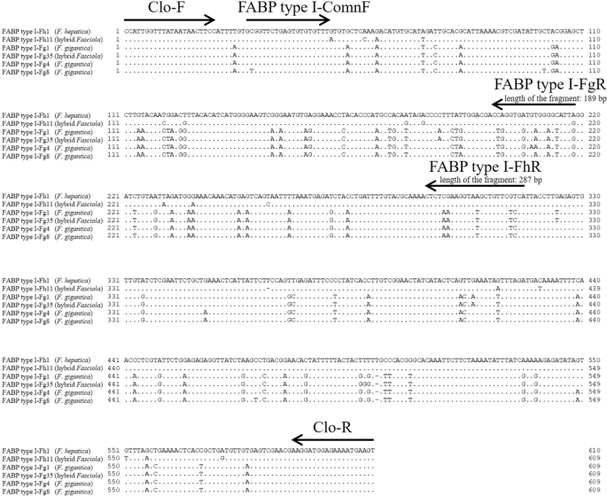
Another inner primer set, Clo-F (5′-CCATTGGTTTATAATAACTTCC-3′) and Clo-R (5′-ACTTCATTTTCTCCATCCTT-3′), which could amplify an intron of FABP type I, was designed to examine nucleotide variations between the primer regions (F. hepatica: 567 or 568 bp; F. gigantica: 566 or 567 bp) (Fig. 1). Sequence determination between Clo-F and Clo-R was performed for approximately 5% of F. hepatica and F. gigantica as well as 10 hybrid flukes selected from each country, and flukes with different nad1 haplotypes were selected as much as possible to ensure variations in the samples (Additional file 1: Table S1). PCRs were performed in a 25 μl reaction mixture containing 2 μl template DNA, 0.4 mM of each dNTP, 0.3 μM of each primer (Clo-F and Clo-R), 1 U of KOD FX Neo (Toyobo, Osaka, Japan), and the manufacturer’s supplied reaction buffer. Thermal conditions included an initial denaturation step at 94 °C for 120 s, followed by 35 cycles of 98 °C for 10 s, 50 °C for 30 s, and 68 °C for 30 s. PCR products were purified using the NucleoSpin Gel and PCR Clean-up kit (Macherey-Nagel), cloned into the pUC118 Hinc II/BAP vector (Takara Bio), and sequenced. Two clones were analyzed for F. hepatica and F. gigantica, whereas four clones (two for F. hepatica genotype and two for F. gigantica genotype) were analyzed for the hybrid Fasciola fluke (Additional file 1: Table S1). The obtained sequences were aligned to construct a maximum likelihood (ML) tree using MEGA 10.0.5 software [21]. For ML tree construction, all sites were selected in the gaps/missing data treatment, and the T92 + I model was used.
Alignment of partial FABP type I gene to generate primers. Six representative genotypes of FABP type I were analyzed using Clo-F and Clo-R. A dot in the alignment indicates that the sequence was identical to that of FABP type I-Fh1 (Fasciola hepatica). The arrows indicate the position and direction of the primers. The horizontal bars represent the alignment gap. Three primers (FABP type I-ComnF, FABP type I-FhR, and FABP type I-FgR) were designed for the multiplex PCR
Multiplex PCR
A primer set for multiplex PCR was designed using the resulting sequences of Clo-F and Clo-R. FABP type I-ComnF (5-′GCGGTTCTGAGTGTGTGTTT-3′) is a common primer for F. hepatica and F. gigantica, whereas FABP type I-FhR (5′-TGACGAACAGCTTACCTTCGAG-3′) and FABP type I-FgR (5′-CAATACTCCTCACCACCCAG-3′) are specific to F. hepatica (length of the amplicon: 287 bp) and F. gigantica (189 bp), respectively (Fig. 1). PCR amplification was performed in 10 μl reaction mixtures containing 0.5 µl template DNA, 0.1 µM of each dNTP, 0.2 µM of each primer, 0.01 U of Go Taq DNA Polymerase (Promega, Madison, WI, USA), and the manufacturer’s supplied reaction buffer. The PCR conditions included an initial denaturation step at 95 °C for 120 s, followed by 35 cycles of 95 °C for 30 s, 55 °C for 30 s, and 72 °C for 60 s, and a final extension step at 72 °C for 5 min. PCR amplicons were electrophoresed on 1.8% agarose gels and visualized using ethidium bromide staining. Multiplex PCR was then applied to all 1312 flukes (Table 1).
Results and discussion
The nucleotide sequences of PCR amplicons generated by FABP type I-2F and R for F. hepatica (1951 bp) and F. gigantica (1961 bp) were deposited in the DNA data bank of Japan (DDBJ) under accession numbers LC718926 and LC718927, respectively. The shorter nucleotide sequences of 24 F. hepatica, 31 F. gigantica, and 20 hybrid Fasciola flukes amplified using Clo-F and Clo-R were determined by cloning analysis (Additional file 1: Table S1). As a result, 10 genotypes (FABP type I-Fh1 to Fh10) were detected from F. hepatica, and 34 genotypes (FABP type I-Fg1 to Fg34) were detected from F. gigantica. Moreover, 12 F. hepatica (FABP type I-Fh1 and from FABP type I-Fh11 to Fh21) and 11 F. gigantica genotypes (FABP type I-Fg1 and from FABP type I-Fg35 to Fg44) were found in the hybrid Fasciola flukes. They were deposited in the DDBJ under accession numbers LC718928–LC718992 (Additional file 1: Table S1). FABP type I-Fh1 was detected in both F. hepatica and the hybrid Fasciola (Additional file 1: Table S1). FABP type I-Fg1 was found in both F. gigantica and the hybrid flukes (Additional file 1: Table S1). These observations may indicate an ancestor-descendant relationship. However, geographical distribution of the FABP type I genotypes among the 11 countries was not clear (Fig. 2).
Maximum-likelihood tree of FABP type I genotypes. Sequences obtained with Clo-F and Clo-R primers were used in the tree. Bootstrap values > 60% are shown for the tree node. No suitable outgroups were available in the DNA databank. Abbreviations of the names of countries where each genotype was detected are mentioned on the tree. AFG: Afghanistan; DZA: Algeria; PER: Peru; ESP: Spain; IDN: Indonesia; MYS: Malaysia; NGA: Nigeria; PAK: Pakistan; UGA: Uganda; JPN: Japan; BGD: Bangladesh. Red, blue, and green indicate the regions where F. hepatica, F. gigantica, and hybrid Fasciola used in the present study were collected
The FABP type I genotypes obtained in this study were clearly divided into the clades of F. hepatica and F. gigantica (Fig. 2). Therefore, the nucleotide variations of FABP type I are sufficient to distinguish F. hepatica and F. gigantica genotypes and therefore can be regarded as a useful molecular discrimination marker. The species-specific primers developed for multiplex PCR successfully generated specific fragments of F. hepatica (approximately 290 bp) and F. gigantica (approximately 190 bp), whereas hybrid flukes had both fragment patterns (Fig. 1 and 3).
Multiplex PCR for FABP type I Fasciola hepatica (Fh type) from 1. Afghanistan, 2. Algeria, 3. Peru, and 4. Spain. F. gigantica (Fg type) from 5. Indonesia, 6. Malaysia, 7. Nigeria, 8. Pakistan, and 9. Uganda. Hybrid Fasciola flukes (Fh/Fg type) from 10, 11. Japan and 12, 13. Bangladesh. M: 100-bp DNA ladder
No mutation was found in the FABP type I-ComnF primer region of F. hepatica, whereas a single-nucleotide mutation was found in the four F. gigantica genotypes (Fig. 4a and b). Similarly, no mutation was detected in the FABP type I-FhR region, but one to two nucleotide substitutions were observed in five F. gigantica genotypes in the FABP type I-FgR region (Fig. 4c and d). However, these mutations did not interfere with the DNA amplification of the multiplex PCR in this study because no ambiguous or variant fragments were detected (Table 1).
Nucleotide variations in the primer region of the multiplex PCR. a Nucleotide variations of FABP type I-ComnF region. Fasciola hepatica genotypes. b Nucleotide variations of FABP type I-ComnF region. F. gigantica genotypes. c Nucleotide variations of FABP type I-FhR. d Nucleotide variations of FABP type I-FgR
Previous studies observed discrepancies in 7, 19, 6, and 27 F. hepatica isolates from Afghanistan, Algeria, Peru, and Spain, respectively. They displayed the Fg or Fh/Fg type in the pepck (Table 1). However, in this study, all of them displayed Fh fragment patterns in the multiplex PCR for FABP type I, and there was no discrepancy when compared with the results of pold (Table 1). Moreover, the 15 F. gigantica from Nigeria showed Fg type in the multiplex PCR for FABP type I, which coincided with the results of pepck, even though they displayed an Fh/Fg-like fragment pattern in the pold (Table 1). Therefore, the novel multiplex PCR for FABP type I proved to be a useful marker to replace pepck and pold.
Conclusions
We successfully developed a novel multiplex PCR based on FABP type I using 1312 Fasciola flukes from 11 countries. Although discrimination errors occurring in pepck and pold were completely resolved by fragment analysis of FABP type I (Table 1), the robustness of the species-specific primer should be examined continuously in the future using a larger number of Fasciola flukes worldwide as nucleotide variations were detected in the primer regions.
Availability of data and materials
The nucleotide sequences obtained in this study are available under accession nos. LC718926 to LC718992.
Abbreviations
- FABP type I:
-
Fatty acid binding protein type I
- PCR:
-
Polymerase chain reaction
- PCR-RFLP:
-
PCR-restriction fragment length polymorphism
- pepck :
-
Nuclear phosphoenolpyruvate carboxykinase
- pold :
-
Polymerase delta
- mRNA:
-
Messenger RNA
References
Kaplan RM. Fasciola hepatica: a review of the economic impact in cattle and considerations for control. Vet Ther. 2001;2:40–50.
Mas-Coma S, Bargues MD, Valero MA. Fascioliasis and other plant-borne trematode zoonoses. Int J Parasitol. 2005;35:1255–78.
Hayashi K, Ichikawa-Seki M, Mohanta UK, Shoriki T, Chaichanasak P, Itagaki T. Hybrid origin of Asian aspermic Fasciola flukes is confirmed by analyzing two single-copy genes, pepck and pold. J Vet Med Sci. 2018;80:98–102. https://doi.org/10.1292/jvms.17-0406.
Terasaki K, Noda Y, Shibahara T, Itagaki T. Morphological comparisons and hypotheses on the origin of polyploids in parthenogenetic Fasciola sp. J Parasitol. 2000;86:724–9. https://doi.org/10.1645/0022-3395(2000)086[0724:MCAHOT]2.0.CO;2.
Terasaki K, Itagaki T, Shibahara T, Noda Y, Moriyama-Gonda N. Comparative study of the reproductive organs of Fasciola groups by optical microscope. J Vet Med Sci. 2001;63:735–42. https://doi.org/10.1292/jvms.63.735.
Ichikawa-Seki M, Peng M, Hayashi K, Shoriki T, Mohanta UK, Shibahara T, et al. Nuclear and mitochondrial DNA analysis reveals that hybridization between Fasciola hepatica and Fasciola gigantica occurred in China. Parasitology. 2017;144:206–13. https://doi.org/10.1017/S003118201600161X.
Shoriki T, Ichikawa-Seki M, Suganuma K, Naito I, Hayashi K, Nakao M, et al. Novel methods for the molecular discrimination of Fasciola spp on the basis of nuclear protein-coding genes. Parasitol Int. 2016;65:180–3. https://doi.org/10.1016/j.parint.2015.12.002.
Thang TN, Hakim H, Rahimi RR, Ichikawa-Seki M. Molecular analysis reveals expansion of Fasciola hepatica distribution from Afghanistan to China. Parasitol Int. 2019;72:101930. https://doi.org/10.1016/j.parint.2019.101930.
Laatamna AE, Tashiro M, Zokbi Z, Chibout Y, Megrane S, Mebarka F, et al. Molecular characterization and phylogenetic analysis of Fasciola hepatica from high- plateau and steppe areas in Algeria. Parasitol Int. 2021;80:102234. https://doi.org/10.1016/j.parint.2020.102234.
Kasahara S, Ohari Y, Jin S, Calvopina M, Takagi H, Sugiyama H, et al. Molecular characterization revealed Fasciola specimens in Ecuador are all Fasciola hepatica, none at all of Fasciola gigantica or parthenogenic Fasciola species. Parasitol Int. 2021;80:102215. https://doi.org/10.1016/j.parint.2020.102215.
Thang TN, Vázquez-Prieto S, Vilas R, Paniagua E, Ubeira FM, Ichikawa-Seki M. Genetic diversity of Fasciola hepatica in Spain and Peru. Parasitol Int. 2020;76:102100. https://doi.org/10.1016/j.parint.2020.102100.
Ichikawa-Seki M, Tokashiki M, Opara MN, Iroh G, Hayashi K, Kumar UM, et al. Molecular characterization and phylogenetic analysis of Fasciola gigantica from Nigeria. Parasitol Int. 2017;66:893–7. https://doi.org/10.1016/j.parint.2016.10.010.
Morphew RM, Wilkinson TJ, Mackintosh N, Jahndel V, Paterson S, McVeigh P, et al. Exploring and expanding the fatty-acid-binding protein superfamily in Fasciola species. J Proteome Res. 2016;15:3308–21. https://doi.org/10.1021/acs.jproteome.6b00331.
Ichikawa-Seki M, Ortiz P, Cabrera M, Hobán C, Itagaki T. Molecular characterization and phylogenetic analysis of Fasciola hepatica from Peru. Parasitol Int. 2016;65:171–4. https://doi.org/10.1016/j.parint.2015.11.010.
Hayashi K, Ichikawa-Seki M, Allamanda P, Wibowo PE, Mohanta UK, Guswanto A, et al. Molecular characterization and phylogenetic analysis of Fasciola gigantica from western Java. Indonesia Parasitol Int. 2016;65:424–7.
Ichikawa-Seki M, Shiroma T, Kariya T, Nakao R, Ohari Y, Hayashi K, et al. Molecular characterization of Fasciola flukes obtained from wild sika deer and domestic cattle in Hokkaido. Japan Parasitol Int. 2017;66:519–21. https://doi.org/10.1016/j.parint.2017.04.005.
Rehman ZU, Tashibu A, Tashiro M, Rashid I, Ali Q, Zahid O, et al. Molecular characterization and phylogenetic analyses of Fasciola gigantica of buffaloes and goats in Punjab. Pakistan Parasitol Int. 2021;82:102288. https://doi.org/10.1016/j.parint.2021.102288.
Vudriko P, Echodu R, Tashiro M, Oka N, Hayashi K, Ichikawa-Seki M. Population structure, molecular characterization, and phylogenetic analysis of Fasciola gigantica from two locations in Uganda. Infect Genet Evol. 2022;104:105359. https://doi.org/10.1016/j.meegid.2022.105359.
Mohanta UK, Ichikawa-Seki M, Shoriki T, Katakura K, Itagaki T. Characteristics and molecular phylogeny of Fasciola flukes from Bangladesh, determined based on spermatogenesis and nuclear and mitochondrial DNA analyses. Parasitol Res. 2014;113:2493–501.
Ichikawa-Seki M, Hayashi K, Tashiro M, Khadijah S. Dispersal direction of Malaysian Fasciola gigantica from neighboring Southeast Asian countries inferred using mitochondrial DNA analysis. Infect Genet Evol. 2022;105:105373.
Kumar S, Stecher G, Li M, Knyaz C, Tamura K. MEGA X: Molecular evolutionary genetics analysis across computing platforms. Mol Biol Evol. 2018;35:1547–9. https://doi.org/10.1093/molbev/msy096.
Acknowledgements
Not applicable.
Funding
This work was supported by a Grant-in-Aid for Scientific Research (C) from MEXT KAKENHI (grant number 21K05954) and the Tohoku Initiative for Fostering Global Researchers for Interdisciplinary Sciences (TI FRIS) of MEXT's Strategic Professional Development Program for Young Researchers.
Author information
Authors and Affiliations
Contributions
All authors have made substantial contributions to the study conception; EO, molecular analysis, and drafting of the manuscript; MT molecular analyses; PO, sampling; UKM, sampling; MI designed the study and substantially revised the manuscript. All authors read and approved the final manuscript.
Corresponding author
Ethics declarations
Ethics approval and consent to participate
Not applicable.
Consent for publication
Not applicable.
Competing interests
The authors declare that they have no competing interests.
Additional information
Publisher's Note
Springer Nature remains neutral with regard to jurisdictional claims in published maps and institutional affiliations.
Supplementary Information
Additional file 1: Table S1.
Fatty acid binding protein type I (FABP type I) genotypes of Fasciola flukes used in the present study.
Rights and permissions
Open Access This article is licensed under a Creative Commons Attribution 4.0 International License, which permits use, sharing, adaptation, distribution and reproduction in any medium or format, as long as you give appropriate credit to the original author(s) and the source, provide a link to the Creative Commons licence, and indicate if changes were made. The images or other third party material in this article are included in the article's Creative Commons licence, unless indicated otherwise in a credit line to the material. If material is not included in the article's Creative Commons licence and your intended use is not permitted by statutory regulation or exceeds the permitted use, you will need to obtain permission directly from the copyright holder. To view a copy of this licence, visit http://creativecommons.org/licenses/by/4.0/. The Creative Commons Public Domain Dedication waiver (http://creativecommons.org/publicdomain/zero/1.0/) applies to the data made available in this article, unless otherwise stated in a credit line to the data.
About this article
Cite this article
Okamoto, E., Tashiro, M., Ortiz, P. et al. Development of novel DNA marker for species discrimination of Fasciola flukes based on the fatty acid binding protein type I gene. Parasites Vectors 15, 379 (2022). https://doi.org/10.1186/s13071-022-05538-7
Received:
Accepted:
Published:
DOI: https://doi.org/10.1186/s13071-022-05538-7


