
Correction to: J Nanobiotechnol (2021) 19:229 https://doi.org/10.1186/s12951-021-00978-2
Following publication of the original article [1], some errors were identified in the original article.
-
1.
The CLSM image of live and dead bacteria for the Ag-MOF-Vanc group after the 16 h treatment had been improperly inserted. The correct image of Fig. 5 and its caption is given in this erratum.
Fig. 5 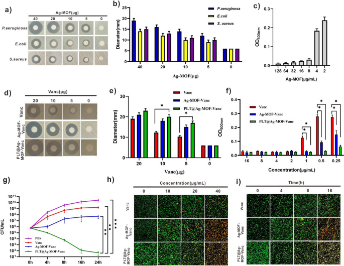
In vitro antibacterial effect of Ag-MOF-Vanc. a Inhibition zones and b corresponding inhibition zone diameters of Ag-MOF against different bacteria. c Concentration effects of Ag-MOF on the growth of MRSA. d Inhibition zones, e corresponding inhibition zone diameters and f concentration effects of Vanc, Ag-MOF-Vanc and PLT@Ag-MOF-Vanc against MRSA. g CFU of MRSA treated with 0.5 μg/mL of different drugs. h CLSM imaging of death/live staining after exposing MRSA to varying concentrations of Vanc, Ag-MOF-Vanc or PLT@Ag-MOF-Vanc. i CLSM imaging of death/live staining after MRSA exposure to Vanc, Ag-MOF-Vanc or PLT@Ag-MOF-Vanc with different incubation time. Scale bar: 20 μm. Data are presented as means ± SD (n = 3).*p < 0.05, **p < 0.01, ***p < 0.001
-
2.
In the paragraphs of “Synthesis of Ag-MOF-Vanc” and “Determination of EE and LE”, the authors miswrote the amount of vancomycin added into synthesis. The correct description is given below:
We dissolved 0.5 mg Ag-MOF in 1 mL ddH2O; 0.4 mg vancomycin was added, stirred overnight at room temperature by magnetic force, and Ag-MOF-Vanc was obtained by centrifugation.
-
3.
The animal models were established with Kunming mice, and the “Kunming rats” mentioned in some sentences were caused by translation errors.

These errors do not affect the conclusions of this research. The authors apologize for not noticing these errors before publication, and for any inconvenience caused.
The original article has been revised.
Reference
Huang R, Cai GQ, Li J, Li X-S, Liu H-T, Shang X-L, Zhou J-D, Nie X-M, Gui R. Platelet membrane-camouflaged silver metal-organic framework drug system against infections caused by methicillin-resistant Staphylococcus aureus. J Nanobiotechnol. 2021;19:229. https://doi.org/10.1186/s12951-021-00978-2.
Author information
Authors and Affiliations
Corresponding authors
Additional information
Publisher's Note
Springer Nature remains neutral with regard to jurisdictional claims in published maps and institutional affiliations.
Rights and permissions
Open Access This article is licensed under a Creative Commons Attribution 4.0 International License, which permits use, sharing, adaptation, distribution and reproduction in any medium or format, as long as you give appropriate credit to the original author(s) and the source, provide a link to the Creative Commons licence, and indicate if changes were made. The images or other third party material in this article are included in the article's Creative Commons licence, unless indicated otherwise in a credit line to the material. If material is not included in the article's Creative Commons licence and your intended use is not permitted by statutory regulation or exceeds the permitted use, you will need to obtain permission directly from the copyright holder. To view a copy of this licence, visit http://creativecommons.org/licenses/by/4.0/. The Creative Commons Public Domain Dedication waiver (http://creativecommons.org/publicdomain/zero/1.0/) applies to the data made available in this article, unless otherwise stated in a credit line to the data.
About this article

Cite this article
Huang, R., Cai, GQ., Li, J. et al. Correction to: Platelet membrane-camouflaged silver metal-organic framework drug system against infections caused by methicillin-resistant Staphylococcus aureus. J Nanobiotechnol 19, 278 (2021). https://doi.org/10.1186/s12951-021-01009-w
Published:
DOI: https://doi.org/10.1186/s12951-021-01009-w