

Abstract
Collagen, the most abundant protein in the mammalian extracellular matrix, is critical for skin structure and function, with Type I and Type III collagens being particularly important. Collagen degradation in skin is accelerated by aging and UV exposure, leading to structural and functional impairments. Exogenous collagen supplementation has been shown to restore skin structure and function. Traditional collagen extraction from animal tissues is limited by safety and quality concerns, while recombinant human collagen offers improved safety but faces challenges in solubility and production. This study aimed to construct a chimeric collagen derived from both Type I and Type III collagens and achieve its soluble expression in E. coli. Through translational pausing technology, a recombinant chimeric human collagen containing functional domains of both collagen types was successfully constructed and expressed with a yield of 1.36 g/L. A cysteine-rich C-propeptide domain was fused to enhance assembly and stability. The “dual-function” collagen significantly promoted fibroblast proliferation, migration, and adhesion, while stimulating endogenous Type I and Type III collagen production. Using the C. elegans model, the recombinant protein extended lifespan and enhanced oxidative aging resistance. Skin imaging confirmed its penetration into the dermis, and human skin efficacy tests demonstrated its ability to reduce periorbital wrinkles and crow’s feet. This recombinant “dual” human collagen promotes endogenous collagen synthesis, accelerates skin repair, reduces aging signs, and shows no observed side effects, offering promising potential for anti-aging applications.
Clinical trial number
Not applicable.
Introduction
Collagen is the most abundant and widely distributed protein in mammals and is found in the extracellular matrix. In human skin, collagen makes up more than 70% of the total dry weight of the skin [1,2,3]. Type I and type III collagen are important fibril-forming collagens in the skin that support various cellular functions such as proliferation, migration, and adhesion [4,5,6]. In the dermis, type I and type III collagen interact with each other and coexist on the same collagen fibers to support skin structure and to plump and firm the skin [2, 7, 8]. The formation of a triple helix is the structural basis for the biological functions of collagen. The cysteine-rich C-propeptide domain at the collagen C-terminus enables the formation of disulfide bonds between molecules, thereby facilitating the assembly of collagen into trimers [9,10,11].
Collagen degradation in the skin occurs due to aging, UV exposure and other pathologic or physiologic processes, while its synthesis rate decreases with aging, resulting in a continuous loss of collagen from the skin [12, 13]. Supplementation with exogenous collagen is a viable measure to combat skin aging [14, 15].
Traditionally, collagen has primarily been derived from animal tissues, such as bovine and porcine sources. However, the use of collagen from animal sources is accompanied by a series of challenges, including immunogenicity, risks of disease transmission, inconsistent quality, and limited supply [16, 17]. For instance, prion diseases, such as bovine spongiform encephalopathy (BSE), pose significant restrictions on the use of bovine collagen in infected regions [18]. Additionally, traditional methods for processing animal-derived collagen, such as thermal treatment, often fail to preserve its structural and functional integrity, further complicating its application [19].
In recent years, advancements in biotechnology have facilitated the development of recombinant human collagen through recombinant DNA technology. This innovative approach enables precise control over the molecular structure of collagen, allowing the synthesis of materials that closely resemble natural human tissues. Compared to animal-derived collagen, recombinant collagen exhibits superior mechanical strength, a controllable degradation rate, and enhanced biological activity. For example, studies have demonstrated that recombinant collagen significantly promotes cell proliferation and migration [20]. These characteristics make recombinant collagen an ideal candidate for clinical applications.
Recombinant collagen has broad application prospects across multiple fields. In the medical aesthetics industry, it can be used to develop anti-aging skincare products and medical aesthetic materials to improve skin elasticity and reduce wrinkles. In regenerative medicine, recombinant collagen can serve as a biomaterial for tissue engineering and wound repair, promoting tissue regeneration and repair [21]. With further technological advancements, recombinant collagen has the potential to become a highly efficient, safe, and sustainable biomaterial, meeting diverse clinical and industrial demands [22].
However, owing to the high molecular weight of full-length type I or type III collagen, it is difficult to obtain soluble recombinant proteins in prokaryotic host cells. Characteristic repetitive amino acid sequences (Gly-X-Y) in collagen proteins may also be chosen as target recombinant proteins [23, 24]. However, the stability and bioactivity of recombinant collagen-like proteins with characteristic repetitive Gly-X-Y sequences are not satisfactory. Therefore, purposeful selection and optimization of functional sequences in collagen is a key prerequisite for designing soluble recombinant collagen with increased stability and bioactivity.
Our previous studies have shown that the speed of protein synthesis during translation on the ribosome is not constant. Protein translation slows at certain amino acid sites, known as a ‘translational pause’ [25, 26]. According to this theory, some synonymous codons could be used for the recombinant protein sequence to control the translation rate of the target protein and enable the protein to fold out of the conformation correctly [27]. By using degenerate codons of less abundant tRNAs, the translation speed of a segment can be artificially slowed to assist in protein spatial folding. Based on these previous studies, it was hypothesized that regulating the speed of protein synthesis by designing suitable pause points might be a feasible way to improve the soluble expression of collagen in Escherichia coli (E. coli) and enable the recombinant collagen to form the correct structure to exhibit better biological activity.
In this study, recombinant collagen (Dual-collagen, DuCol) creatively integrates functional active regions from both Type I and Type III collagens, achieving large-scale soluble expression in the cytoplasm of E. coli with a yield of 1.36 g/L. This DuCol could promote the proliferation, migration and adhesion of fibroblasts. The DuCol protein permeates into the dermis of the mouse skin to increase the number of collagen fibers. Furthermore, the antiaging potency of recombinant DuCol was tested in C. elegans and in human volunteers.
Materials and methods
Cell culture
The mouse embryonic fibroblast NIH/3T3 cell line was purchased from the China Center for Type Culture Collection (CCTCC, Wuhan, China). The cells were cultured in Dulbecco’s modified Eagle medium (ThermoFisher Scientific, Waltham, MA) supplemented with 10% fetal bovine serum (FBS; Beyotime Biotechnology Corp., Shanghai, China), 50 IU/mL penicillin and 50 μg/ml streptomycin in 5% CO2 at 37 °C.
Construction of chimeric collagen and its expression plasmid
The DNA sequences of COL1A1 2,848 to 3,225 nt (designated s1), COL3A1 1,780 to 2,187 nt (s2) and 3,577 to 3,597 nt (s3) were optimized according to translation pause theory [27, 28]. The s1 sequence encodes a type I collagen domain with 126 amino acid (aa) residues, whereas s2 encodes a type III collagen domain with 136 aa residues, and s3 encodes a C-pro sequence with 7 aa residues (GenBank: PQ496881). The recombinant collagen covered a cell adhesion-related domain of type I collagen and cell proliferation- and migration-related domains of type III collagen. Several nucleotides were replaced by synonymous codons to ensure efficient translation and correct folding of the protein products (GenBank: PQ496882). The optimized recombinant sequence was artificially synthesized (Synbio Technologies Co. Ltd. SuZhou, China) and then inserted into the pET-28a (+) plasmid using the restriction endonuclease sites NdeI (5’) and BamHI (3’).
Protein expression and purification
Escherichia coli BL21 (DE3) (TIANGEN Biotech Co., Ltd. Beijing, China) transformed with recombinant plasmids were cultured in Luria–Bertani (LB) medium and induced with 1 mM IPTG at 37 °C for 4 h. The bacteria were then collected and sonicated to obtain the soluble total proteins. The soluble DuCol was purified using Ni Bestarose FF (Biorigin Biotechnology Co., Ltd., Shanghai, China) and eluted with elution buffer (20 mM Tris, 150 mM NaCl, and 250 mM imidazole at pH 8.0). The eluate was then desalted using an equilibration buffer (20 mM PB, pH 7.4) over a G-25 desalting column.
SDS‒PAGE and western blot
SDS‒PAGE and western blot analysis were performed as described previously [29]. Briefly, samples with equal amounts of protein were resolved via SDS‒PAGE. The gels were stained with Coomassie Brilliant Blue Rapid Stain Solution (Beyotime) to analyze the expression of protein products. For western blotting, proteins were transferred to NC membranes following the manufacturer’s recommended methods and then incubated with anti-type I or type III collagen antibodies (BIOSS, Beijing, China). The blots were detected by enhanced chemiluminescence reagents (Thermo Fisher Scientific, Waltham, MA, USA).
Cell proliferation assay
A cell proliferation assay was performed via a Cell Counting Kit-8 (CCK-8) assay as follows. NIH/3T3 cells were seeded into 96-well plates at a concentration of 3,000 cells per well and incubated overnight. The total medium was replaced with medium containing 2% serum and different concentrations of DuCol. After 24 h of culture, 10 μL of CCK8 reagent (Beyotime) was added to each well, and the samples were then incubated at 37 °C for 2 h. The cell proliferation rates were calculated from the absorbance of the samples at 450 nm, which was determined on a Spark 10 M (TECAN, Männedorf, Switzerland).
Cell scratch assay
For the scratch assay, NIH/3T3 cells were seeded into 24-well plates and incubated at 37 °C to form a confluent monolayer. The cell monolayer was then scraped in a straight line with a pipet tip to create a ‘scratch’. The cell debris was removed by washing the cells once with 1 mL of growth medium and then replaced with 2 mL of medium containing 5 μg/mL control collagen (HY-NP101, MedChemExpress, Monmouth, NJ, USA) or DuCol. The scratch fields were marked, and photos of the scratches were taken. After 24 h of culture, photos of the scratches in the same fields were acquired. Images were taken under an MF52-N microscope (Mshot, Guangzhou, China), and the distance changes in the scratches were quantitatively analyzed using the MShot image analysis system.
Cell adhesion assay
Assessment of cell adhesion to collagen was conducted as follows. The plates were coated with 5 μg/mL type I collagen from bovine skin (HY-NP101, MedChemExpress, Monmouth, NJ, USA) or DuCol before the cells were seeded. 3T3 cells were seeded onto precoated 24-well plates and then incubated at 37 °C for 4 h. After the wells were gently washed three times with cold PBS, add 200 μL of 4% paraformaldehyde fixation solution and incubate at room temperature for 20 minutes. Wash twice with PBS. Add 200 μL of 0.1% crystal violet staining solution and incubate at room temperature for 20 minutes. Wash twice with PBS, photos of the cells were taken for counting. The data used for quantification analysis were normalized to those of the control group.
Worm strains and maintenance
The C. elegans wild-type strain Bristol N2 was supplied by the Caenorhabditis Genetics Center (CGC, University of Minnesota, Minneapolis, MN). Worms were raised and maintained at 20 °C on nematode growth media (NGM) seeded with E. coli OP50 bacteria as a food source starting from the first day of hatching, except where indicated. The rCC solutions (0.4 mg/mL in ultrapure water) were mixed with E. coli OP50 (v/v, 5:95). The mixture was seeded in an NGM (φ6 cm) plate and allowed to dry for 48 h at room temperature. Water was substituted for DuCol in the control group.
Lifespan assay
Lifespan assays were performed according to the protocol described previously [30, 31]. On Day 0 of the experiment, synchronized L4 larvae were transferred to an NGM plate with or without DuCol using a platinum wire. L4 larvae (50 worms per plate for 3 plates per treatment and control) were then transferred to fresh treatment plates daily throughout the reproductive period and thereafter approximately every other day to maintain the drug concentration. The dead and surviving age-synchronized worms were counted daily (starting from Day 0) until all individuals had died. Worms that did not respond to a mechanical stimulus were scored as dead. Nematodes that died after internal hatching or escaped the plates were excluded from the statistical data. The mean lifespan was calculated as the sum of the lifespan of all the nematodes divided by the total number of nematodes. The maximum lifespan was the number of days corresponding to the last dead nematode on each plate. All lifespan assays were carried out at 20 °C unless stated otherwise.
Mobility assay
Nematode mobility was estimated based on locomotion in three phases of the life cycle. Thirty individual nematodes were randomly selected for mobility analysis, with ten individuals tested at each of three time points (Days 4, 10, and 16). The locomotion phenotypes of the worms were judged by introducing a mechanical stimulus (a platinum wire) and were classified into three motility grades: Motion A, worms that moved smoothly without touch; Motion B, worms that responded to touch but with somewhat uncoordinated motion and moved slowly; and Motion C, worms that were unable to move independently but moved their heads or tails upon stimulation.
Stress resistance assays
At least 180 worms treated or not treated with DuCol for 5 d were used to comprise three independent biological replicates (administered from the egg stage). The worms were exposed to various stresses until all the worms died. These assays were performed according to a method described in previous research with minor modifications [32, 33]. For the H2O2-induced oxidative stress assay, the worms were transferred to freshly prepared NGM containing 0.1% of 10% H2O2. The viability of the worms was examined every 30 min. For the paraquat-induced oxidative stress assay, survival was monitored every 24 h after the worms were subjected to plates containing 10 mM paraquat.
Animals
Healthy BALB/c mice were purchased from Vital River Laboratories (China). All animals used in this study were male adults (6–8 weeks, 20 ± 2 g body weight). Mice were kept in a specific pathogen-free environment with freely available food and water and a 12 h light-dark cycle. In this study, five mice were used at each time point, each of which received a topical dose of 10 mg/ml of DuCol on a shaved back on the left side of the thoracic spine and a saline control area on the right.
SHG-TPEF skin imaging
An iFluorTM 594 (AAT Bioquest, Pleasanton, CA, USA) was used to fluorescently label recombinant collagen, and SDS‒PAGE was used to detect DuCol labeling. The back hair was shaved from the mice, which were treated with iFluor-DuCol (10 mg/ml) for 0, 0.5, 1, 1.5, 2, 2.5 and 3 h at room temperature while avoiding light. At the end of the experiment, all mice were anesthetized with isoflurane and fixed on a mouse plate while the back skin was fixed on holder. The full-thickness skin tissue was scanned by a two-photon scanning confocal microscope (FV1000-MPE, Olympus, Japan) [34]. Second harmonic generation (SHG) imaging and two-photon fluorescence imaging both collect signals in the reverse direction. The excitation wavelength for SHG was 850 nm, whereas the fluorescence excitation wavelength for the sample was 543 nm. The excitation light is focused onto the sample by the objective lens, and the resulting inverted SHG signal and fluorescence signal are collected by the same objective lens (magnification 25x, aperture 1.05). The tissue was scanned through the full Z-axis layer by layer, with a step size of 5 µm. The images were rendered and processed using Imaris 7.4.2 software, with green representing SHG imaging and red representing fluorescence imaging.
Transdermal absorption detection
Using a transdermal absorption instrument (TP6, Tianjin Jingtuo Instrument Technology), a semipermeable membrane or mouse skin was used as the transdermal medium, which was fixed between the DuCol chamber and the receiving pool of the transdermal absorption chamber. Next, 10 mg of DuCol dissolved in saline was added, and the DuCol chamber was sealed with tin foil. The receiving pool was filled with a 0.9% NaCl solution (pH 7.4) and stirred at 37 °C and 300 rpm. DuCol was taken from the receiving pool for detection at 1, 2, 3, 4, 6, 8, 10, and 24 h. A BCA protein detection kit (Beyotime) was used to detect protein content.
Immunohistochemical staining
DuCol was applied to the exposed skin on the back of the mouse; the unabsorbed DuCol was removed from the surface of the skin after 1 h, and full-thickness skin was collected to prepare 10 μm frozen sections. The immunohistochemical kit (Zhongshan Golden Bridge, Beijing, China) uses H2O2 to eliminate endogenous H2O2 enzyme activity in the tissue, reducing nonspecific staining. Antigen retrieval was performed using a trypsin solution. After the samples were blocked with a 5% BSA-PBS solution for 30 min, primary antibodies (Hexiucheng, Beijing, China) were added to the slices, which were incubated at 4 °C for 16 h and then incubated with secondary antibodies. After the target protein appeared brown (due to DAB color development solution), the cell nuclei were counterstained with hematoxylin. The stained sections were observed using an Olympus B×51microscope.
Human skin efficacy tests
DuCol was prepared as a liquid essence for human skin efficacy tests. The bacterial endotoxin test results in Table S1. A total of 33 healthy Chinese women (Table 1) from nearby communities were recruited as volunteers by Guangzhou Landproof Testing Technology Co., Ltd. A single-blind, randomized antiaging trial was carried out, and the physiology parameters of the facial skin of each volunteer on Day 0 were determined as the control. DuCol essence (1–1.5 ml) was applied twice (morning and night) to the entire face after face cleaning. During 56 d of testing, three volunteers withdrew from the test for personal reasons, leaving 30 volunteers to complete the full test at Days 14, 28 and 56.
The detection of transepidermal water loss (TEWL) was performed with a VapoMeter SWL5142 (Delfin Technologies Ltd., Kuopio, Finland). The water content of the stratum corneum of the skin was measured using a CM825 corneometer (Courage + Khazaka electronic GmbH, Köln, Germany). The detection of skin glossiness was performed using a SkinGlossMeter SGM2008 (Delfin). Skin elasticity was measured using a Cutometer MPA580 (Courage + Khazaka). The Primos Clinical Research System (Canfield Scientific, Inc., Parsippany, USA) was used for analysis of wrinkle number, length, area and volume.
GAPDH
GAPDH (Glyceraldehyde-3-Phosphate Dehydrogenase) was selected as the reference gene due to its stable expression levels across a variety of cell types [35].
Data analysis and statistics
The data represent the mean ± s.e.m. of the results obtained from at least three independent experiments. The statistical significance of differences between experimental groups was assessed using two-tailed Student’s t tests in GraphPad Prism 8.0. The lifespan analysis of C. elegans was performed via the Kaplan–Meier method, and the results were compared via the log-rank (Mantel‒Cox) method [36]. Values of p < 0.05 were considered statistically significant.
Results
Construction and expression of DuCol
To obtain DuCol, selected DNA fragments of type I (3,228 ~ 3,608 bp) and type III (1,782 ~ 2,540 bp) collagen were inserted into the pET-28a vector. To best mimic the structure of natural human-derived collagen, the C-propeptide region was also included in the recombinant plasmid (Fig. 1A). To optimize the expression of DuCol, synonymous codon substitutions were used on the original amino acid sequence of DuCol. The amino acid translation speed was calculated using the RiboTempo web server (https://www.translatome.net/RiboTempo/) (Fig. 1B). After optimization via the translation pause method, an ~ 26 kD protein product was successfully expressed after induction with IPTG (Fig. 1C and D). The total protein expression of the optimized sequence was greater than that of the original sequence, and its solubility was much higher than that of the original sequence. Only after long exposure can some soluble proteins be observed in the supernatant of the original sequence, and the expressed protein was confirmed to be collagen by western blotting (Fig. 1E). Following the optimization of the fermentation process, DuCol was successfully produced at the 500 L scale. The expression level of DuCol reached 1.36 g/L when the bacterial optical density (OD) attained 30.8. Post-purification, the total recovery rate of DuCol was measured at 63.66%. The C-propeptide plays a key role in the trimeric assembly of collagen. To confirm whether successful folding occurred, comparative checks were performed on reduced and nonreduced protein samples via SDS‒PAGE. The dimer and trimer bands appeared in the nonreduced samples, indicating that the added C-propeptide successfully assisted DuCol in folding. As the induction time increased, the expression level of the target protein increased, and the formation of dimers and trimers in the nonreduced samples gradually increased, whereas no dimers or trimers formed in the reduced samples (Fig. 1F).

Construction and expression of DuCol. A) assembly of the DuCol (pEt28a) vector (plasmid). B) translation rate profiles of recombinant collagen and its pause-optimized version. C) comparison of the soluble proteins expressed from the two collagen coding sequences. The arrow indicates DuCol. (lane 1, marker; Lane 2, supernatant of the original collagen sequence; Lane 3, supernatant of the optimized DuCol sequence.) D) expression and purification of DuCol. The arrow indicates DuCol. (lane 1, marker; Lane 2 and Lane 3, total proteins of uninduced and induced bacteria, respectively; Lane 4, supernatant of bacterial lysate; Lane 5, pellet of bacterial lysate; Lane 6, the flow-through fraction; Lane 7, the washed fraction; Lane 8, the collected target protein.) E) Western blot detection of DuCol. (lane 1, total protein of the original sequence; Lane 2, total protein of the optimized sequence; Lane 3, soluble protein of the original sequence; Lane 4, soluble protein of the optimized sequence. ST, short exposure; LT, long exposure). F) expression of DuCol (lanes 1 and 7 are markers, lanes 2 to 6 are nonreduced DuCol purified from E. coli induced from 2 to 6 h, and lanes 8 to 12 are reduced DuCol from bacteria induced from 2 to 6 h)
DuCol promotes cell proliferation, migration and adhesion
DuCol contains a cell adhesion-related domain of type I collagen and cell proliferation- and migration-related domains of type III collagen. Therefore, it was also necessary to identify whether DuCol could promote cell proliferation, migration and adhesion. Cell proliferation was significantly promoted when the final concentration of DuCol in the culture medium exceeded 1 µg/mL (Fig. 2A). For the cell migration and cell adhesion assays, DuCol was compared with control collagen at 5 μg/mL. After culture for 24 h, the wound healing rate of DuCol-treated cells was greater than 60%, which was significantly higher than that of control collagen-treated cells (Fig. 2B and C). Cell adhesion to collagen was then performed, and the results showed that the cells adhered more strongly to DuCol than to control collagen (Fig. 2D and E). These results indicate that the DuCol expression construct simultaneously possesses the adhesive properties of type I collagen and the proliferative and migratory capabilities of type III collagen.

Recombinant collagen promotes cell proliferation, migration and adhesion. A) cell viability of NIH-3T3 cells assessed by the CCK-8 assay, the control is untreated (n = 5). B) cell migration is presented as scratch images after the administration of recombinant collagen, the control collagen is type I collagen from bovine skin. C) quantification of cell migration rates derived from the scratch assay images (n = 5). D) cell adhesion was assessed after the administration of recombinant collagen, the control is untreated, the control collagen is type I collagen from bovine skin (n = 5). E) quantification of cell adhesion derived from assay images. Data are presented as means ± SD. *p < 0.05, **p < 0.01, ***p < 0.001 vs. control
DuCol promotes expression of type I & III collagen in fibroblasts
As an extracellular matrix protein, collagen also affects the synthesis and secretory capacity of fibroblasts. After the cells were treated with various concentrations of DuCol, the expression of endogenous type I and III collagens in the fibroblasts was detected via western blotting. DuCol was able to induce the expression of both endogenous type I and type III collagen in fibroblasts in a concentration-dependent manner (Fig. 3A). The expression of type III collagen was 1.7 times from its control under 12.5 µg/mL DuCol treatment and exceeded by 3.3 times that of the control group under 200 µg/mL DuCol treatment. Moreover, the expression of type I collagen was 3.4 times greater than that of the control when the cells were treated with 50 µg/mL DuCol (Fig. 3B).
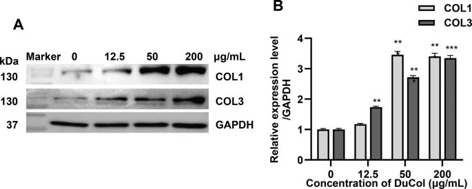
Recombinant DuCol promotes the expression of endogenous type I & III collagen in fibroblasts. A) expression of type I collagen and type III collagen after the administration of DuCol, as detected via western blotting. B) quantification of type I collagen and type III collagen via western blotting (n = 3). Data are presented as means ± SD. *p < 0.05, **p < 0.01, ***p < 0.001 vs. control
DuCol prolongs the lifespan of C. elegans
As a model organism, C. elegans has been used to test the effects of different substances on lifespan. When treated with DuCol, the lifespan and locomotion of C. elegans were recorded for up to one month. Fig. 4A shows that the survival time of nematodes treated with DuCol was significantly greater than that of the control group (CK group). In the lifespan test, supplementation with DuCol increased the mean lifespan of C. elegans by 21.13% (Table S2) and increased the maximum lifespan of C. elegans by 12.33%. Moreover, DuCol treatment improved the movement of C. elegans. On Day 10, the DuCol group had a lower percentage of individuals needing touch stimulation to move than did the control group, but the difference was not statistically significant (p > 0.05). On Day 16, the rate of spontaneous movement in the DuCol-treated nematode group was significantly greater than that in the control group (Fig. 4B).

Recombinant DuCol prolonged the lifespan of C. elegans. A) lifespan assay of C. elegans after DuCol treatment. B) statistical analysis of nematode movement rate after DuCol treatment. C) survival curve of C. elegans after the addition of DuCol under H2O2-induced oxidative stress. D) survival curve of C. elegans after the addition of DuCol under paraquat-induced oxidative stress
Preliminary research showed a positive association between the increase in stress tolerance and the extension of lifespan [37]. The survival curve of C. elegans in the DuCol treatment group shifted to the right under oxidative stress (p < 0.01). Compared with that of the control group, the average lifespan increased by 37.34% under H2O2-induced oxidative stress (Fig. 4C) and by 30.00% under paraquat-induced oxidative stress (Fig. 4D). These experiments suggest that DuCol significantly increased the resistance of C. elegans to oxidative stress, with statistical analysis of the lifespan data presented in Table S2.
DuCol permeates the dermis of the skin to increase collagen fiber synthesis
To investigate the transdermal absorption efficiency of the recombinant bioactive protein DuCol, a mouse skin model combined with two-photon microscopy was employed. Following the application of labeled DuCol to the surface of the mouse skin, the fluorescence signal gradually increased from 0.5 to 3 h, indicating rapid penetration of DuCol from the epidermis to the dermis, with a peak observed at 1.5 h (Fig. 5A and Fig. S1A and S1B). Concurrently, the fluorescence signal of endogenous collagen fibers in the dermis significantly intensified and peaked within 1 h, suggesting that DuCol may increase the synthesis of endogenous collagen fibers (Fig. 5Band C). Two-photon microscopy revealed that the DuCol signal progressively localized near hair follicles after 2 h, whereas the fluorescence signal in the dermis diminished after 3 h (Fig. 5D).


Following fluorescent labeling of DuCol, its transdermal absorption behavior in mouse skin was investigated. A) temporal changes in the fluorescence signals of collagen fibers and DuCol in the dermis, Z = 18 ~ 21 μm. B) quantitative statistical analysis of DuCol fluorescence signals (n = 5). C) quantitative statistical analysis of endogenous collagen fiber fluorescence signals (n = 5). D) fluorescence signal distribution of recombinant collagen in the epidermal layer. White arrows, epidermal cells; green arrows, hair follicles; yellow arrows, hair shafts. E) detection of fluorescence signals and second harmonic generation (SHG) signals in tissue sections. F) concentration of DuCol in the receptor chamber when a semipermeable membrane was used as the diffusion medium. G) concentration of DuCol in the receptor chamber when mouse skin was used as the diffusion medium. H) detection of DuCol in the receptor chamber via SDS‒PAGE after transdermal absorption through mouse skin. I) detection of DuCol in skin tissue via western blotting. J) distribution of DuCol in skin tissue detected by immunohistochemistry. Yellow arrow, samples attached to collagen fibers; red arrows, samples in the intercellular spaces of epidermal cells; black arrows, samples in hair follicle cells and fibroblasts. Data are presented as means ± SD. *p < 0.05, **p < 0.01, ***p < 0.001 vs. control
These findings were corroborated by imaging of mouse skin tissue sections, which aligned with the in vivo observations (Fig. 5E). Franz cell diffusion assays demonstrated that DuCol was unable to diffuse freely through the semipermeable membrane into the receptor chamber within 10 h, although a minimal amount of DuCol was detected after 24 h (Fig. 5F). In isolated mouse skin, the protein concentration in the receptor chamber increased after 1 h but decreased to a comparable level by 2.5 h (Fig. 5G).
However, SDS‒PAGE analysis failed to detect distinct DuCol-like protein bands, indicating limited transdermal absorption (Fig. 5H). Based on these findings, it is hypothesized that DuCol may traverse the skin via an active transdermal absorption pathway, a notion supported by western blot analysis, which confirmed the presence of DuCol in the total lysate of skin tissue (Fig. 5I). Immunohistochemical staining further revealed that DuCol was distributed within the intercellular spaces of epidermal cells, collagen fibers in the dermis, hair follicle cells, and fibroblasts (Fig. 5J). In conclusion, DuCol can permeate the skin through intercellular spaces in the epidermis and hair follicle tissues, accumulating in proximity to fibroblasts and even entering these cells.
DuCol increases the moisture content of the skin and reduces the appearance of wrinkles
A total of 30 volunteers were recruited for functional tests of DuCol. Transepidermal water loss (TEWL) is an important parameter for measuring skin moisture and barrier function. After 56 d of continuous use of DuCol, the TEWL index decreased from 13.4 g/h•m2 before use to 11.6 g/h•m2 (Fig. 6A). Moreover, on Day 14 of use, the water content of the stratum corneum in the skin of the volunteers had increased significantly and continued to increase over 56 d (Fig. 6B). The glossiness and elasticity of skin also increased with the use of DuCol over time (Fig. S2A and S2B). The appearance of crow’s feet and undereye wrinkles are signs of aging in facial skin. The test results confirmed that the number of crow’s feet was already significantly reduced after 14 d of DuCol use, but there was no further reduction over the next 6 weeks (Fig. 6C). Furthermore, the volume, area and length of crow’s feet were also significantly reduced to the lowest level after 14 d of DuCol use (Fig. 6D, Fig. S2C and S2D). The results for the undereye wrinkles were similar to those for the crow’s feet wrinkles. The number (Fig. 6E), volume, area and length (Fig. 6F, Fig. S2E and S2F, respectively) of undereye wrinkles were reduced to a minimum level after 14 d of DuCol use.
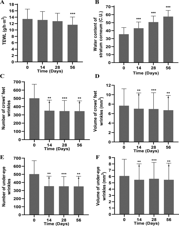
DuCol increases the moisture content of the skin and reduces the appearance of wrinkles. A) statistics on transdermal water loss (TEWL) after using DuCol (n = 30). B) statistics on the moisture content of the stratum corneum after using DuCol (n = 30). C) statistics on the number of crow’s feet after using DuCol (n = 30). D) statistics on the volume of crow’s feet after using DuCol (n = 30). E) statistics on the number of undereye wrinkles after using DuCol (n = 30). F) statistics on the volume of undereye wrinkles after using DuCol (n = 30). Data are presented as means ± SD. *p < 0.05, **p < 0.01, ***p < 0.001 vs. 0 day
Discussion
Type I and III collagens are the major protein components of the skin. They assemble into collagen fibers in the form of triple-helix structures, which are critical for maintaining the structural integrity of the dermis. These fibers not only provide mechanical support to the skin but also play a significant role in key biological processes such as cell proliferation, migration, and adhesion. As age advances or exposure to environmental factors occurs, the synthesis of collagen decreases, leading to reduced skin elasticity and the formation of wrinkles [38]. Therefore, the development of recombinant collagens that mimic the functions of native collagens holds significant clinical and commercial potential.
In this study, we successfully constructed recombinant chimeric collagen protein DuCol by fusing the cell adhesion domain of type I collagen with the cell proliferation/migration domain of type III collagen. Through optimization of translational pausing technology, we achieved high-yield soluble expression of DuCol in E. coli, with yields reaching 1.36 g/L. This strategy significantly improved protein expression efficiency and bioactivity. Additionally, we fused a cysteine-rich C-terminal propeptide domain to the C-terminus of DuCol, further enhancing its stability and self-assembly capability.
Cell biology experiments demonstrated that DuCol retains the ability of native collagens to promote cell proliferation, migration, and adhesion, which is of great significance for wound healing and tissue repair [39]. Notably, compared to conventional collagens, DuCol exhibits higher bioactivity. This phenomenon may be attributed to its unique C-terminal propeptide domain, which optimizes interactions with cell receptors through conformational changes, thereby triggering more effective cellular responses. Experimental results further showed that DuCol significantly upregulated the expression of endogenous type I and III collagens in fibroblasts. This effect may be mediated by the binding of DuCol to integrins on the cell membrane, subsequently activating downstream signaling pathways such as the FAK/Src or PI3K/AKT pathways, thereby promoting collagen synthesis and secretion [40]. This bidirectional regulatory mechanism not only enhances skin repair capacity but also provides a theoretical basis for the development of novel anti-aging products.
Furthermore, using the Caenorhabditis elegans model, we observed that DuCol significantly extended the lifespan and enhanced locomotion of the worms. This result suggests that DuCol may delay the aging process by promoting collagen synthesis and improving cellular functions. Combining existing research, this anti-aging effect may be associated with DuCol’s ability to activate antioxidant pathways and reduce reactive oxygen species (ROS) accumulation [41].
In terms of skincare applications, our research revealed that DuCol exhibits significant transdermal absorption capacity. Through fluorescence labeling and in vivo detection techniques, we found that DuCol penetrates the epidermis and accumulates in the dermis, significantly increasing the number of native collagen fibers. These findings indicate that DuCol may enter the skin through intercellular spaces in the epidermis and hair follicle tissues, where it accumulates near fibroblasts, thereby exerting its localized effects. This discovery provides direct evidence of the transdermal absorption mechanism of recombinant collagens and lays a foundation for the development of highly efficient skin repair products.
Human trials further validated the skincare benefits of DuCol. The results demonstrated that DuCol significantly increased skin hydration and reduced wrinkle formation, with no adverse reactions observed. These findings are consistent with previous studies, highlighting the potential of recombinant collagens in improving skin condition and combating aging. However, this study has certain limitations. For instance, the specific signaling pathways underlying the interaction between DuCol and integrins remain to be fully elucidated. Future research may employ molecular dynamics simulations and signaling pathway inhibition experiments to further investigate the mechanisms of action. Additionally, while the human trials demonstrated preliminary efficacy, the long-term safety and efficacy of DuCol require further validation through large-scale clinical trials.
Conclusions
In this study, we modified the original collagen sequence based on the translational pause theory and successfully constructed and expressed soluble DuCol containing type I and type III collagen fragments in prokaryotic cells, with a yield as high as 1.36 g/L. DuCol can significantly promote cell proliferation, migration and adhesion. Moreover, DuCol can also significantly increase the content of type I and type III collagen in the body. In animal models, we found that the application of DuCol can prolong the lifespan of nematodes. In addition, we also found that the expressed DuCol can penetrate the dermis and enter fibroblasts through active diffusion. DuCol can also effectively increase the moisture content of the skin, thereby reducing the appearance of wrinkles. These findings provide a scientific basis for the application of DuCol in skin repair, anti-aging, and regenerative medicine. Future studies will further optimize its performance and explore its potential application in complex skincare formulations.
Data Availability
Data is provided within the manuscript or supplementary information files.
Abbreviations
- BSA:
-
Bovine Serum Albumin
- CCK-8:
-
Cell Counting Kit-8
- COL1A1:
-
Collagen Type I Alpha 1 Chain
- COL3A1:
-
Collagen Type III Alpha 1 Chain
- C-pro:
-
C-Propeptide
- C. Elegans :
-
Caenorhabditis elegans
- DuCol:
-
Dual Collagen
- E. coli :
-
Escherichia coli
- FBS:
-
Fetal Bovine Serum
- IPTG:
-
Isopropyl β-D-1-thiogalactopyranoside
- LB:
-
Luria–Bertani medium
- NC membranes:
-
Nitrocellulose membrane
- NGM:
-
Nematode Growth Media
- PBS:
-
Phosphate Buffered Saline
- SDS‒PAGE:
-
Sodium Dodecyl Sulfate Polyacrylamide Gel Electrophoresis
- SHG-TPEF:
-
Second Harmonic Generation and Two-Photon Excitation Fluorescence Imaging
- TEWL:
-
Transepidermal Water Loss
- TPEF:
-
Two-Photon Excitation Fluorescence
References
Shoulders MD, Raines RT. Collagen structure and stability. Annu Rev biochem. 2009;78:929–58.
Nimni ME, H RD. Molecular structure and functions of collagen. In: Collagen. CRC Press; 2018. p. 1–78.
Ricard-Blum S. The collagen family. Cold Spring Harb Perspect Biol. 2011;3(1):a004978.
Bella J, Hulmes DJ. Fibrillar Collagens. Subcell Biochem. 2017;82:457–90.
Kuivaniemi H, Tromp G. Type III collagen (COL3A1): gene and protein structure, tissue distribution, and associated diseases. Gene. 2019;707:151–71.
Wang J. Characterization of recombinant humanized collagen type III and its influence on cell behavior and phenotype. Collagen And leather. 2022;5(1):33.
Fleischmajer R, et al. Type I and type III collagen interactions during fibrillogenesis. Ann N Y Acad Sci. 1990;580:161–75.
Liu X, H W, Byrne M, Krane S, Jaenisch R. Type III collagen is crucial for collagen I fibrillogenesis and for normal cardiovascular development. Proc Natl Acad Sci USA. 1997.
DiChiara AS, et al. A cysteine-based molecular code informs collagen C-propeptide assembly. Nat Commun. 2018;9(1):4206.
Kotch FW, Raines RT. Self-assembly of synthetic collagen triple helices. Proc Natl Acad Sci U S A. 2006;103(9):3028–33.
Mouw JK, et al. Extracellular matrix assembly: A multiscale deconstruction. Nat Rev Mol Cell Biol. 2014;15(12):771–85.
Kim M, H P. Molecular mechanisms of skin aging and rejuvenation. 2016.
Pashley DH, et al. Collagen degradation by host-derived enzymes during aging. J Dent Res. 2004;83(3):216–21.
Baroni Edo R, et al. Influence of aging on the quality of the skin of white women: the role of collagen. Acta Cir Bras. 2012;27(10):736–40.
Bonte F, et al. Skin changes during ageing. Subcell Biochem. 2019;91:249–80.
Bai M, et al. The influence of tag sequence on recombinant humanized collagen (rhCol) and the evaluation of rhCol on Schwann cell behaviors. Regenerative Biomater. 2023;10.
You S, et al. Recombinant humanized collagen remodels endometrial immune microenvironment of chronic endometritis through macrophage immunomodulation. Regenerative Biomater. 2023;10.
Gould LJ. Topical collagen-based biomaterials for chronic wounds: rationale and clinical application. Adv Wound Care. 2016;5(1):19–31.
Yang H, et al. Effect of heat treatment on the enzymatic stability of grass carp skin collagen and its ability to form fibrils in vitro. J Educ Chang Sci Food Agric. 2014;95(2):329–36.
Shuai X, et al. Recombination humanized type III collagen promotes oral ulcer healing. Oral Dis. 2023;30(3):1286–95.
Cao L, et al. Tissue engineering applications of recombinant human collagen: a review of recent progress. Front Bioeng Biotechnol. 2024;12.
Guo X, et al. Status and developmental trends in recombinant collagen preparation technology. Regener Biomater. 2024;11.
Bulleid NJ, et al. Recombinant expression systems for the production of collagen. Biochem Soc Trans. 2000;28(4):350–53.
Wang T, J L, Premkumar J, Poh CL, Win Naing M. Production of recombinant collagen: state of the art and challenges. Eng Biol. 2017.
Chen W, et al. Rational design of translational pausing without altering the amino acid sequence dramatically promotes soluble protein expression: a strategic demonstration. J Biotechnol. 2014;189:104–13.
Huang W, et al. Steady-state structural fluctuation is a predictor of the necessity of pausing-mediated co-translational folding for small proteins. Biochem Biophys Res Commun. 2018;498(1):186–92.
Zhang G, et al. Transient ribosomal attenuation coordinates protein synthesis and co-translational folding. Nat Struct Mol Biol. 2009;16(3):274–80.
Zhang G, Ignatova Z. Generic algorithm to predict the speed of translational elongation: implications for protein biogenesis. PLoS One. 2009;4(4):e 5036.
Schagger H. Tricine-SDS-PAGE. Nat Protoc. 2006;1(1):16–22.
Park HH, et al. Survival assays using Caenorhabditis elegans. Mol Cells. 2017;40(2):90–99.
Stroustrup N, et al. The caenorhabditis elegans lifespan machine. Nat Methods. 2013;10(7):665–70.
Honda Y, Honda S. Oxidative stress and life span determination in the nematode Caenorhabditis elegans. Ann N Y Acad Sci. 2002;959:466–74.
Jr, V. Oxidative stress and ageing in Caenorhabditis elegans. 1993.
Sun Y, et al. In vivo visualization of collagen transdermal absorption by second-harmonic generation and two-photon excited fluorescence microscopy. Front Chem. 2022;10:925931.
Majima T, et al. ACL transection influences mRNA levels for collagen type I and TNF-α in MCL scar. J Orthop Res. 2006;20(3):520–25.
Rich JT, et al. A practical guide to understanding Kaplan-Meier curves. Otolaryngology-Head Neck Surg. 2010;143(3):331–36.
Lithgow GJ, et al. Thermotolerance and extended life-span conferred by single-gene mutations and induced by thermal stress. Proc Natl Acad Sci USA. 1995;92(16):7540–44.
Jadach B, et al. Use of collagen in cosmetic products. Curr Issues Mol Biol. 2024;46(3):2043–70.
Liu T, J H, Lei H, Chen Y, Liu L, Jia L, Gu J, Kang H, Shi J, He J, Song Y, Tang Y, Fan D. Recombinant collagen for the repair of skin wounds and photo-aging damage. Regen Biomater. 2023.
Yuan Z, et al. Collagen remodeling-mediated signaling pathways and their impact on tumor therapy. J Biol Chem. 2025;301(3).
Lyu J-L, et al. Protective effect of djulis (Chenopodium formosanum) extract against UV- and AGEs-induced skin aging via alleviating oxidative stress and collagen degradation. Molecules. 2022;27(7).
Acknowledgements
We thank Dr. Xiaohui Wang and Dr. Lei Huang for correcting the grammar.
Funding
This research was supported by National Key Research and Development Program of China (2024YFC3406900) and the Key Areas Research and Development Programs’ Green biomanufacturing’ of Guangdong Provincial Department of Science and Technology (2022B1111070007 & 2024B1111160006).
Author information
Authors and Affiliations
Contributions
S.X. and H.S.organized the research. S.X. and W. C. conceived and designed the research. W. C., N. W., X. X, H. L., Y. W., Y. N., and C. C. performed the experiments. C. G. and A. H. analyzed the data. W. C., N. W., X. X., and S. X. wrote the manuscript. All the authors reviewed and approved the final version of the manuscript.
Corresponding authors
Ethics declarations
Ethics declarations
The China Academy of Chinese Medical Sciences Experimental Research Center Ethics Review Committee reviewed and approved the protocol for the animal experiments (Approval No. ERCCACMS21–2504–02). The design and testing processes of this study were approved by the Ethics Committee of Guangdong Daily Chemical Chamber of Commerce (GDIRB [2020] 6–1) and was conducted following the World Medical Association Declaration of Helsinki. All volunteers were informed and provided written informed consent to participate in the study, and the volunteer information was kept strictly confidential.
Consent for publication
Not applicable.
Competing interests
The authors declare no competing interests.
Additional information
Publisher’s Note
Springer Nature remains neutral with regard to jurisdictional claims in published maps and institutional affiliations.
Electronic supplementary material
Below is the link to the electronic supplementary material.
Rights and permissions
Open Access This article is licensed under a Creative Commons Attribution-NonCommercial-NoDerivatives 4.0 International License, which permits any non-commercial use, sharing, distribution and reproduction in any medium or format, as long as you give appropriate credit to the original author(s) and the source, provide a link to the Creative Commons licence, and indicate if you modified the licensed material. You do not have permission under this licence to share adapted material derived from this article or parts of it. The images or other third party material in this article are included in the article’s Creative Commons licence, unless indicated otherwise in a credit line to the material. If material is not included in the article’s Creative Commons licence and your intended use is not permitted by statutory regulation or exceeds the permitted use, you will need to obtain permission directly from the copyright holder. To view a copy of this licence, visit http://creativecommons.org/licenses/by-nc-nd/4.0/.
About this article

Cite this article
Chen, W., Wang, N., Xie, X. et al. Recombinant type I & III chimeric collagen significantly enhances the vitality of fibroblasts and decelerates the skin aging process. BMC Biotechnol 25, 116 (2025). https://doi.org/10.1186/s12896-025-01050-9
Received:
Accepted:
Published:
Version of record:
DOI: https://doi.org/10.1186/s12896-025-01050-9