
Abstract
Laparoscopy has become a common tool in modern gynaecological surgery. Almost 30 years have passed since the first laparoscopic appendectomy was performed by Semm in 1983. Basic standards are missing though laparoscopic interventions are performed worldwide. The objective of this paper was to report on our experience in laparoscopic surgery and education of young trainees. During 16 years of laparoscopic surgery, we have performed about 15,000 interventions. Inspired by the possibility of videotaping operative sequences, we built up an internal school of laparoscopy. As a function of the result of steady work and education in laparoscopic surgery, we have worked out a common security standard which is to be considered at any intervention performed at our centre. We call this standard ‘The 12 golden rules’. We now report for the first time on our security aspects the 12 golden rules publicly.
The development of modern endoscopic surgery mainly took place in the second half of the last century. Focusing on its origins, it soon becomes clear that its key achievements cannot be traced back to one person or study group only. Moreover, development of modern endoscopic surgery results of a unique mixture of ideas and interdisciplinary as well as international collaboration. Pointing out just a few landmarks of its development, J.B. McKernan and W.B. Saye opened a new chapter of modern medicine when performing the first laparoscopic cholecystectomy in the USA in 1988 [1]. Both of them were influenced and encouraged by the experience of the German gynaecologist Kurt Semm. He developed and built many endoscopic operation instruments on his own and was the first to perform a laparoscopic appendectomy in 1983 [2]. The urologist E.A. Wickham was one of the first characters to introduce this new technique in Britain as was Philippe Mouret in France [3].
Today, laparoscopic techniques have become a common tool in modern surgery. This method allows performing surgery even for malignancy including prostate, uterine, rectal and cervical carcinoma [4–6]. Former limitations in laparoscopic surgery such as the loss of several degrees of freedom have been bridged by the latest robotic pelvic surgery instruments [7].
Concerning the risks of laparoscopic surgery it certainly meant a journey into the unknown in its early days. Injuries like burnings, bleeding and destruction of tissue due to underdeveloped surgical equipment were not uncommon [8–11]. Times have changed. Today, we are facing different problems. Though we have the necessary technology, we have a worldwide lack of laparoscopic specialists. Besides that, only a few centres can afford expensive technology. Nevertheless, today, like in the past, every access in the pelvis and upper abdomen means a journey into the unknown [12]. Severe adhesions, the spread of carcinoma tissue and vessels in loco non-typical can complicate every operation. Proceeding laparoscopically or not sticks to the surgeon alone, his education and cleverness [12, 13].
These facts clarify the special need of a qualified education of any surgeon who performs laparoscopy [14]. Basic standards are missing, and only some centres provide their young colleagues with training facilities such as the pelvic trainer or video reviews of certain operations. In the majority of smaller hospitals, the only way to laparoscopy often remains the auto didactical one. Recent publications dealing with laparoscopic training facilities and the education of trainees reflect these problems [15–20].
Our aim was to develop basic security standards for minimal invasive surgery which should be considered by any one working in this field.
In our university centre, we perform laparoscopy since the early 1990. So far, from 1992 to today, we have been performing approximately 15,000 laparoscopic interventions. At the moment, we are performing about 1,000 laparoscopic operations per year. Our operative spectrum ranges from diagnostic laparoscopic interventions across total and vaginal-assisted hysterectomies up to oncologic interventions including pelvic and aortal lymphonodectomies. We have built up an internal school of laparoscopy where we discuss and review all important operations being videotaped before. Over the last 10 years, videotaping of minimal invasive surgery has been done and has supplied our school with material of more than 1,000 recorded operations. Furthermore, we encourage our young colleagues to work with the pelvic trainer, and a timetable created for the young colleagues allows a continuous rotation in the operation room. Here, they work together with one of our experienced specialists. Before rotating into the operation room, our young colleagues have to study, review and discuss a variety of the films mentioned above.
Due to complications in operations and adverse events of laparoscopy, we have created a security standard for laparoscopy. This standard is to be considered by any surgeon who performs laparoscopy in our centre. Now, we want to present this standard to the public.
The 12 golden rules
-
1.
The optic always has to be held in the correct position:
12 o′clock on the screen is identical to 12 o′clock on the object! (Fig. 1a, b)
Fig. 1 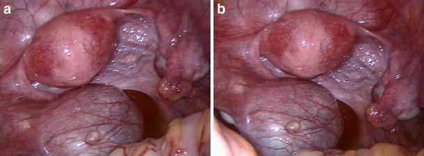
a The picture shows the incorrect position of the optic. b Here, the optic is held in the correct position
-
2.
The action of surgery should always be in the centre of the picture:
Hold the optic in the way the surgeon feels comfortable! (Fig. 2a, b)
Fig. 2 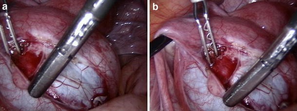
a The picture shows the incorrect position of the optic. The action of surgery is not located in the centre of the picture. b Correct position of the optic. The action of surgery is in the centre of the picture
-
3.
The optic has to be clean and the picture on the screen has to be clear and focused on:
The picture should be clear and in focus! (Fig. 3a, b)
Fig. 3 
a The picture shows the incorrect adjustment and positioning of the camera! The surgeon is unable to judge the situs correctly. b Correct adjustment and positioning of the camera. This picture enables the surgeon to access easily to the pelvic organs
-
4.
Regarding details, the optic has to be verged and zoomed on to the object; achieving overview, the optic and the object have to be refrained, respectively; the object has to be zoomed out:
Vary the distance between optic and object accordingly and use the zoom function of the camera! (Fig. 4a, b)
Fig. 4 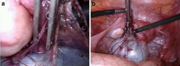
a In order to examine details of the object the camera has to get close to the object respectively zoomed on to the object. b Overview of the pelvis is obtained by refraining optic and object respectively using the zoom out function
-
5.
Optic and instruments are manipulated in a steady and calm fashion:
Avoid any abrupt and quick movements. You are not on a ship!
-
6.
If the position of optic and instruments is satisfying, do not try to improve the position:
Never change a good position unnecessarily!
-
7.
There is no surgeon and assistant but always two surgeons who replace and substitute each other:
interact actively with the other surgeon and replace his instrument if necessary!
-
8.
The instruments of the surgeon should not cross each other:
Hold your instruments parallel and never apart from the perspective! (Fig. 5a, b)
Fig. 5 
a The instruments cross each other. Remaining in this position complicates the operation. b Here, the instruments are held parallel which is the correct approach to the situs
-
9.
Performing operative laparoscopy, both surgeons always have access to the abdomen by two trocars:
Both hands are active! (Fig. 6a, b)
Fig. 6 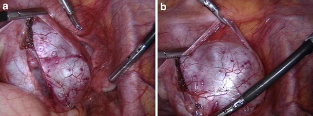
a Only one instrument is working. This may prolong and complicate the operation. b We strongly suggest using all of the trocars actively
-
10.
Preventive coagulation of small blood vessels allows anatomical surgery:
Haemorrhage is your enemy disturbing your optic and view!
-
11.
Avoid using the suction device! In the retroperitoneal space, the anatomical structures are mostly well preserved:
The retroperitonium is your friend!
-
12.
Dissection is done along the embryologic plains using traction and counter traction:
Follow the “spider web”! (Fig. 7)
Fig. 7 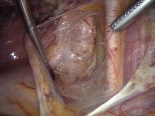
The “spider web” of the retroperitoneum shows the surgeon that he has reached the correct layer


Gaining experience in a two-dimensional operating field was and still remains one of the big challenges a surgeon has to face. Reported learning curves for laparoscopic operating time easily overcome 150–200 cases according to the expansion of the operation [4, 13]. This certainly means a distress to the patient as well as to the surgeon and secondly to the financial budget of each hospital. Qualified surgical intervention requires a qualified education.
Concerning the access to the abdomen and pelvis, we refer to the nine security steps presented by Mettler et al. [21]. Our aim was to create a security standard working inside the abdomen. Severe complications often occur during the training period [14]. Presenting the 12 golden rules, we aspire decreasing the rate of complication in minimal invasive surgery. Nevertheless, apart from the beginning of the training period, the 12 golden rules are also helpful during more complex laparoscopic operations.
In conclusion, the 12 golden rules are helpful to develop minimal invasive surgical skills, to decrease the rate of complication, to improve the patient’s health and to protect the beginner developing frustration during operation.
We would like to put these guidelines up for discussion, and we are grateful for any comment.
References
McKernan JB (1991) Laparoscopic cholecystectomy. AmSurg 57(5):309–312
Semm K (1983) Endoscopic appendectomy. Endoscopy. 15(2):59–64
Mouret P (1996) How I developed laparoscopic cholecystectomy. AnnAcadMedSingapore. 25(5):744–747
Eden CG, Neill MG, Louie-Johnsun MW (2008) The first 1000 cases of laparoscopic radical prostatectomy in the UK: Evidence of multiple `learning curves´. BJU Int, in press
Querleu D, Leblanc E, Ferron G, Narducci F (2006) Laparoscopic surgery in gynecological oncology. Eur J Surg Oncol 32(8):853–858
Darzi A, Mackay S (2002) Recent advances in minimal access surgery. BMJ 324:31–34
Wexner SD, Bergamaschi R, Lacy A, Udo J, Brölmann H, Kennedy RH, John H (2008) The current status of robotic pelvic surgery: results of a multinational interdisciplinary consensus conference. Surg Endosc 23(2):438–442
Altgassen C, Kuss S, Berger U, Löning M, Diedrich K, Schneider A (2006) Complications in laparoscopic myomectomy. Surg Endosc 20(4):614–618
Donnez O, Jadoul P, Squifflet J, Donnez J (2008) A series of 3190 laparoscopic hysterectomies for benign disease from 1990 to 2006: evaluation of complications compared with vaginal and abdominal procedures. BJOG 116(4):492–500
Hur HC, Guido RS, Mansuria SM, Hacker MR, Sanfilippo JS, Lee TT (2007) Incidence and patient characteristics of vaginal cuff dehiscence after different modes of hysterectomies. J Minim Invasive Gynecol 14(3):311–317
Semm K (1983) Endoscopic intraabdominal surgery in gynecology. WienKlinWochenschr 95(11):353–367
Johnson N, Barlow D, Lethaby A, Tavender E, Curr E, Garry R (2005) Surgical approach to hysterectomy for benign gynaecological disease. Cochrane Database Syst Rev 1:CD003677
Fraser SA, Feldman LS, Stanbridge D, Frie GM (2005) Characterizing the learning curvefor a basic laparoscopic drill. Surg Endosc 19:1572–1578
Nussbaum MS (2002) Surgical endoscopy training is integral to general surgery residency and should be integrated inti residency and fellowships abandoned. Semin Laparosc Surg 9:212–215
Huirne JAF, Kennedy R, Stolzenberg JU, Brölmann HAM (2008) What is the impact of surgical expertise and how to get it? Gynecol Surg 5:265–267
Molinas CR, deWin G, Ritter O, Keckstein J, Miserez M, Campo R (2008) Feasibility and construct validity of a novel laparoscopic skills testing and training model. Gynecol Surg 5:281–290
Hiemstra E, Kolkman W, Jansen FW (2008) Skills training in minimally invasive surgery in Dutch obstetrics and gynecology residency curriculum. Gynecol Surg 5:321–325
Gallagher AG, McClure N, McGuigan J, Ritchie K, Sheehy NP (1998) An ergonomic analysis of the fulcrum effect in the acquisition of endoscopic skills. Endoscopy 30:617–620
Munz Y, Kumar BD, Moorthy K, Bann S, Darzi A (2004) Laparoscopic virtual reality and box trainers: is one superior to the other? SurgEndosc 18:485–494
Aggarwal R, Tully A, Grantcharov T, Larsen CR, Miskry T, Farthing A, Darzi A (2006) Virtual reality simulation trainingcan improve technical skills during laparoscopic salpingectomy for ectopic pregnancy. BJOG 113:1382–1387
Mettler L, Wallwiener D (2002) Endoskopische Abdominalchirurgie in der Gynäkologie Schattauer Verlag
Ethical standards
I declare that all the operations done and pictures taken comply with the current law of Germany.
Author information
Authors and Affiliations
Corresponding author
Rights and permissions
About this article
Cite this article
Beyer, D.A., Hornemann, A., Banz, C. et al. Security aspects of modern endoscopic surgery—the 12 golden rules. Gynecol Surg 6, 351–355 (2009). https://doi.org/10.1007/s10397-009-0478-2
Received:
Accepted:
Published:
Issue Date:
DOI: https://doi.org/10.1007/s10397-009-0478-2