Avoid common mistakes on your manuscript.
The ductus venosus (DV) carries oxygen-rich blood from the umbilical vein (UV) to the heart, bypassing the fetal liver. In 1:500 to 1:2500 pregnancies, the DV is either missing or it drains at an atypical site. The types of DV abnormalities are grouped according to the drainage site (intra- or extrahepatic) and whether they bypass the liver (1,2). The presence of a DV abnormality increases the risk of other fetal defects, especially cardiac and genetic abnormalities (3).
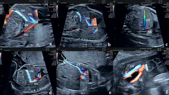
In the image, color Doppler is used to demonstrate normal DV anatomy and five typical abnormalities:
Figure 1a, Normal DV anatomy. DV enters the inferior vena cava (IVC) close to the right atrium
Figure 1b, DV appears to be absent. The umbilical vein drains entirely into the liver without direct connection to the systemic venous circulation. This abnormality generally has a good outcome
Figure 1c, UV drains directly into the IVC at the level of the liver. A focal narrowing of the UV close to its entry into the IVC is present which causes “aliasing” in the Doppler studies. If the narrowing is present, the development of the portal venous system is generally normal
Figure 1d, This abnormality is similar to that in fig. 1c. However, the narrowing is absent. This allows for the blood to enter the IVC easily bypassing the liver. This results in underperfusion and underdevelopment of the portal venous system
Figure 1e, The UV courses over the anterior surface of the liver and enters the right atrium directly without an intervening DV
Figure 1f, The UV is located at the level of the urinary bladder and enters the systemic venous circulation either in the IVC or in the iliac vein
References
Achiron R, Kivilevitch Z (2016) Fetal umbilical–portal–systemic venous shunt: in-utero classification and clinical significance. Ultrasound Obst Gyn 47:739–747. https://doi.org/10.1002/uog.14906
Chaoui R, Heling K, Karl K (2014) Ultrasound of the Fetal Veins Part 1: the intrahepatic venous system. Ultraschall Med 35:208–228. https://doi.org/10.1055/s-0034-1366316
Strizek B, Zamprakou A, Gottschalk I et al (2017) Prenatal diagnosis of agenesis of ductus venosus: a retrospective study of anatomic variants, associated anomalies and impact on postnatal outcome. Ultraschall Med 40:333–339. https://doi.org/10.1055/s-0043-115109
Funding
Open Access funding enabled and organized by Projekt DEAL.
Author information
Authors and Affiliations
Contributions
K. O. K. contributed to project conception and development, image collection, and manuscript writing and editing. R. C. was involved in manuscript writing and editing. M. H. contributed to manuscript writing and editing.
Corresponding author
Ethics declarations
Conflicts of interest
None.
Additional information
Publisher's Note
Springer Nature remains neutral with regard to jurisdictional claims in published maps and institutional affiliations.
Rights and permissions
Open Access This article is licensed under a Creative Commons Attribution 4.0 International License, which permits use, sharing, adaptation, distribution and reproduction in any medium or format, as long as you give appropriate credit to the original author(s) and the source, provide a link to the Creative Commons licence, and indicate if changes were made. The images or other third party material in this article are included in the article's Creative Commons licence, unless indicated otherwise in a credit line to the material. If material is not included in the article's Creative Commons licence and your intended use is not permitted by statutory regulation or exceeds the permitted use, you will need to obtain permission directly from the copyright holder. To view a copy of this licence, visit http://creativecommons.org/licenses/by/4.0/.
About this article
Cite this article
Kagan, K.O., Chaoui, R. & Hoopmann, M. Absent or atypical drainage of the ductus venosus. Arch Gynecol Obstet 307, 633–634 (2023). https://doi.org/10.1007/s00404-022-06828-2
Received:
Accepted:
Published:
Issue Date:
DOI: https://doi.org/10.1007/s00404-022-06828-2


