Abstract
Stag beetles, recognized as common saproxylic insects, are valued for their vibrant coloration and distinctive morphology. These beetles play a crucial ecological role in decomposition and nutrient cycling, serving as a vital functional component in ecosystem functioning. Although previous studies have confirmed that stag beetles are predominantly fungivores, the fluctuations in their intestinal fungal communities at different developmental stages remain poorly understood. In the current study, high-throughput sequencing was employed to investigate the dynamic changes within intestinal fungal communities at various developmental stages in the stag beetle Dorcus hopei. Results showed that microbial diversity was higher during the larval stage than during the pupal and adult stages. Furthermore, significant differences were identified in the composition of the intestinal fungal communities across the larval, pupal, and adult stages, suggesting that developmental transitions may be crucial factors contributing to variations in fungal community composition and diversity. Dominant genera included Candida, Scheffersomyces, Phaeoacremonium, and Trichosporon. Functional predictions indicated a greater diversity and relative abundance of endosymbiotic fungi in the larval gut, suggesting a potential dependency of larvae on beneficial gut fungi for nutrient acquisition. Additionally, the application of abundance-based β-null deviation and niche width analyses revealed that the adult gut exerted a stronger selection pressure on its fungal community, favoring certain taxa. This selection process culminates in a more robust co-occurrence network of fungal communities within the adult gut, thereby enhancing their adaptability to environmental fluctuations. This study advances our understanding of the intestinal fungal community structure in stag beetles, providing a crucial theoretical foundation for the development of saproxylic beetle resources, biomass energy utilization, plastic degradation strategies, and beetle conservation efforts.
Introduction
Beetles, belonging to the order Coleoptera, represent one of the most abundant and diverse groups of insects, accounting for approximately 25% of all known animal species [1, 2]. Within Coleoptera, members of the family Lucanidae are distinguished not only for their striking morphological features, such as enlarged mandibles that resemble deer antlers, but also for their pivotal role in forest ecosystems as saproxylic decomposers of woody debris [3,4,5]. Lucanid beetles also possess a highly specialized and complex gut system, which is home to a vast array of microbes [6, 7] and plays a pivotal role in facilitating insect adaptation to various environments and food resources, including the acquisition of essential amino acids, vitamins, and sterols [8,9,10,11]. Stag beetles, as prominent representatives of the Lucanidae family, exemplify a profound symbiosis between insects and their gut microbiota. During their different developmental stages, stag beetles utilize intestinal microbes (both bacteria and fungi) to maintain health and digest food, especially otherwise indigestible macromolecular substances [12, 13]. This symbiotic relationship highlights the potential of these microbes in nutrient recycling, as well as industrial applications such as plastic degradation and enzyme production [14,15,16]. Understanding the dynamics of gut microbes is essential for devising informed conservation strategies, which are crucial for maintaining beetle diversity and ecosystem resilience in response to climate change.
With the microbial communities residing in insect guts, fungi have garnered increasing attention due to their significant impact on insect physiology and ecology [8, 9]. Gut fungi serve as integral components of the digestive systems of various insect species, facilitating the decomposition of refractory macromolecular organic matter in food and providing essential nutrients necessary for host survival [17]. Additionally, gut fungi enhance symbiotic relationships between insects and their microbial partners, impacting host development, fitness, and ecological interactions. They also produce various enzymes, such as cellulases, peroxidases, laccases, xylanases, and xyloglucanases, which help in the degradation of cellulose, lignin, and hemicellulose, thus playing a pivotal mutualistic role in stag beetle guts [18,19,20,21]. By facilitating the decomposition and assimilation of complex plant-derived carbohydrates and nitrogenous substances, which insects typically find difficult to decompose, gut fungi can help overcome dietary limitations associated with nutrient-poor food sources, thus supporting beetle development and reproduction [22]. Furthermore, these gut-residing fungi contribute to ecosystem nutrient cycling by transforming low-nutrition food sources into bioavailable nutrients [17].
While previous research has successfully reared the fungivorous stag beetle Dorcus rectus (Motschulsky) in controlled laboratory environments [23], whether Dorcus hopei can complete its life cycle on a fungal diet in laboratory settings has not been reported. Examination of the interactions between fungivorous diets and different life stages of D. hopei could help elucidate the distribution and abundance of this insect and provide insights into the ecological and physiological importance of fungal microbes in D. hopei biology. This study selected the giant stag beetle at different developmental stages (1st, 2nd, and 3rd instar larval, pupa, callow adult, mature adult) under fungivorous conditions as the research subject. High-throughput Illumina MiSeq sequencing was employed to investigate the diversity and composition of the intestinal fungal community, exploring its potential role in the growth and development of D. hopei. Specifically, we aimed to (1) reveal the dynamic changes in fungal communities throughout the developmental stages of D. hopei under artificial diets, (2) characterize fungal community diversity in adults and larvae fed two distinct diets, and (3) explore the distribution and assembly of core fungi within the D. hopei gut.
Materials and Methods
Sample Collection and Rearing Conditions
The D. hopei stag beetles were sourced from the Mu-Ye Insect Company (Lishui, Zhejiang, China) and subsequently reared in the laboratory. In total, 72 specimens were artificially reared under optimum conditions (temperature: 22–25 °C). The larvae were fed an artificial diet containing fermented wood shavings mixed with fungal mycelia from Pleurotus geesteranus in pudding boxes (fermented Quercus acutissima wood), which was continuously replaced and rehydrated. Adults were fed beetle jelly. At different developmental stages, including larvae (1st (L1), 2nd (L2), and 3rd (L3) instars, with 13 individuals for each instar), pupae (Pu (12 individuals)), callow adults (CAd (eight individuals)), and mature adults (MAd (13 individuals)), specimens were collected for analysis.
Sample Dissection and Microbial DNA Extraction
Before dissection, all samples were disinfected for 3 min with 70% ethanol, followed by washing with distilled water to clean the surface. The samples were then treated with tenfold diluted phosphate-buffered saline (PBS) (total 500 mL, NaCl 1.37 M, KCl 26.8 mM, Na2HPO4 81.0 mM, KH2PO4 17.6 mM, pH 7.2–7.4) on a horizontal clean bench [24]. The midgut and hindgut were removed using sterile fine-tip forceps and placed into 2 mL of Lysing Matrix E under sterile conditions to avoid contamination. Microbial DNA from the gut of D. hopei was extracted using a Fast DNA® SPIN for Soil Kit (MP Biomedicals, Solon, OH, USA), following the provided instructions, then stored at − 20 °C before use.
Polymerase Chain Reaction (PCR) Amplification
Purified DNA from each sample was utilized as a template for amplification, with the primer pairs ITS1F (5′-CTTGGTCATTTAGAGGAAGTAA-3′) [25] and ITS2R (5′-GCTGCGTTCTTCATCGATGC-3′) [26] used for PCR amplification of fungal internal transcribed spacer (ITS) fragments. The ITS region is recommended as the universal DNA bar-code marker for fungal identification [27]. The PCR procedures were carried out in 20-μL reaction mixtures containing 2 μL of 10 × buffer, 2 μL of 2.5 mM dNTPs, 0.8 μL of each primer, 0.2 μL of TaKaRa rTaq DNA polymerase, 0.2 μL of bovine serum albumin (BSA), 10 ng of template DNA, and deionized water (to 20 μL). The thermal cycling for PCR included an initial step at 95 °C for 3 min, followed by 27 cycles at 95 °C for 30 s, 55 °C for 30 s, and 72 °C for 45 s, with a final extension at 72 °C for 10 min. Negative controls without DNA template were included to check for potential contamination. Amplification products were detected by 2% agarose gel electrophoresis, and subsequent sequencing of the amplified PCR products was conducted by Majorbio (Shanghai, China).
Processing of Sequencing Data
The raw data were processed for preliminary analysis using QIIME v.1.9 [28]. Sequences with the same barcode primer were first grouped into one sample. The sequences obtained from high-throughput sequencing were spliced, the number of overlapping bases was set to be no less than 20, and the error rate of base pairing was 0. After splicing, low-quality sequences (average quality score < 30, length < 250 bp) were removed, ensuring a q-value of sample sequences above 25. The high-quality sequences were saved to seqs.fna, and chimeras were removed with USEARCH (v1. 8.0). Subsequently, the sequences were subjected to cluster analysis, and high-quality sequences were clustered into operational taxonomic units (OTUs) based on 97% similarity using the de novo approach. Representative ITS sequences from these OTUs were then classified taxonomically using the UNITE v.8.0 (http://unite.ut.ee) database [29]. To equally rarefy samples, randomly selected subsets of 3000 sequences (lowest sequence read depth; repetition 20 times) per sample were used to compare fungal community composition and diversity for all samples.
Statistical Analysis
Fungal alpha diversity and relative abundance of dominant genera among life stages were based on one-way analysis of variance (ANOVA) using the SPSS v20.0 (Chicago, IL, USA) [30]. Differences in fungal community composition between different stages and sexes were analyzed by non-metric multidimensional scaling (NMDS) and analysis of similarity (ANOSIM; permutations = 999) using the vegan package in R (v4.3.0) [31,32,33]. The contribution of fungal OTUs to differences among stages was analyzed using SIMPER in the vegan package in R (v4.3.0) [32]. Biomarkers of intestinal fungi in different developmental stages were identified by linear discriminant analysis (LDA) effect size (LEfSe) [33]. Indicator analysis was performed using the labdsv package in R (v4.3.0). The FUNGuild method was used to predict fungal community functions, with only those showing high confidence (i.e., highly probable and possible) selected for further analysis, visualized using the Sigmaplot (v12.5) [34]. The abundance-based beta-null model was used to differentiate the relative significance of deterministic and stochastic processes based on null deviation (NDV) [35]. Co-occurrence network analyses were conducted using the dplyr, igraph, and Hmisc packages in R (v4.3.0) and visualized in Gephi (v0.9.2) [36].
Results
Intestinal Fungal Alpha Diversity
In this study, a total of 3,717,848 high-quality sequences were obtained after processing 72 fungal samples, ranging from 3105 to 110,938 sequences per sample. Venn diagram analysis identified 2357 fungal OTUs, including 631, 713, and 405 OTUs in larvae, pupae, and adults, respectively. Additionally, unique OTUs were identified at each developmental stage, with adults containing 210 unique OTUs, representing 15.3% of total OTUs identified in adults, larvae containing a higher proportion of unique OTUs, accounting for 28.2%, and pupae exhibiting the highest proportion of unique OTUs at 36.3%. Venn diagram analysis was used to compare the similarities and differences between the communities at different developmental stages (Fig. S1).
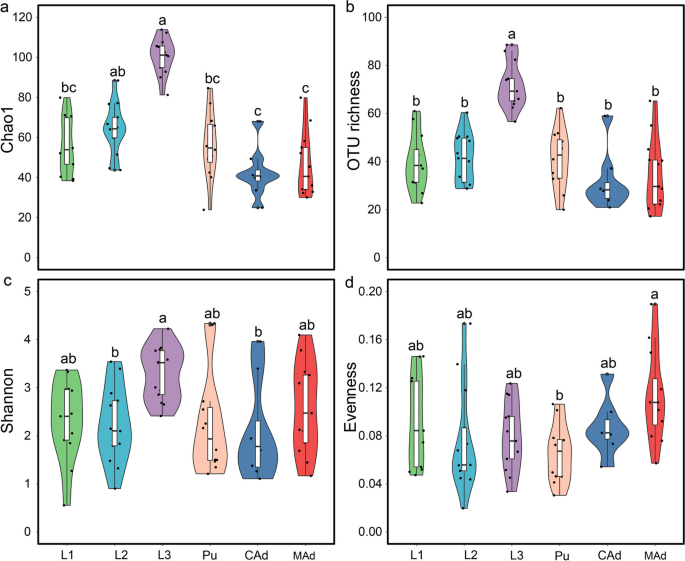
The D. hopei stag beetle undergoes complete metamorphosis, leading to significant differences in diet and function between larvae and adults. In this study, we analyzed four alpha diversity indices (OTU richness, Shannon, Evenness, and Chao1) to evaluate variations in gut fungal diversity across six developmental stages and among insects subjected to two distinct diets. Results showed that fungal alpha diversity initially increased, then decreased, and subsequently increased again, with the highest diversity observed in 3rd instar larvae and the lowest in callow adults (Fig. 1).
Intestinal fungal alpha diversity at different developmental stages, including Chao1 (a), OTU richness (b), Shannon (c), Evenness (d). Different letters above bars represent significant differences based on Tukey’s HSD comparisons (P < 0.05). 1st instar (L1), 2nd instar (L2), 3rd instar (L3), pupa (Pu), callow adult (CAd), and mature adult (MAd)
Intestinal Fungal Community Structure
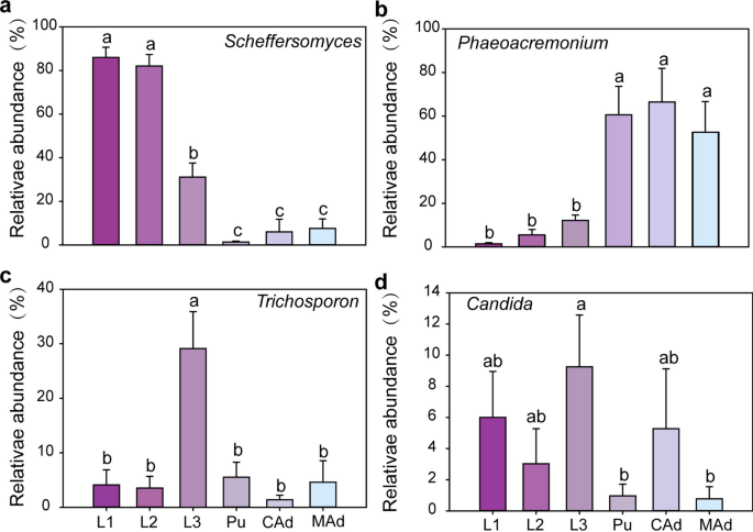
The observed variations in beta diversity were attributable to differences in the community structure of gut fungi. The dominant genera, including Scheffersomyces (38.23%), Phaeoacremonium (30.43%), Trichosporon (8.55%), and Candida (4.19%), showed marked differences in their relative abundances across larvae, pupae, and adults (one-way ANOVA: P < 0.05). Similarly, significant differences were noted among the 1st instar, 2nd instar, 3rd instar, callow adult, and mature adult stages (one-way ANOVA: P < 0.05).
The relative abundance of Scheffersomyces was significantly higher in 1st and 2nd instar larvae compared to the other developmental stages. Phaeoacremonium displayed significantly higher relative abundance during the pupal and adult stages than during the larval stage. The peak relative abundance of Trichosporon was highest in 3rd instar larvae compared to other stages. Furthermore, Candida was also significantly higher in the 3rd instar stage compared to the other developmental stages (Fig. 2).
Relative abundance of fungal taxa at the genus level in the gut of D. hopei at different life stages, respectively are Scheffersomyces (a), Phaeoacremonium (b), Trichosporon (c), Candida (d). Bars represent mean, error bars denote standard deviation, and letters above bars represent significant differences from one-way ANOVA (P < 0.05). 1st instar (L1), 2nd instar (L2), 3rd instar (L3), pupa (Pu), callow adult (CAd), and mature adult (MAd)
The NMDS and ANOSIM analyses revealed distinct fungal community structures across the various developmental stages of D. hopei, with significant differences observed in larvae, pupae, and adults (ANOSIM: P = 0.001; Table 1; Fig. 3). Notably, the fungal communities in the 1st instar larvae differed markedly from those in the 2nd (P = 0.039) and 3rd instar larvae (P = 0.001) (Fig. 3c). In contrast, based on Tukey’s HSD test for multiple comparisons, the community composition between callow and mature adults showed a similar fungal structure (Fig. 3b; Table 1).
The neutral abundance-based β-null model was applied to differentiate the relative importance of deterministic and stochastic processes in the assembly of intestinal fungal communities at different developmental stages (Fig. S2a). Results showed an increasing trend in NDV, suggesting a greater influence of deterministic processes across developmental stages. Additionally, habitat niche breadth values of intestinal fungal communities demonstrated a marked decrease with advancing developmental stages (Fig. S2b).
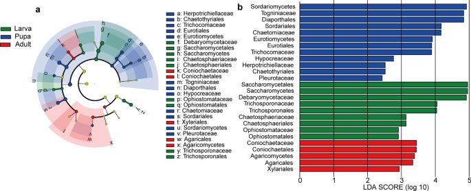
LEfSe analysis was conducted to identify fungal taxa exhibiting differential abundance at different developmental stages in D. hopei at the phylum to family levels (Fig. 4a). Analysis was limited to gut microbial taxa with an LDA score exceeding 2.0 among different groups (Fig. 4b). The constructed cladogram illustrated the phylogenetic distribution of dominant microbial taxa from the phylum to family level across different life stages (Fig. 4a). Notably, nine microbial taxa (one class, four orders, and four families) were enriched in the larval stage, nine microbial taxa (two classes, three orders, and four families) were enriched in the pupal stage, and seven microbial taxa (one class, four orders, and two families) were enriched in the adult stage (Fig. 4).
LEfSe analysis of intestinal fungi at different developmental stages. Cladogram showing phylogenetic distribution of microbial communities across different life stages (a). Yellow nodes represent microbial taxa with no significant difference between different life stages; other color nodes represent microbial taxa significantly enriched at those life stages. Identified phylotype biomarkers ranked by effect size (alpha < 0.05) at different stages (b). Phylotype biomarkers were considered significantly abundant when samples from larval, pupal, and adult stages were compared. Larvae include 1st instar, 2nd instar, and 3rd instar larvae. Adults include callow and mature adults
Indicator species also varied across the different development stages, including one species (Scheffersomyces henanensis) in the 1st instar stage, one species (Ophiostoma protea-sedis) in the 2nd instar stage, seven species (e.g., Trichosporon veenhuisii, Chloridium virescens var. chlamydosporum) in the 3rd instar stage, seven species (e.g., Cryptococcus podzolicus, Humicola nigrescens) in the pupal stage, one species (Trametes versicolor) in the callow adult stage, and one species (Penicillium citrinum) in the mature adult stage (Table S1). SIMPER analysis indicated that Scheffersomyces and Phaeoacremonium were the primary contributors to the variance in intestinal fungal community composition among the different developmental stages (Table S2).
Intestinal Saprotrophs, Pathogens, and Endosymbionts
FUNGuild analysis was employed to categorize gut fungi within D. hopei as plant saprotrophs, animal pathogens, or endosymbionts. Results indicated an increase in the diversity of animal pathogens during the larval stages, with a subsequent decline from the pupal to adult stages, as well as an initial increase and later decrease in the relative abundance of animal pathogens. The peak in both diversity and relative abundance of animal pathogens was observed in the third instar stage, showing significant differences compared to the other developmental stages. In contrast, the 1st, 2nd, and 3rd instar larvae displayed significantly higher diversity and relative abundance of saprophytic bacteria compared to the other life stages. These findings suggest that gut fungi in larvae may play a more critical role in facilitating host food digestion than in pupae and adults, as evidenced by the increased diversity and relative abundance of endosymbiotic fungi within their gut (Fig. S3).
Intestinal Fungal Network Analysis
Co-occurrence network analysis was performed to clarify the interactions among intestinal fungal taxa in D. hopei during the six life stages. Generally, most links in the intestinal network were related to Ascomycota (Fig. S4). The fungal networks in larvae contained more nodes and edges than other stages, demonstrating a higher density of interactions within gut microbial communities in the larval stages (Table S3). Networks for pupal and adult stages, however, demonstrated higher average connectivity and graph density, along with shorter network diameters and average path lengths, suggesting more complex community structures in the guts of pupae and adults compared to the simpler structures observed in the larval stages.
Discussion
Various factors, including diet, developmental stage, and host habitat, influence the structure of the gut microbiome [8, 36, 37]. Insects, in response to varying environmental conditions, have evolved symbiotic relationships with distinct microorganisms at different stages of their life cycle [17]. In the present study, we compared the microbial community composition in D. hopei across different life stages using Illumina MiSeq high-throughput sequencing. Diet is a well-established driving force affecting changes in the host’s gut microbiota [38]. In this study, the D. hopei larvae were fed fermented wood enriched with P. geesteranus, whereas adult stages were fed a jelly-based diet. This variation in diet emerged as a significant determinant of gut microbiota diversity, corroborating the findings of earlier research [7]. Prior research has highlighted the complex interplay between dietary components and gut microbes in insects, with dietary shifts leading to rapid changes in gut microbial composition [39]. Moreover, investigations across diverse insect species have revealed that host-specific factors, such as digestive physiology and immune responses, can exert selective pressure on gut microbial communities, independent of dietary influences [40]. Our results demonstrated no significant differences in the gut fungal community between the third instar larvae and earlier larval stages, nor between the callow adult and mature adult stages. These findings may be attributed to two possible factors: first, the consistency in diet and living environment; second, the lesser quantities of beetle jelly food ingested by callow adults compared to mature adults, resulting in insufficient supplementation of both endogenous and exogenous microorganisms. Furthermore, the metamorphosis experienced by holometabolous insects during the pupal stage, which involves significant restructuring of the intestine and other organs and affects gut microbiota attachment [41, 42], likely plays a role in the observed differences in gut fungal community composition across the larval, pupal, and adult stages of D. hopei.
Scheffersomyces, Trichosporon, Candida, and Phaeoacremonium were identified as the dominant genera in the gut fungal community of D. hopei across various developmental stages (Fig. 2). Scheffersomyces exhibited a high and consistent relative abundance during the larval stages, yet its presence significantly diminished in the adult stages (Fig. 2). This genus is also found in other insects, such as Ceruchus and Sinodendron, suggesting its potential involvement in lignin, hemicellulose, and cellulose degradation [20, 43]. Furthermore, Trichosporon and Candida are known to ferment cellulose and d-xylose, facilitating the absorption of nitrates, xylose, and cellulose, thereby aiding in larval digestion and nutrient uptake [24, 44] (Fig. 2).
In this study, the relative abundance of endophytic fungi was higher in the larval stage than in other developmental stages (Fig. S3). Prior research has indicated that endophytes contribute digestive enzymes and enhance nutritional quality by providing essential amino acids, vitamins, and sterols to their insect host [45]. Additionally, endosymbiotic fungi assist in the degradation of polymeric structural compounds, such as lignocellulose, and in the synthesis of nutrients [46, 47]. Two hypotheses are proposed to elucidate these observations. First, fermented wood, as opposed to beetle jelly, encompasses a more complex composition and a broader spectrum of microorganisms, resulting in larvae ingesting additional microorganisms from fermented wood during feeding. Second, as fermented sawdust is primarily composed of macromolecular substances such as lignocellulose and hemicellulose, compared to simple substances such as agar and brown sugar in beetle jelly, larvae exhibit increased diversity and relative abundance of endosymbiotic fungi in their gut (Fig. S3). Consequently, variations in dominant fungal genera within the gut of D. hopei across different developmental stages may result from differences in dietary intake. Such diversity in fungal communities is essential, facilitating access to a wider array of nutrients, thus supporting growth, development, and reproductive success at each developmental stage.
Our results also showed that gut fungal diversity within D. hopei first increased and then decreased across developmental stages, consistent with previous research [7, 17]. This pattern may be attributed to the third instar larvae undergoing rapid growth and development, necessitating increased nutrient intake from their diet in preparation for pupation, while the post-eclosion phase in adults requires reduced food consumption. Additionally, microbial community diversity in the fermented sawdust consumed by larvae is higher than that in the jelly consumed by adults, thereby potentially contributing to higher microbial colonization and fungal diversity in the larval gut. Earlier studies have indicated that gut microbial community assembly is controlled by both stochastic and deterministic processes [48, 49], with the gut environment acting as a selective “island” for microbial communities, favoring the formation of unique compositions [35, 50]. In the current study, analysis of gut fungal communities revealed higher NDVs in adults compared to larvae and pupae, indicating a stronger influence of deterministic processes on gut fungal selection [51]. Gut selection is a crucial factor affecting the composition and diversity of microbial communities, favoring certain microbes while excluding others, thereby leading to changes in microbial community composition and diversity. Consequently, the enhanced selective capacity in adults compared to larvae and pupae may significantly impact the gut fungal community structure and reduce diversity in D. hopei adults.
Co-occurrence network analysis indicated that the fungal community within the gut of adult D. hopei displayed increased complexity, with enhanced network stability, elevated connectivity, and reduced path lengths (Table S3), suggesting more efficient metabolic and informational exchange pathways among fungal taxa and better adaptability and resistance to environmental fluctuations [52]. Adults demonstrate superior adaptability and resilience to unfavorable conditions compared to larvae, highlighting a developmental shift towards a more selectively curated and stable gut fungal community. Results showed that the predominant associations within the co-occurrence network were between Ascomycota and Basidiomycota, which were pivotal in structuring the gut fungal community across developmental stages (Fig. S4). These fungal taxa produce various enzymes in insects, such as xylanase and carboxymethyl cellulase, which impact habitat adaptation by facilitating plant polymer digestion [53, 54].
Taken together, our study elucidated the structure of the intestinal fungal community across different life stages of the stag beetle, D. hopei, demonstrating significant differences in fungal composition among larvae, pupae, and adults. These findings enhance our understanding of gut microbes in D. hopei and their potential interactions with the host. Notably, fungi such as Scheffersomyces, Trichosporon, Candida, and Phaeoacremonium, prevalent in the gut of the beetle, play crucial roles in the degradation of cellulose, lignin, and hemicellulose, potentially supporting growth, development, and reproductive success of the host. These insights contribute to a deeper understanding of the potential roles of gut microbes in host development and provide theoretical support for the sustainable utilization and protection of saprophytic insects from a microbial perspective. However, this study has several limitations, including the need for more detailed analysis of fungal content in diet to assess its impact on the composition of the gut fungal community. Future research should investigate gut microbiota variations in wild populations to understand the impact of natural conditions on the microbial community structure and composition.
Data Availability
All raw data were submitted to the Sequence Read Archive (SRA) of NCBI under accession number PRJNA1063643.
References
Grove SJ, Stork NE (2000) An inordinate fondness for beetles. Invert Syst 14:733. https://doi.org/10.1071/IT00023
Zhang S-Q, Che L-H, Li Y et al (2018) Evolutionary history of Coleoptera revealed by extensive sampling of genes and species. Nat Commun 9:205. https://doi.org/10.1038/s41467-017-02644-4
Songvorawit N, Butcher BA, Chaisuekul C (2017) Decaying wood preference of stag beetles (Coleoptera: Lucanidae) in a tropical dry-evergreen forest. Environ Entomol 46:1322–1328. https://doi.org/10.1093/ee/nvx143
Huang T-I (2018) Diversity and ecology of stag beetles (Lucanidae). In: Ulyshen MD (ed) Saproxylic Insects. Springer International Publishing, Cham, pp 149–165
Chen D, Cao L, Zhao J et al (2020) Geographic patterns of Lucanus (Coleoptera: Lucanidae) species diversity and environmental determinants in China. Ecol Evol 10:13190–13197. https://doi.org/10.1002/ece3.6911
Wan X, Jiang Y, Cao YY et al (2020) Divergence in gut bacterial community structure between male and female stag beetles Odontolabis fallaciosa (Coleoptera, Lucanidae). Animals 10:2352. https://doi.org/10.3390/ani10122352
Wang MM, Xiang XJ, Wan X (2020) Divergence in gut bacterial community among life stages of the rainbow stag beetle Phalacrognathus muelleri (Coleptera: Lucanidae). Insects 11:719. https://doi.org/10.3390/insects11100719
Engel P, Moran NA (2013) The gut microbiota of insects — diversity in structure and function. FEMS Microbiol Rev 37:699–735. https://doi.org/10.1111/1574-6976.12025
Jing T-Z, Qi F-H, Wang Z-Y (2020) Most dominant roles of insect gut bacteria: digestion, detoxification, or essential nutrient provision? Microbiome 8:38. https://doi.org/10.1186/s40168-020-00823-y
Dar MA, Shaikh AF, Pawar KD et al (2021) Evaluation of cellulose degrading bacteria isolated from the gut-system of cotton bollworm, Helicoverpa armigera and their potential values in biomass conversion. PeerJ 9:e11254. https://doi.org/10.7717/peerj.11254
Douglas AE (2015) Multiorganismal insects: diversity and function of resident microorganisms. Annu Rev Entomol 60:17–34. https://doi.org/10.1146/annurev-ento-010814-020822
Schloss PD, Delalibera I, Handelsman J, Raffa KF (2006) Bacteria associated with the guts of two wood-boring beetles: Anoplophora glabripennis and Saperda vestita (Cerambycidae). Environ Entomol 35:625–629. https://doi.org/10.1603/0046-225X-35.3.625
Grünwald S, Pilhofer M, Höll W (2010) Microbial associations in gut systems of wood- and bark-inhabiting longhorned beetles (Coleoptera: Cerambycidae). Syst Appl Microbiol 33:25–34. https://doi.org/10.1016/j.syapm.2009.10.002
Hongoh Y, Sharma VK, Prakash T et al (2008) Complete genome of the uncultured Termite Group 1 bacteria in a single host protist cell. Proc Natl Acad Sci USA 105:5555–5560. https://doi.org/10.1073/pnas.0801389105
Kubota K, Watanabe K, Zhu X-J et al (2020) Evolutionary relationship between platycerus stag beetles and their mycangium-associated yeast Symbionts. Front Microbiol 11:1436. https://doi.org/10.3389/fmicb.2020.01436
Ayaz Z, Çiftçi DC (2021) An effective method for the monitoring of stag beetle (Lucanus cervus (Linnaeus, 1758) (Coleoptera: Lucanidae)) larvae. J Entomol Res Soc 1:69–81. https://doi.org/10.51963/jers.v23i1.1968
Hu X, Li M, Chen H (2015) Community structure of gut fungi during different developmental stages of the Chinese white pine beetle (Dendroctonus armandi). Sci Rep 5:8411. https://doi.org/10.1038/srep08411
Andlar M, Rezić T, Marđetko N et al (2018) Lignocellulose degradation: an overview of fungi and fungal enzymes involved in lignocellulose degradation. Eng Life Sci 18:768–778. https://doi.org/10.1002/elsc.201800039
Fan J-W, Shelomi M (2023) Symbiotic yeasts of prosopocoilus astacoides blanchardi (Parry, 1873) (Coleoptera: Lucanidae). Coleopt Bull 77(4):527–536. https://doi.org/10.1649/0010-065X-77.4.527
Roets F, Oberlander KC (2020) Symbiotic yeasts from the mycangium, larval gut and woody substrate of an African stag beetle Xiphodontus antilope (Coleoptera: Lucanidae). Antonie Van Leeuwenhoek 113:1123–1134. https://doi.org/10.1007/s10482-020-01418-1
Solomon KV, Haitjema CH, Henske JK et al (2016) Early-branching gut fungi possess a large, comprehensive array of biomass-degrading enzymes. Science 351:1192–1195. https://doi.org/10.1126/science.aad1431
Kaczmarczyk-Ziemba A, Wagner GK, Grzywnowicz K et al (2019) The microbiome profiling of fungivorous black tinder fungus beetle Bolitophagus reticulatus reveals the insight into bacterial communities associated with larvae and adults. PeerJ 7:e6852. https://doi.org/10.7717/peerj.6852
Tanahashi M, Matsushita N, Togashi K (2009) Are stag beetles fungivorous? J Insect Physiol 55:983–988. https://doi.org/10.1016/j.jinsphys.2009.07.002
Morales-Jiménez J, Zúñiga G, Ramírez-Saad HC, Hernández-Rodríguez C (2012) Gut-associated bacteria throughout the life cycle of the bark beetle Dendroctonus rhizophagus thomas and bright (Curculionidae: Scolytinae) and their cellulolytic activities. Microb Ecol 64:268–278. https://doi.org/10.1007/s00248-011-9999-0
Gardes M, Bruns TD (1993) ITS primers with enhanced specificity for basidiomycetes - application to the identification of mycorrhizae and rusts. Mol Ecol 2:113–118. https://doi.org/10.1111/j.1365-294X.1993.tb00005.x
White T J, Bruns T, Lee S, Taylor J (1990) Amplification and direct sequencing offungal ribosomal RNA Genes for phylogenetics. PCR protocols: a guide tomethods and applications. Academic Press, Inc, Cambridge, pp 315–322
Mohd Yusoff MZ, Hu A, Feng C et al (2013) Influence of pretreated activated sludge for electricity generation in microbial fuel cell application. Bioresour Technol 145:90–96. https://doi.org/10.1016/j.biortech.2013.03.003
Caporaso JG, Kuczynski J, Stombaugh J et al (2010) QIIME allows analysis of high-throughput community sequencing data. Nat Methods 7:335–336. https://doi.org/10.1038/nmeth.f.303
Edgar RC (2010) Search and clustering orders of magnitude faster than BLAST. Bioinformatics 26:2460–2461. https://doi.org/10.1093/bioinformatics/btq461
Xiang X, He D, He J-S et al (2017) Ammonia-oxidizing bacteria rather than archaea respond to short-term urea amendment in an alpine grassland. Soil Biol Biochem 107:218–225. https://doi.org/10.1016/j.soilbio.2017.01.012
Anderson MJ (2008) A new method for non-parametric multivariate analysis of variance. Austr Ecol 26:32–46. https://doi.org/10.1111/j.1442-9993.2001.tb00081.x
Gibert C, Escarguel G (2019) PER-SIMPER—a new tool for inferring community assembly processes from taxon occurrences. Global Ecol Biogeogr 28:374–385. https://doi.org/10.1111/geb.12859
Segata N, Izard J, Waldron L et al (2011) Metagenomic biomarker discovery and explanation. Genome Biol 12:R60. https://doi.org/10.1186/gb-2011-12-6-r60
Nguyen NH, Song Z, Bates ST et al (2016) FUNGuild: an open annotation tool for parsing fungal community datasets by ecological guild. Fungal Ecol 20:241–248. https://doi.org/10.1016/j.funeco.2015.06.006
Luan L, Liang C, Chen L et al (2020) Coupling bacterial community assembly to microbial metabolism across soil profiles. mSystems 5:e00298-20. https://doi.org/10.1128/mSystems.00298-20
Fan K, Weisenhorn P, Gilbert JA et al (2018) Soil pH correlates with the co-occurrence and assemblage process of diazotrophic communities in rhizosphere and bulk soils of wheat fields. Soil Biol Biochem 121:185–192. https://doi.org/10.1016/j.soilbio.2018.03.017
Jang S, Kikuchi Y (2020) Impact of the insect gut microbiota on ecology, evolution, and industry. Curr Opin Insect Sci 41:33–39. https://doi.org/10.1016/j.cois.2020.06.004
Tokuda G, Mikaelyan A, Fukui C, et al (2018) Fiber-associated spirochetes are major agents of hemicellulose degradation in the hindgut of wood-feeding higher termites. Proc Natl Acad Sci USA 115: https://doi.org/10.1073/pnas.1810550115
Yun J-H, Roh SW, Whon TW et al (2014) Insect gut bacterial diversity determined by environmental habitat, diet, developmental stage, and phylogeny of host. Appl Environ Microbiol 80:5254–5264. https://doi.org/10.1128/AEM.01226-14
Hammer TJ, Janzen DH, Hallwachs W et al (2017) Caterpillars lack a resident gut microbiome. Proc Natl Acad Sci USA 114:9641–9646. https://doi.org/10.1073/pnas.1707186114
Pérez-Cobas AE, Maiques E, Angelova A, et al (2015) Diet shapes the gut microbiota of the omnivorous cockroach Blattella germanica. FEMS Microbiol Ecol 91. https://doi.org/10.1093/femsec/fiv022
Chouaia B, Goda N, Mazza G et al (2019) Developmental stages and gut microenvironments influence gut microbiota dynamics in the invasive beetle Popillia japonica Newman (Coleoptera: Scarabaeidae). Environ Microbiol 21:4343–4359. https://doi.org/10.1111/1462-2920.14797
Tanahashi M, Kubota K, Matsushita N, Togashi K (2010) Discovery of mycangia and the associated xylose-fermenting yeasts in stag beetles (Coleoptera: Lucanidae). Naturwissenschaften 97:311–317. https://doi.org/10.1007/s00114-009-0643-5
Essakhi S, Mugnai L, Crous PW et al (2008) Molecular and phenotypic characterisation of novel Phaeoacremonium species isolated from esca diseased grapevines. Persoonia 21:119–134. https://doi.org/10.3767/003158508X374385
Kunthiphun S, Endoh R, Takashima M et al (2016) Trichosporon heliocopridis sp. nov., a urease-negative basidiomycetous yeast associated with dung beetles (Heliocopris bucephalus Fabricius). Int J of Syst Evol Microbiol 66:1180–1186. https://doi.org/10.1099/ijsem.0.000850
Douglas AE (2017) The B vitamin nutrition of insects: the contributions of diet, microbiome and horizontally acquired genes. Curr Opin in Insect Sci 23:65–69. https://doi.org/10.1016/j.cois.2017.07.012
Douglas AE (2016) How multi-partner endosymbioses function. Nat Rev Microbiol 14:731–743. https://doi.org/10.1038/nrmicro.2016.151
Stegen JC, Lin X, Fredrickson JK et al (2013) Quantifying community assembly processes and identifying features that impose them. ISME J 7:2069–2079. https://doi.org/10.1038/ismej.2013.93
Stegen JC, Lin X, Konopka AE, Fredrickson JK (2012) Stochastic and deterministic assembly processes in subsurface microbial communities. ISME J 6:1653–1664. https://doi.org/10.1038/ismej.2012.22
Yang J, Yu Z, Wang B, Ndayisenga F (2021) Gut region induces gastrointestinal microbiota community shift in Ujimqin sheep (Ovis aries): from a multi-domain perspective. Environ Microbiol 23:7603–7616. https://doi.org/10.1111/1462-2920.15782
Yan Q, Li J, Yu Y et al (2016) Environmental filtering decreases with fish development for the assembly of gut microbiota. Environ Microbiol 18:4739–4754. https://doi.org/10.1111/1462-2920.13365
Zhou J, Deng Y, Luo F et al (2010) Functional molecular ecological networks. mBio 1:e00169-10. https://doi.org/10.1128/mBio.00169-10
Wijayawardene NN, Hyde KD, Lumbsch HT et al (2018) Outline of Ascomycota: 2017. Fungal Divers 88:167–263. https://doi.org/10.1007/s13225-018-0394-8
Suárez-Moo P, Cruz-Rosales M, Ibarra-Laclette E et al (2020) Diversity and composition of the gut Microbiota in the developmental stages of the dung beetle Copris incertus say (Coleoptera, Scarabaeidae). Front Microbiol 11:1698. https://doi.org/10.3389/fmicb.2020.01698
Acknowledgements
We thank Miss Xiaolu Li (Anhui University) for help with feeding the Dorcus hopei beetles in the early stages. We are also grateful to Miss Jingru Zhao, Miss Meng Yana, and Mr. Yuannuo Wu (Anhui University) for data analysis and helpful suggestions. We thank Abbas (Anhui University) for their critical reading of the manuscript and helpful suggestions.
Funding
This work was supported by the National Natural Science Foundation of China (31872276).
Author information
Authors and Affiliations
Contributions
Xia Wan and Xingjia Xiang designed the experiments. Xiaoyan Bin, Pan Wang, and Yagang Shen collected the samples and conducted all experiments. Xiaoyan Bin analyzed the data and wrote the manuscript. Muhmmad Jafir reviewed and edited the manuscript. Xia Wan improved the manuscript. All authors read and approved the final manuscript.
Corresponding author
Ethics declarations
Conflict of Interest
The authors declare no competing interests.
Supplementary Information
Below is the link to the electronic supplementary material.
Rights and permissions
Open Access This article is licensed under a Creative Commons Attribution 4.0 International License, which permits use, sharing, adaptation, distribution and reproduction in any medium or format, as long as you give appropriate credit to the original author(s) and the source, provide a link to the Creative Commons licence, and indicate if changes were made. The images or other third party material in this article are included in the article's Creative Commons licence, unless indicated otherwise in a credit line to the material. If material is not included in the article's Creative Commons licence and your intended use is not permitted by statutory regulation or exceeds the permitted use, you will need to obtain permission directly from the copyright holder. To view a copy of this licence, visit http://creativecommons.org/licenses/by/4.0/.
About this article
Cite this article
Bin, X., Wang, P., Shen, Y. et al. Investigation of Fungal Community Structure in the Gut of the Stag Beetle Dorcus hopei (Coleoptera; Lucanidae): Comparisons Among Developmental Stages. Microb Ecol 87, 70 (2024). https://doi.org/10.1007/s00248-024-02379-y
Received:
Accepted:
Published:
DOI: https://doi.org/10.1007/s00248-024-02379-y


