
Abstract
Electrophysiological measurements of single synapses are challenging given the size of a single synapse relative to a patch pipette. In addition, one has to take into account the limitations of microscopes in that they need to provide acceptable visualization of a single synapse for patching. However, despite these limitations, researchers have successfully measured single synaptic function along dendrites. The purpose of this chapter is to introduce the techniques that can be implemented to measure single synaptic function. Included in this chapter are such techniques as localized perfusion, localized electrical stimulation, photostimulation, and imaging. These techniques are designed with the assumption that multiple excitatory synapses do not contact a single spine, but rather only one synapse per spine. Whereas this assumption is supported by some empirical data [1], other data suggest otherwise [2], meaning that a complete understanding of the anatomical region is necessary before beginning single synapse experiments.
Access this chapter
Tax calculation will be finalised at checkout
Purchases are for personal use only
Notes
- 1.
A loose patch (Fig. 4) is formed in the absence of the conventional gigaseal, which is necessary for all other patch clamp techniques (e.g., cell-attached, inside out, outside out, whole cell). Instead, in a loose patch clamp, a loose seal is formed with a low electrical resistance (2–11 MΩ). Typically, the micropipettes used for patch-clamp experiments have resistances of 2–5 MΩ. Therefore, watching the resistance of the micropipette while approaching the membrane can indicate when a loose patch is forming (the resistance will increase). As the distance between the micropipette and the membrane decreases, the resistance increases. By observing the changes in resistance, the experimenter can avoid coming too close to the membrane and thus forming a seal. Instead, the micropipette should stay far enough away from the membrane so that a gigaseal is not formed leaving the membrane intact (no suction is used). Since the loose patch method is a noninvasive patch, the EPSCs are visualized as outward and not inward currents. This patch-clamp technique is useful in that the micropipette can be removed from the membrane sampling multiple locations without any damage to the cell.
Fig. 4 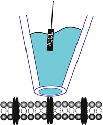
A loose patch. A micropipette is positioned just above the cell membrane so that a gigaseal is not formed. Notice that the membrane is not deformed in a loose patch as is the case in a cell-attached patch in which negative pressure elicits a gigaseal (see Fig. 1.2)
- 2.
Two-photon laser scanning microscopy is a fluorescent imaging technique that enables deep tissue penetration, reduced light diffraction in the tissue and limits phototoxicity [16]. It works by exciting a fluorophore (a fluorescent chemical compound that reemits light when excited) using two photons of low energy (i.e., two photons with half of the wavelength needed to excite the fluorophore). The simultaneous absorption of the two photons by the fluorophore results in fluorescence emission. What gives this imaging technique the advantages listed above is that the probability of two photons simultaneously exciting a fluorophore is very low. Therefore, the two photons must be concentrated spatially and temporally at the focal plane. Only then at the focal plane are fluorophores excited, thus drastically reducing background noise.

References
Schikorski T, Stevens CF (1997) Quantitative ultrastructural analysis of hippocampal excitatory synapses. J Neurosci 17(15):5858–5867
Toni N, Buchs PA, Nikonenko I, Bron CR, Muller D (1999) LTP promotes formation of multiple spine synapses between a single axon terminal and a dendrite. Nature 402(6760):421–425
Bekkers JM, Stevens CF (1995) Quantal analysis of EPSCs recorded from small numbers of synapses in hippocampal cultures. J Neurophysiol 73(3):1145–1156
Kraszewski K, Grantyn R (1992) Unitary, quantal and miniature GABA-activated synaptic chloride currents in cultured neurons from the rat superior colliculus. Neuroscience 47(3):555–570
Veselovsky NS, Engert F, Lux HD (1996) Fast local superfusion technique. Pflugers Arch 432(2):351–354
Chen G, Harata NC, Tsien RW (2004) Paired-pulse depression of unitary quantal amplitude at single hippocampal synapses. Proc Natl Acad Sci U S A 101(4):1063–1068
Kirischuk S, Veselovsky N, Grantyn R (1999) Relationship between presynaptic calcium transients and postsynaptic currents at single gamma-aminobutyric acid (GABA)ergic boutons. Proc Natl Acad Sci U S A 96(13):7520–7525
Matsuzaki M, Honkura N, Ellis-Davies GC, Kasai H (2004) Structural basis of long-term potentiation in single dendritic spines. Nature 429(6993):761–766
Lee MC, Yasuda R, Ehlers MD (2010) Metaplasticity at single glutamatergic synapses. Neuron 66(6):859–870
Lee SJ, Escobedo-Lozoya Y, Szatmari EM, Yasuda R (2009) Activation of CaMKII in single dendritic spines during long-term potentiation. Nature 458(7236):299–304
Forti L, Bossi M, Bergamaschi A, Villa A, Malgaroli A (1997) Loose-patch recordings of single quanta at individual hippocampal synapses. Nature 388(6645):874–878
Mainen ZF, Malinow R, Svoboda K (1999) Synaptic calcium transients in single spines indicate that NMDA receptors are not saturated. Nature 399(6732):151–155
McAllister AK, Stevens CF (2000) Nonsaturation of AMPA and NMDA receptors at hippocampal synapses. Proc Natl Acad Sci U S A 97(11):6173–6178
Umemiya M, Senda M, Murphy TH (1999) Behaviour of NMDA and AMPA receptor-mediated miniature EPSCs at rat cortical neuron synapses identified by calcium imaging. J Physiol 521(Pt 1):113–122
Oertner TG, Sabatini BL, Nimchinsky EA, Svoboda K (2002) Facilitation at single synapses probed with optical quantal analysis. Nat Neurosci 5(7):657–664
Denk W, Strickler JH, Webb WW (1990) Two-photon laser scanning fluorescence microscopy. Science 248(4951):73–76
Aravanis AM, Pyle JL, Tsien RW (2003) Single synaptic vesicles fusing transiently and successively without loss of identity. Nature 423(6940):643–647
Prange O, Murphy TH (1999) Analysis of multiquantal transmitter release from single cultured cortical neuron terminals. J Neurophysiol 81(4):1810–1817
Nauen DW (2011) Methods of measuring activity at individual synapses: a review of techniques and the findings they have made possible. J Neurosci Methods 194(2):195–205
Author information
Authors and Affiliations
Corresponding author
Rights and permissions
Copyright information
© 2016 Springer Science+Business Media New York
About this protocol
Cite this protocol
Graziane, N., Dong, Y. (2016). Measurement of a Single Synapse. In: Electrophysiological Analysis of Synaptic Transmission. Neuromethods, vol 112. Humana Press, New York, NY. https://doi.org/10.1007/978-1-4939-3274-0_18
Download citation
DOI: https://doi.org/10.1007/978-1-4939-3274-0_18
Publisher Name: Humana Press, New York, NY
Print ISBN: 978-1-4939-3273-3
Online ISBN: 978-1-4939-3274-0
eBook Packages: Springer Protocols