
Abstract
The clinical use of human induced pluripotent stem cells (hiPSCs) and the development of patients-specific gene and cell therapies rely on the development of fast, reliable, and integration-free methods of derivation of pluripotent stem cells from somatic tissues. Here we describe an integration-free protocol for the rapid derivation of hiPSCs from dermal fibroblasts using modified mRNAs. This method is inexpensive, highly efficient, and makes use of reagents that are xeno-free and chemically defined and can therefore be adopted by any Good Manufacturing Practice (GMP) facility.
1 Introduction
Derivation of human induced pluripotent stem cells (hiPSCs) from virtually any adult tissue provides numerous opportunities for development of therapeutic strategies in regenerative medicine, cell therapies, and drug discovery applications. Substantial progress has been made in the derivation and optimization of hiPSCs since its technology has first been described in 2007 (1). Moreover and in contrast to human embryonic stem cell (hESC) lines, hiPSCs provide an unlimited cell source without the destruction of human embryos, thus presenting a promising, unlimited, and alternative supply of undifferentiated pluripotent cells. Despite this, the promise of hiPSCs in regenerative medicine relies on overcoming several hurdles. In particular, clinical application of hiPSCs derivatives necessitates a protocol of derivation with minimal risk of integration of exogenous DNA as random integration can lead to insertional mutagenesis with unpredictable effects on the quality of the cells and the potential safety after transplantation (2). Although non-integrative DNA-based methods of inducing pluripotency including episomal vectors (3) and minicircles (4) have been developed, it is difficult to exclude the possibility of integration of very small fragments of DNA. Other proposed non-integrative methods like protein-based reprogramming (5) or Sendai virus (6) are very inefficient (protein based) or require extensive passaging to remove residual viral expression (Sendai virus).
More importantly, the clinical application of hiPSCs requires that their production has to meet GMP-compatible standards but also that the derivation of specified cell derivatives (used for transplantation) from pluripotent stem cells is performed under GMP conditions. Previous regulatory oversight suggests that two methods may be acceptable for this purpose: (1) Derivation of cells and cell products under GMP requirements and (2) Conversion of cells or cell products derived under research-grade conditions to GMP quality standards (http://www.fda.gov/downloads/drugs/guidancecomplianceregulatoryinformation/guidances/ucm070273.pdf). Here, we developed a standardized platform of human iPSC derivation by making use only of chemically defined matrices and animal-free reagents that are already in use in GMP facilities. This protocol is integration-free, fast, efficient, and easily adoptable by any GMP facility.
2 Materials
Prepare all solutions using ultrapure RNase/DNase-free water and analytical grade reagents. All reagents and equipment are handled under sterile and GMP-compliant conditions.
2.1 Culture of Human Fibroblasts
-
CELLstart (Life Technologies).
-
TrypLE Express (Life Technologies).
-
Serum-free Fibroblasts Medium (e.g., FibroLife from Life Technologies).
-
DBPS with calcium and magnesium (Life Technologies).
2.2 Derivation of Induced Pluripotent Stem Cells
-
Modified mRNAs encoding for OCT4, SOX2, KLF4, cMYC, LIN28A, and GFP (100 ng/μl) (TriLink)
Preparation of mRNA cocktail and single-use aliquots
-
Thaw individual mRNAs on ice (Note 1 ). Combine mRNA factors into a 1.5 ml RNase-free microcentrifuge tube on ice as follows: 385 μl OCT4 mRNA + 119 μl SOX2 mRNA + 156 μl KLF4 mRNA + 148 μl cMYC mRNA + 83 μl LIN28A mRNA + 110 μl nGFP mRNA. Mix well and aliquot 50 μl of the mRNA cocktail into 20 individual sterile, 1.5 ml RNase-free microcentrifuge tubes. Store aliquots at −70 °C (Note 2 ).
-
-
Opti-MEM basal medium (Life Technologies).
-
RNAiMAX (Life Technologies).
-
Pluriton Basal Medium (Stemgent).
Preparation of aliquots
-
Thaw Pluriton Basal Medium overnight at 4 °C. Pipet 40 ml aliquots of Pluriton Basal Medium into seven 50 ml conical tubes (280 ml total). Store aliquots at −20 °C (Note 3 ).
-
-
Pluriton Supplement (Stemgent)
Preparation of single-use aliquots
-
Thaw 200 μl of Pluriton Supplement on ice. Pipet 4 μl of Pluriton Supplement into 50 sterile, low protein-binding microcentrifuge tubes and store at −70 °C (Note 4 ).
-
-
B18R interferon inhibitor (eBioscience)
Preparation of single-use aliquots
-
Thaw 40 μg of B18R protein (0.5 mg/ml stock concentration, 80 μl total volume) on ice. Pipet 4 μl of B18R protein into 20 sterile, low protein-binding microcentrifuge tubes and store at −70 °C (Note 4 ).
-
-
CELLstart (Life Technologies).
-
StainAlive DyLight 488 Mouse anti-Human TRA-1-60 (Stemgent).
-
StainAlive DyLight 488 Mouse anti-Human TRA-1-81 (Stemgent).
2.3 Culture and Cryopreservation of Induced Pluripotent Stem Cells
-
TeSR2 Basal Medium (Stem Cell Technologies).
-
TeSR2 5X Supplement (Stem Cell Technologies).
-
TeSR2 250X Supplement (Stem Cell Technologies).
Preparation of Complete TeSR2 medium
-
Nutristem (Stemgent).
-
CELLstart (Life Technologies).
-
Accutase (Innovative Cell Technologies).
-
Bambanker (Wako).
-
Liquid nitrogen.
-
Cryovials.
2.4 Characterization of GMP-Compliant hiPSCs
-
Gene expression analysis of pluripotency markers.
-
Immunocytochemistry of pluripotency markers.
-
EB formation and spontaneous in vitro differentiation.
-
Gene expression analysis and immunocytochemistry of differentiation markers.
-
Teratoma formation.
2.5 Equipment
-
Pipette-aid.
-
Serological pipettes (2, 5, and 10 ml).
-
2, 10, 200, and 1,000 μl pipettors.
-
6-well tissue culture plates.
-
100 mm dishes.
-
15 ml and 50 ml conical centrifuge tubes.
-
Syringe filters (0.22 μm).
-
Triple gas tissue culture incubator set at 37 °C, 5 % CO2, 5 % O2.
-
Tissue culture incubator set at 37 °C, 5 % CO2.
-
Tissue culture centrifuge.
-
Phase-contrast microscope.
-
Fluorescent microscope.
-
Stereo microscope.
-
Dissection scope.
-
Hemocytometer.
-
Coverslips.
-
70 % Ethanol.
-
Cell scrapers.
-
Low protein-binding microcentrifuge tubes.
-
RNase-free aerosol-barrier tips.
-
Cell lifters.
-
Needles (20 Gauge).
-
Glass pipettes.
-
Vertical laminar flow hood certified for Level II handling of biological materials and GMP compliant.
-
Vortexer.
-
Table centrifuge.
3 Methods
Carry out all procedures in a sterile tissue culture hood under GMP-compliant conditions unless indicated otherwise. This protocol describes mRNA-based reprogramming of fibroblast cells that are cultured in 4 wells of 6-well tissue culture plate (Fig. 1). Three wells are used to reprogram target fibroblasts at different starting densities while the remaining well serves as positive control and is used to reprogram BJ fibroblasts. Reprogramming experiments are performed in a triple gas tissue culture incubator set at 37 °C, 5 % CO2, 5 % O2 as it has been shown to increase reprogramming efficiencies (7).

Overview of feeder-free derivation of mRNA-induced pluripotent stem cells under GMP-compliant conditions. Morphology tracking of reprogrammed human fibroblasts during the course of reprogramming and passaging. Fibroblasts show early epithelioid morphology and small cluster formation that lead into small hES cell-like colonies. Small colonies grow in size and become mature iPSC colonies
3.1 Coating 6-Well Tissue Culture Plate
-
1.
Dilute CELLstart 1:50 in DPBS.
-
2.
Add 750 μl diluted CELLstart to 4 wells of a 6-well tissue culture plate (final volume is 0.078 ml/cm2).
-
3.
Incubate in the triple gas incubator for 2 h (Note 7 ).
-
4.
Aspirate CELLstart. The plate is now ready for plating of cells.
3.2 Culturing and Plating of Target Fibroblasts
-
1.
Aspirate culture medium from fibroblasts (Note 8 ).
-
2.
Add 0.05 % Trypsin/EDTA to the cells.
-
3.
Rinse twice with DPBS
-
4.
Incubate the cells for 3–5 min in the triple gas incubator.
-
5.
Add 2× (volume) fibroblast culture medium to the cells to dilute the Trypsin/EDTA.
-
6.
Transfer the harvested cell solution into a 15 ml conical tube with a serological pipette.
-
7.
Centrifuge the cells for 4 min at 200 × g.
-
8.
Remove the supernatant and resuspend the cell pellet in 5 ml of fibroblast culture medium.
-
9.
Count the cells with the hemocytometer and calculate the plating cell density (Note 9 ).
-
10.
Plate the cells in three independent wells of a CELLstart-coated 6-well tissue culture plate at densities of 1 × 10⁴, 2.5 × 10⁴, and 5 × 10⁴ in 2 ml culture media. Plate BJ fibroblasts at a density of 1 × 10⁴ into the fourth well of the CELLstart coated 6-well tissue culture plate.
-
11.
Incubate the cells overnight in the triple gas incubator (Note 10 ).
3.3 Transfect Cells and Start Reprogramming
-
1.
Add 10 ml of Pluriton Medium to a sterile 100 mm dish (Note 11 ).
-
2.
Incubate Medium for 2 h in triple gas incubator (Note 12 ).
-
3.
Just prior to use, thaw one aliquot of Pluriton Supplement and B18R protein on ice (Note 13 ).
-
4.
Add 4 μl of each aliquot to the equilibrated Pluriton Medium to generate Complete Pluriton Medium.
-
5.
Aspirate cell medium from all 4 wells of 6-well tissue culture plate and add 2 ml of Complete Pluriton Medium (Note 14 ).
-
6.
Incubate the cells at least 2 h in the trigas incubator prior to transfection (Note 15 ).
-
7.
Thaw one 50 μl aliquot of mRNA cocktail on ice.
-
8.
Add 200 μl of basal Opti-MEM to the tube containing the mRNA cocktail and gently mix up and down 4–5 times (=tube 1) (Note 16 ).
-
9.
In a second sterile, RNase-free 1.5 ml microcentrifuge tube, add 225 μl of Opti-MEM (=tube 2).
-
10.
Add 25 μl of RNAiMAX to tube 2 directly into the Opti-MEM.
-
11.
Using a 1,000 μl RNase-free, aerosol-barrier pipet tip, pipet gently but thoroughly to mix and immediately transfer diluted RNAiMAX to tube 1 containing the diluted mRNA cocktail.
-
12.
Briefly vortex the mRNA transfection complex and spin down with a table centrifuge.
-
13.
Incubate the mRNA transfection complex at room temperature for 15 min for mRNA-lipids complex formation (Note 17 ).
-
14.
During the incubation time, equilibrate fresh Pluriton Medium to be used after transfection as follows: add 10 ml of Pluriton Medium to a sterile 100 mm dish and put into trigas incubator for equilibration.
-
15.
Remove equilibrated cells in 6-well tissue culture plate from trigas incubator and in drop-wise fashion, add 120 μl of the mRNA transfection complex to each of the 4 wells (Note 18 ).
-
16.
Gently rock the 6-well tissue culture plate from side to side and front to back to distribute mRNA transfection complex.
-
17.
Put back into the trigas incubator and incubate for at least 3 h (Note 19 ).
-
18.
After 3 h of incubation, thaw one aliquot of Pluriton Supplement and B18R protein on ice.
-
19.
Take out the 100 mm dish with pre-equilibrated Pluriton medium (see step 14) and add 4 μl of both aliquots to generate Complete Pluriton Medium.
-
20.
Take out transfected cells (see step 17), aspirate cell medium from all 4 wells of 6-well tissue plate and add 2 ml of fresh Complete Pluriton Medium.
-
21.
Incubate cells overnight in trigas incubator.
-
22.
Repeat transfection procedure, medium equilibration, and medium change for 12–18 consecutive days as described in steps 7–21 (Note 20 ).
3.4 Assess Cell Proliferation Rate and Density of Target Cells
-
1.
One of three wells should have the optimal cell density throughout the reprogramming experiment (see Fig. 2 for optimal cell density). Monitor cell death and proliferation rate every other day under a phase contrast microscope.
Fig. 2 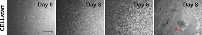
Derivation of iPSCs on GMP compatible CELLstart matrix. Red arrow indicated emerging iPSC colony
-
2.
In the first week of reprogramming, positive GFP signal helps to assess transfection efficiency (should be between 80 and 90 %) (Note 21 ).
-
3.
From the second week onwards, positive GFP signal aids in identifying early colony formation (Fig. 2) (tightly packed colonies are more difficult to transfect, thus appear as dark round “holes” under a fluorescent microscope) (Note 22 ).
-
4.
Balance between cell death and cell proliferation of target cells should be approximately the same compared to the positive control (fourth well with BJ fibroblasts) (Note 23 ).

3.5 Identify Emerging Colonies
-
1.
Around day 8 the first colonies may appear and will grow in size. Between day 12 and 16 the iPSC colonies should emerge and be clearly visible.
-
2.
Primary colonies can be identified with live staining of TRA-1-60 or TRA-1-81 (Note 24 ) as described below.
-
3.
Add 4 ml of Pluriton Medium to a 15 ml conical centrifuge tube.
-
4.
Add 40 μl of either StainAlive DyLight 488 TRA-1-60 or TRA-1-81 antibody to the conical tube (Note 25 ).
-
5.
Aspirate medium from all 4 wells of 6-well tissue culture plate containing cells.
-
6.
Add 1 ml of diluted antibody to each of the 4 wells.
-
7.
Incubate cells in Triple gas incubator for 30 min.
-
8.
Aspirate Pluriton Medium containing the antibody from each well and wash with Pluriton Medium twice.
-
9.
Add 2 ml of Complete Pluriton Medium (with B18R and Pluriton Supplement) to each of the 4 wells.
-
10.
Examine colony formation under a fluorescent microscope with the appropriate wavelength (Fig. 3) (Note 26 ).
Fig. 3 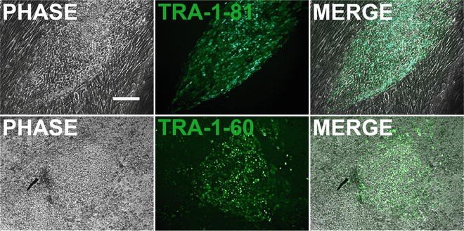
TRA-1-60 and TRA-1-81 live immunostaining during reprogramming used for colony identification. Scale bar = 150 μm
-
11.
Mark primary colonies for manual dissection.

3.6 Manual Dissection of Primary Colonies
-
1.
Dilute CELLstart 1:50 in DPBS.
-
2.
Add 250 μl diluted CELLstart to 8 wells of a 12-well tissue culture plate (final volume is 0.078 ml/cm2).
-
3.
Incubate in a cell culture incubator (37 °C and 5 % CO2) for 2 h (Note 7 ).
-
4.
Aspirate CELLstart. The plate is now ready for plating of cells.
-
5.
Add 10 ml of Pluriton Medium to a sterile 100 mm dish.
-
6.
Incubate Medium for 1 h in cell culture incubator.
-
7.
Thaw one aliquot of Pluriton Supplement and B18R protein on ice.
-
8.
Add 4 μl of both aliquots to the pre-equilibrated Pluriton Medium to generate Complete Pluriton Medium.
-
9.
Add 1 ml of Complete Pluriton Medium to 8 wells of a 12-well tissue culture plate coated with CELLstart.
-
10.
Return 12-well tissue culture plate to cell culture incubator.
-
11.
Take out 6-well tissue culture containing primary iPSC colonies.
-
12.
Using a stereo microscope, locate iPSC colonies based on morphology and based on positive TRA-1-60 or TRA-1-81 signal (see Section 3.5).
-
13.
Using a glass picking tool, gently separate the colonies from the surrounding fibroblasts.
-
14.
Using the same tool, gently cut each colony into 6–8 pieces (Note 27 ).
-
15.
Using either the same tool or a cell lifter, gently detach the colony pieces from the bottom of the tissue culture plate.
-
16.
Using a pipet with a sterile 20 μl pipet tip, transfer the detached colony pieces into an individual well of a 12-well tissue culture plate filled with freshly prepared Complete Pluriton Medium (see step 10).
-
17.
Repeat picking and transferring of primary colonies until 8 individual colonies are picked (Note 28 ).
-
18.
Return both plates (6-well tissue culture plate with primary iPSC colonies and 12-well tissue culture plate with freshly picked colonies) to cell culture incubator. Shake 12-well tissue culture plate back and forth and left and right a few times inside the incubator to distribute colony pieces.
-
19.
Let pieces attach overnight.
-
20.
Change culture medium daily with Pluriton Medium containing Pluriton Supplement (Note 29 ).
-
21.
When cells are 80–90 % confluent, manually passage into a fresh cell culture plate by repeating steps 1–19.
-
22.
Cells are now ready to be transitioned to a new cell culture medium environment.
3.7 Transition of Culture Medium
-
1.
After manual passaging of iPSC colonies to passage 2, gradually transition culture medium from Pluriton Medium containing Pluriton Supplement to a 1:1 mix of Nutristem and Complete TeSR2 Medium.
-
2.
With each daily media change, convert culture from Pluriton Medium to Complete TeSR2/Nutristem as follows: 1:0, 0.8:0.2, 0.5:0.5, 0.2:0.8, 0:1 (ratio of volumes of culture media) (Note 30 ).
-
3.
Cells are now fully converted to GMP-compliant conditions.
3.8 Characterization of GMP-Compliant iPSCs
Cells can now be characterized for their pluripotent character by applying several assays including gene expression analysis of pluripotency markers, immunocytochemistry of pluripotency markers, embryoid body (EB) formation and spontaneous in vitro differentiation, gene expression analysis and immunocytochemistry of differentiation markers, and teratoma formation.
4 Notes
-
1.
Keep mRNAs on ice at all times and avoid repeated opening and closing of vials containing mRNAs. Use RNaseZAP (Life Technologies) or similar RNase contamination solution to wipe down surface of work area. Modified mRNAs should be made in a GMP-compliant environment and in vitro transcribed as previously described (8).
-
2.
Aliquots can be stored up to 3 months. Refreezing aliquots is not recommended. Aliquots are prepared to reprogram 4 wells at a time. Each aliquot will be used for each day of transfection. The molar stoichiometry of all mRNA molecules is 3:1:1:1:1:1 for OCT4, SOX2, KLF4, cMYC, LIN28A, nGFP. nGFP is used to track transfection efficiency over the course of reprogramming and aids to identify early forming colonies.
-
3.
The remaining 220 ml of Pluriton Basal Medium can be stored at 4 °C for use during the first week of reprogramming.
-
4.
Aliquots can be stored up to 3 months. Refreezing aliquots is not recommended. Aliquots are prepared to reprogram 4 wells at a time. Each aliquot will be used for each day of transfection.
-
5.
TeSR2 5× Supplement and 250× Supplement can be dispensed into working aliquots and stored at −20 °C. Frozen aliquots can be stored up to 6 months. Thawed aliquots should be used within 1 day to prepare Complete TeSR2 media. Refreezing aliquots is not recommended.
-
6.
Complete TeSR2 is stable up to 2 weeks or when frozen at −20 °C for up to 6 months. Frozen aliquots of Complete TeSR2 can be thawed at room temperature (15–25 °C) or overnight at 4 °C.
-
7.
Optimal results are achieved when coated 6-well tissue culture plates are used the same day or 1 day after. If precoating the day before, the plates must be stored at 4 °C and wrapped with Parafilm to avoid drying.
-
8.
It is recommended to have target fibroblast in culture prior passaging. Target cells can be directly plated from frozen stocks, though attaching efficiencies are generally lower. It is crucial that the passage number of target cells to be reprogrammed is between 1 and 5.
-
9.
Calculated cell density is crucial for the success of reprogramming. It is recommended to count twice and calculate the average of both results. Also, the initial cell density to be used for counting should not exceed 5 × 10⁵ cells/ml.
-
10.
Rock 6-well tissue culture plate in the Trigas incubator back and forth and left and right to evenly distribute the target fibroblast cells.
-
11.
Due to a high risk of contamination we recommend keeping a full stack of 100 mm dishes at a designated and sterile area to be used only for the reprogramming experiment.
-
12.
Pluriton Medium has to be equilibrated at low O2 tensions and to 37 °C prior use. Conical tubes for equilibration are not recommended since the smaller surface increases incubation time for complete equilibration.
-
13.
We recommend keeping all aliquots (mRNA cocktail, B18R protein, Pluriton Supplement) at one designated area at −20 °C. All aliquots will be used on a daily basis.
-
14.
Incubation of small volumes of culture medium is not recommended due to evaporation. A total of 8 ml (4 × 2 ml) is needed. The remaining volume can be discarded.
-
15.
Cells have to be pretreated with the B18R protein (200 ng/ml) as it is required prior the first transfection on Day 0 to pre-suppress the cells’ interferon response. Subsequent culture and transfections on the following days will be carried out in Complete Pluriton Medium that is already supplemented with the B18R protein.
-
16.
Incubate Opti-MEM at room temperature prior to use.
-
17.
It is essential that the tube containing the mRNA transfection complex be not disturbed during incubation.
-
18.
The addition of the whole mRNA transfection should be applied dropwise to guarantee an even distribution of the complex and avoid citotoxicity.
-
19.
Shake 6-well tissue culture plate again back and forth and left and right as the mRNA transfection complex may have concentrated towards the middle of each well. A minimum of 3 h incubation is recommended. Do not incubate for more than 4 h.
-
20.
It is important to transfect the cells at the same time each day (±2 h) in order to maintain sufficient levels of mRNA transcripts to allow for continual expression of the mRNA factors. The transfection procedure must be repeated each day exactly as done on Day 0.
-
21.
Cell cultures should be not more than 70 % confluent in the first week as overgrown cultures significantly decrease transfection efficiencies, thus reducing the amount of mRNA entering the cells.
-
22.
Although we do not recommend passaging cells during a reprogramming experiment, they can be passaged if they are close to 100 % confluence by day 6 or day 7.
-
23.
BJ fibroblast cells (positive control) should not have reached 100 % confluence by day 6 or 7 and therefore do not need to be split. If they have reached confluence than adjust your cell counting prior to the transfection accordingly. Non-reprogrammed fibroblast that grows around early emerging colonies can be removed to allow for colony growth (see Fig. 2).
-
24.
Cell cultures are very confluent and should appear overgrown. It is sometimes difficult to identify derived colonies from fibroblast.
-
25.
This dilution will yield 5 μg/ml solution of StainAlive antibody. If desired one can down to 2.5 μg/ml.
-
26.
Avoid cell cultures to be kept outside of the incubator for more than 15 min.
-
27.
Optimal sizes of clumps should be empirically determined. Avoid single cells passaging for the first 10 passages.
-
28.
For each individual picked colony use a separate glass picking tool and cell lifter to avoid cross contamination between cell clones.
-
29.
Addition of B18R is not necessary since mRNA transfections have stopped.
-
30.
Cells have to be closely monitored throughout the transitioning process. Allow for modifications (e.g., change ratio every other day or increase ratio steps), as each cell line behaves differently. Also, consider extending transition time up to 4 passages if needed.
References
Takahashi K, Tanabe K, Ohnuki M, Narita M, Ichisaka T et al (2007) Induction of pluripotent stem cells from adult human fibroblasts by defined factors. Cell 131:861–872
Hacein-Bey-Abina S, Von Kalle C, Schmidt M, McCormack MP, Wulffraat N et al (2003) LMO2-associated clonal T cell proliferation in two patients after gene therapy for SCID-X1. Science 302:415–419
Dowey SN, Huang X, Chou BK, Ye Z, Cheng L (2012) Generation of integration-free human induced pluripotent stem cells from postnatal blood mononuclear cells by plasmid vector expression. Nat Protoc 7:2013–2021
Jia F, Wilson KD, Sun N, Gupta DM, Huang M et al (2010) A nonviral minicircle vector for deriving human iPS cells. Nat Methods 7:197–199
Cho HJ, Lee CS, Kwon YW, Paek JS, Lee SH et al (2010) Induction of pluripotent stem cells from adult somatic cells by protein-based reprogramming without genetic manipulation. Blood 116:386–395
Merling RK, Sweeney CL, Choi U, De Ravin SS, Myers TG et al (2013) Transgene-free iPSCs generated from small volume peripheral blood nonmobilized CD34+ cells. Blood 121:e98–e107
Yoshida Y, Takahashi K, Okita K, Ichisaka T, Yamanaka S (2009) Hypoxia enhances the generation of induced pluripotent stem cells. Cell Stem Cell 5:237–241
Durruthy Durruthy J, Ramathal C, Sukhwani M, Fang F, Cui J et al (2014) Fate of induced pluripotent stem cells following transplantation to murine seminiferous tubules. Hum Mol Genet 23:3071–3084
Author information
Authors and Affiliations
Corresponding author
Editor information
Editors and Affiliations
Rights and permissions
Copyright information
© 2014 Springer Science+Business Media New York
About this protocol
Cite this protocol
Durruthy, J.D., Sebastiano, V. (2014). Derivation of GMP-Compliant Integration-Free hiPSCs Using Modified mRNAs. In: Turksen, K. (eds) Stem Cells and Good Manufacturing Practices. Methods in Molecular Biology, vol 1283. Humana Press, New York, NY. https://doi.org/10.1007/7651_2014_124
Download citation
DOI: https://doi.org/10.1007/7651_2014_124
Published:
Publisher Name: Humana Press, New York, NY
Print ISBN: 978-1-4939-2434-9
Online ISBN: 978-1-4939-2435-6
eBook Packages: Springer Protocols