Echocardiographic evaluation of left ventricular function using an automated analysis algorithm is feasible for beginners and experts: comparison with invasive and non-invasive methods Philipp NicolAndreas RankLeif-Christopher Engel Original Investigation Open access 13 October 2022 Pages: 65 - 73
Survey results: status report on problems caused by sexual mismatch between sonographer and patient during echocardiography—a 2020 report of the Japanese Society of Echocardiography Mai IwatakiMitsushige MurataKazuhiro Yamamoto Original Investigation 28 October 2022 Pages: 74 - 78
Multi-modality imaging of post-myocardial infarction ventricular septal defect associated to basal inferoseptal pseudoaneurysm Giovanni BarbatiGiovanna ErenteFrancesco Caprioglio Case image in cardiovascular ultrasound 15 September 2021 Pages: 79 - 80
Paravalvular leak vanishing at end-diastole during transcatheter aortic valve replacement Yoji TamakiShingo TsujinagaToshihisa Anzai Case image in cardiovascular ultrasound 06 October 2021 Pages: 81 - 82
Concurrent ST-elevation myocardial infarction and severe valvular regurgitation causing cardiogenic shock in a patient with infective endocarditis: how to manage? R. S. KuipersN. D. FagelR. K. Riezebos Case image in cardiovascular ultrasound 27 October 2021 Pages: 83 - 84
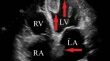
Giant mobile thrombi in both the left ventricle and left atrium Kazuhiro NomuraTakahisa MoronukiKeiji Yamamoto Case image in cardiovascular ultrasound 25 January 2022 Pages: 85 - 86
Non-invasive imaging of ventricular–atrial fistulization secondary to infective rupture of caseous calcification of the mitral annulus Gabriella LocorotondoAlessio AngeliniAntonella Lombardo Case image in cardiovascular ultrasound Open access 28 February 2022 Pages: 87 - 90
Bilateral deep vein thrombosis in pregnancy as first manifestation of an anomalous inferior vena cava Tsukasa KatoWakana SatoHiroyuki Watanabe Case image in cardiovascular ultrasound 29 March 2022 Pages: 91 - 93
An “arboreal” infective pseudoaneurysm following TAVR with “pseudovascular” distribution and morphology Kimberly R. DingRod PartowRamdas G. Pai Case image in cardiovascular ultrasound Open access 06 April 2022 Pages: 94 - 96
A case of midventricular obstruction complicating eosinophilic granulomatosis with polyangiitis followed-up by continuous Doppler imaging Takahiro IkomaToshimitsu KatoMasami Murakami Case image in cardiovascular ultrasound 02 April 2022 Pages: 97 - 98