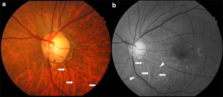
Abstract
Purpose
To investigate whether there is a difference between primary open-angle glaucoma (POAG) patients and control group with regard to choroidal thickness (CT) and the factors influencing CT.
Methods
Ninety eyes of 90 patients who were being followed up with POAG and 72 eyes of 72 healthy subjects matched for age and gender were included. Peripapillary retinal nerve fiber layer thickness (RNFLT), peripapillary CT, lamina cribrosa thickness (LCT), and prelaminar tissue thickness (PTT) were measured with spectral-domain optical coherence tomography (SD-OCT) enhanced depth imaging (EDI) in all patients.
Results
According to multi-variable linear regression analysis results, the factors influencing CT were found as axial length (AL) (B = −22.78, p = 0.002), intraocular pressure (IOP) (B = −7.95, p = 0.001), age (B = −1.77, p = 0.009), and radial pulse rate (B = 1.42, p = 0.015). A statistically significant relationship was not detected between CT and central corneal thickness, mean deviation value of visual field, cup/disk ratio, RNFLT, LCT, PTT. CT was found significantly thinner in glaucoma group (147.5 ± 61.2 μm) compared to control group (167.1 ± 37.3 μm). However, IOP was found significantly higher (p < 0.001) and pulse rate was found significantly lower (p = 0.021) in POAG group. IOP and pulse rate were considered to have affected CT difference between the groups. In advanced and worser stage patients, there were significant positive correlations between CT and RNFLT in inferior and superior quadrants.
Conclusions
In addition to previous studies, IOP and pulse rate were detected to be effective on CT. Further studies are required for determining the whole factors effective on CT and better understanding CT and glaucoma relationship.


References
Resnikoff S, Pascolini D, Mariotti SP, Pokharel GP (2008) Global magnitude of visual impairment caused by uncorrected refractive errors in 2004. Bull World Health Organ 86:63–70
Quigley HA, Broman AT (2006) The number of people with glaucoma worldwide in 2010 and 2020. Br J Ophthalmol 90:262–267
Weinreb RN, Khaw PT (2006) Primary open angle glaucoma. Lancet 363:1711–1720
Garway-Heath DF (2008) Early diagnosis in glaucoma. Prog Brain Res 173:47–57
Flammer J, Orgül S, Costa VP, Orzalesi N, Krieglstein GK, Serra LM, Renard JP, Stefánsson E (2002) The impact of ocular blood flow in glaucoma. Prog Retin Eye Res 21:359–393
Cioffi GA, Wang L, Fortune B, Cull G, Dong J, Bui B, Van Buskirk EM (2004) Chronic ischemia induces regional axonal damage in experimental primate optic neuropathy. Arch Ophthalmol 122:1517–1525
Fuchsjäger-Mayrl G, Wally B, Georgopoulos M, Rainer G, Kircher K, Buehl W, Amoako-Mensah T, Eichler HG, Vass C, Schmetterer L (2004) Ocular blood flow and systemic blood pressure in patients with primary open-angle glaucoma and ocular hypertension. Invest Ophthalmol Vis Sci 45:834–839
Sugiyama K, Gu ZB, Kawase C, Yamamoto T, Kitazawa Y (1999) Optic nerve and peripapillary choroidal microvasculature of the rat eye. Invest Ophthalmol Vis Sci 40:3084–3090
Yin ZQ, Vaegan Millar TJ, Beaumont P, Sarks S (1997) Widespread choroidal insufficiency in primary open-angle glaucoma. J Glaucoma 6:23–32
Spaide RF, Koizumi H, Pozzoni MC (2008) Enhanced depth imaging spectral-domain optical coherence tomography. Am J Ophthalmol 146:496–500
Zeyen T, Roche M, Brigatti L, Caprioli J (1995) Formulas for conversion between Octopus and Humphrey threshold values and indices. Graefes Arch Clin Exp Ophthalmol 233:627–634
Mills RP, Budenz DL, Lee PP, Noecker RJ, Walt JG, Siegartel LR, Evans SJ, Doyle JJ (2006) Categorizing the stage of glaucoma from pre-diagnosis to end-stage disease. Am J Ophthalmol 141:24–30
Li L, Bian A, Zhou Q, Mao J (2013) Peripapillary choroidal thickness in both eyes of glaucoma patients with unilateral visual field loss. Am J Ophthalmol 156:1277–1284
Park HY, Lee NY, Shin HY, Park CK (2014) Analysis of macular and peripapillary choroidal thickness in glaucoma patients by enhanced depth imaging optical coherence tomography. J Glaucoma 23:225–231
Maul EA, Friedman DS, Chang DS, Boland MV, Ramulu PY, Jampel HD, Quigley HA (2011) Choroidal thickness measured by spectral domain optical coherence tomography: factors affecting thickness in glaucoma patients. Ophthalmology 118:1571–1579
Chung HS, Sung KR, Lee KS, Lee JR, Kim S (2014) Relationship between the lamina cribrosa, outer retina, and choroidal thickness as assessed using spectral domain optical coherence tomography. Korean J Ophthalmol 28:234–240
Chakraborty R, Read SA, Collins MJ (2011) Diurnal variations in axial length, choroidal thickness, intraocular pressure and ocular biometrics. Invest Ophthalmol Vis Sci 52:5121–5129
Brown JS, Flitcroft DI, Ying GS, Francis EL, Schmid GF, Quinn GE, Stone RA (2009) In vivo human choroidal thickness measurements: evidence for diurnal fluctuations. Invest Ophthalmol Vis Sci 50:5–12
Hirooka K, Tenkumo K, Fujiwara A, Baba T, Sato S, Shiraga F (2012) Evaluation of peripapillary choroidal thickness in patients with normal-tension glaucoma. BMC Ophthalmol 12:29
Ehrlich JR, Peterson J, Parlitsis G, Kay KY, Kiss S, Radcliffe NM (2011) Peripapillary choroidal thickness in glaucoma measured with optical coherence tomography. Exp Eye Res 92:189–194
Zengin MO, Cinar E, Karahan E, Tuncer I, Yilmaz S, Kocaturk T, Kucukerdonmez C (2015) Choroidal thickness changes in patients with pseudoexfoliation syndrome. Int Ophthalmol 35:513–517
Toprak I, Yaylalı V, Yildirim C (2016) Age-based analysis of choroidal thickness and choroidal vessel diameter in primary open-angle glaucoma. Int Ophthalmol 36:171–177
Acknowledgments
Author contribution
Mehmet Giray Ersoz: Study organization, optic coherence tomography measurement, writing. Duygu Kunak Mart: Optic coherence tomography performing. Emre Ayintap: Writing. Leyla Hazar: Literature investigation. Irfan Botan Guneş: Glaucoma patients recruiting. Seda Karaca Adıyeke: Healthy subjects recruiting. Beysim Doğan: Study orginazation.
Author information
Authors and Affiliations
Corresponding author
Rights and permissions
About this article
Cite this article
Ersoz, M.G., Mart, D.K., Ayintap, E. et al. The factors influencing peripapillary choroidal thickness in primary open-angle glaucoma. Int Ophthalmol 37, 827–833 (2017). https://doi.org/10.1007/s10792-016-0346-9
Received:
Accepted:
Published:
Issue Date:
DOI: https://doi.org/10.1007/s10792-016-0346-9


