
Abstract
The descriptions of seven new and supplemental descriptions of four known species of the genus Acantholaimus (Nematoda: Chromadoridae) from about 5,000 m depth in the abyssal manganese nodule field of the French Claim of the Clarion–Clipperton Fracture Zone (north-eastern tropical Pacific) are given. A. arthrochaeta sp. n. differs from other Acantholaimus species in having jointed cephalic setae. A. barbatus sp. n. is characterized by long cephalic setae and the presence of numerous somatic setae at the level of the pharynx. A. cornutus sp. n. possesses strong onchia and rugae and short cephalic setae. A. robustus sp. n. is characterized by a very large body size, two very large onchia, strongly developed rugae, and cervical setae grouped in threes. A. sieglerae sp. n. is a comparatively small species, though with very large onchia. A. tchesunovi sp. n. can be differentiated from the other species by the lateral differentiation of the body cuticle, consisting of 6–7 longitudinal rows of pores. A. veitkoehlerae sp. n. has a narrow elongate anterior end, two cervical setae, and strong onchia. A. angustus and A. occultus were found about 5,200 km from their type localities in the Peru Basin, south-east Pacific. A. iubilus and A. maks were previously found in different parts of the Atlantic and in the Peru Basin.
Introduction
We present results from studying the nematode collection of the NODINAUT cruise, conducted in 2004, to the French claim of the Clarion–Clipperton Fracture Zone (north-eastern tropical Pacific). This region is considered to be one of the most commercially important nodule areas of the World Ocean (Thiel 2001). The main goal of the cruise was the investigation of the mega-, macro-, and meiofauna of nodule fields. The study of the nematode species community of this area and descriptions of several species from the families Microlaimidae and Benthimermithidae have been published previously (Miljutin and Miljutina 2009a, b; Miljutina et al. 2010, Miljutin et al. 2011).
In the present study, nematodes of the genus Acantholaimus Allgén 1933 were examined. This genus is reported to be very species-rich and abundant in different parts of the deep ocean (Tietjen 1984; Bussau 1993; Vanreusel et al. 2000; Lambshead et al. 2003; De Mesel et al. 2006; Sebastian et al. 2007). Acantholaimus was also one of the most abundant and species-rich genera in the area studied. Of 23 samples examined, Acantholaimus dominated in 4 samples, ranked second in 13 samples, and ranked third in 4. In the samples, the relative abundance of this genus varied from 7.3 to 23.4% (mean value 14.8%), and its density ranged from 7.5 to 33.5 individuals per 10 cm2 (mean 17.3 inds/10 cm2). In total, among 2,513 nematodes examined, 382 individuals (15.2% of all individuals) belonging to 27 putative Acantholaimus species were found. However, most of these species were in juvenile stages, or the number of specimens was insufficient for a new species description. In the end, only 7 putative species, the most abundant ones in the samples, were selected for further description. In addition, we found 4 known species, which were discovered far from the type localities.
Materials and methods
Study area
The scientific cruise “Nodinaut” was conducted by the French RV “L′Atalante” (IFREMER) in the area of French mining claims in the Clarion–Clipperton Fracture Zone (CCFZ) (9–14°N, 130–150°W, ~5,000 m depth) in the summer of 2004 (Table 1).
The seabed in the sampling area is characterized by abyssal hills 100–300 m in height and spaced 5–10 km apart. Most of the area (about 90%) between the seabed hills and their slopes is covered by ferromanganese nodules (2 cm to 15+ cm in diameter). The sediments between the nodules are very fine-grained (<2 μm) silicate oozes (radiolarian and diatomaceous) (Khripounoff et al. 2006).
Nematode sampling and treatment
Most samples were collected using the 8-tube multicorer (tube diameter 100 mm). Two samples were obtained by means of the manipulator of the “Nautile” submersible. On board, the samples were fixed with 10% formalin in seawater.
Nematodes were sorted out, processed into glycerin using Seinhorst’s (1959) method of slow evaporation, and permanently mounted on glycerin--paraffin slides.
The nematodes were examined with the aid of LEICA DMR and LEICA DM2500 microscopes equipped with a drawing apparatus. Initially, all nematodes were identified to family and genus level. Then, species of the genus Acantholaimus with a sufficiently large number of specimens of both sexes were selected for further study and description. Photographs were taken with a LEICA DM2500 microscope equipped with a LEICA EC3 digital camera.
In about half of the Acantholaimus specimens examined, the tails were broken in their thin, cylindrical part. For this reason, in order to minimize errors in the body indices that are traditionally used in taxonomical descriptions of free-living aquatic nematode taxa (a, b, and V), additional indices a′, b′, and V′ were added (see the “List of abbreviations”). In addition, in order to clarify how length of onchia, esophastoma, and male spicules was measured, schemes of these measurements are given in Fig. 1.

Scheme of measurements of onchia and esophastoma (a), and male spicules (b) in specimens of the genus Acantholaimus. Abbreviations: a, b length of onchia, c length of esophastoma, d length of spicule in arc, e length of spicule in chord
All type specimens were deposited in the Muséum National d’Histoire Naturelle, Paris (MNHN).
List of abbreviations
- –:
-
Parameter absent;
- a :
-
Ratio “body length/maximum body diameter”;
- a′:
-
Ratio “body length without tail/maximum body diameter”;
- b :
-
Ratio “body length/length of pharynx”;
- b′:
-
Ratio “body length without tail/length of pharynx”;
- c :
-
Ratio “body length/length of tail”;
- c′:
-
Ratio “length of tail / body diameter at anus”;
- c.b.d.:
-
Corresponding body diameter;
- f :
-
Female;
- i :
-
Intersex;
- L :
-
Total body length;
- L′:
-
Body length without tail;
- m :
-
Male;
- n.a.:
-
Not available for measuring because of poor position or condition of specimen, broken tail or inverted head;
- V :
-
Ratio “distance from anterior end to vulva / total body length” (%);
- V′:
-
Ratio “distance from anterior end to vulva / body length without tail” (%)
Taxonomic descriptions
-
Acantholaimus angustus Bussau 1993 (Figs. 2, 3, 4; Table 2)
Fig. 2 
Acantholaimus angustus, males. a specimen No. 2, total view; b specimen No. 1, spicule; c specimen No. 1, spermatid; d specimen No. 2, spicule; e specimen No. 2, posterior end; f specimen No. 2, postcloacal somatic seta. Scale bars a = 100 μm; b, c, d = 10 μm; e = 50 μm
Fig. 3 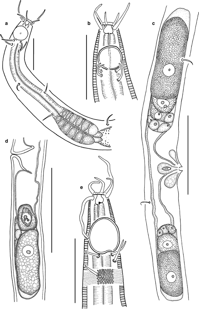
Acantholaimus angustus. a male, specimen No. 2, anterior end; b female, specimen No. 8, head region; c female, specimen No. 7, reproductive system; d female, specimen No. 6, posterior half of reproductive system; e male, specimen No. 1, head region. Scale bars: a, b, e = 20 μm; c, d = 50 μm
Fig. 4 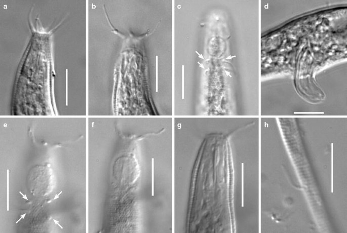
Acantholaimus angustus, micrographs. a female, specimen No. 8, head region; b male, specimen No. 2, head region; c female, specimen No. 10, head region; d male, specimen No. 2, region of spicule; e–g female, specimen No. 7, different optical sections of head region; h specimen No. 7, somatic seta on filiform part of tail. Scale bars = 10 μm. Arrows mark location of cervical setae
Table 2 Acantholaimus angustus Bussau, 1993


Material examined
4 males, 5 females, and 1 intersex possessing well-developed female reproductive system and male spicules with gubernaculum but without testis and vas deferens (Table 1).
Locality
Description
Main measurements: L = 627–910 μm; L′ = 473–604 μm; a = 23.2–47.9; a′ = 17.5–28.2; b = 6.5–9.2; b′ = 4.7–6.7; c = 2.3–4.1; c′ = 8.9–24.6; V = 31.0–47.0%; V′ = 41.6–62.4% (Table 2).
Body slightly spindle-shaped, with narrowed anterior end and filiform posterior end. Somatic setae 6–8-μm-long, numerous, situated along entire body in 4 submedian rows. In postanal region, somatic setae clavate, i.e., with widened distal end; on rest of the body, setae of ordinary form, i.e., edged. Cuticle densely dotted (dots arranged in transverse rows), with lateral fields consisting of much larger dots. Lateral fields beginning at posterior border of amphideal fovea and extending along entire body length except caudal part. In optical cross-section of the cuticle, the dots are discernible as tiny radial struts. Cuticle about 1.0–1.5-μm thick along entire body length except at level of cephalic setae, where it is thinner (less than 1 μm). Lips not visible. 3 rings of head sensilla visible: inner labial papilloid sensilla ca.1 μm long (these sensilla not visible in most specimens), 6 outer labial setae 4–7 μm long, and 4 submedian cephalic setae 9–12 μm long. Outer labial and cephalic setae were cylindrical. Amphideal fovea large (about 1 c.b.d. in width in males), ventrally coiled, single-spiral, with longitudinally oriented oval shape (in males) or round (in females), 9–12 μm width in males and 6–9 μm in females, situated in 0.4–1.0 c.b.d. from anterior end. Two pairs (latero-subdorsal and latero-subventral) of cervical setae 4–10 μm long, located close to posterior part of amphideal fovea. In latero-subventral pair of cervical setae, the distance between anterior and posterior setae is ca. twice the distance between setae of latero-subdorsal pair. Stoma consisting of wide, barrel-shaped cheilostoma and narrow esophastoma. At least two small sclerotized onchia ca. 0.2–0.4 μm long (presumably, dorsal and subventral) visible at anterior end of esophastoma. Esophastoma marked very feebly, walls of esophastomal internal lumen only slightly thicker than in remainder of pharynx. Pharynx muscular, largely cylindrical but widened in its posterior quarter to an oval bulb. This bulb possesses large plasmatic inclusions regularly alternating with muscle cells. Renetta not visible. cardia small, rounded, surrounded by intestine. Tail consisting of proximal conical part and long terminal filiform cylindrical part that constitutes 75–90% of entire tail length. Diameter of cylindrical part of tail in its posterior end ca. 2–3 μm.
Male reproductive system monorchic. Testis directed anteriorly, outstretched, lying to the right of intestine, occupying about 40% of total body length. Spermatids oval, large (ca. 35 × 10 μm), with clearly visible, oblong nuclei. Curved spicules possessing complex cuticular sculpture with thickened cuticular areas in form of longitudinal ridges. Gubernaculum shaped as curved triangular stick with edged proximal end and bifurcated distal end. Supplementary organs not found.
Female reproductive system consisting of two antidromous ovaries (anterior ovary lying to right of intestine, and posterior one lying to left of intestine). Each ovary containing one mature oocyte, from 40 × 15 to 60 × 20 μm in size. One, nearly round spermatozoon ca. 17 μm in diameter visible in uterus of specimen No. 6. Three pairs of vulvar glands seen in some females.
Specimen No. 5 is an intersex with a well developed female reproductive system. However, the male reproductive system is incompletely developed with only spicules present, and the testis and vas deferens are lacking.
Abundance
The density of this species varied from 0 to 7.3 inds/10 cm2 (mean value 2.1 inds/10 cm2). In relative abundance within the nematode community, this species ranked fourth (from 0 to 6.3% in different samples, mean 1.7%).
Remarks
Acantholaimus angustus was initially described by Bussau (1993) in his doctoral thesis. In this work, the author described in total 110 species (97 of them as new) and 11 new genera from the deep-sea nodule-bearing area (Peru Basin, CE Pacific, depth 4,160 m). Unfortunately, the collection of the holotypes was never deposited in a repository institution, and therefore some nematologists consider these species as invalid. Only a few of the species were formally described later (Bussau 1995; Bussau and Vopel 1999). Nevertheless, the value of this doctoral work is very high because of its completeness and care in preparation, and most of the genera and species described therein are easily recognized. Many of Bussau’s species and genera were rediscovered in other regions of the World Ocean (see e.g., Vanreusel et al. 1997; Vanhove et al. 1999; Fonseca et al. 2006, Miljutin and Miljutina 2009b; Miljutina et al. 2010).
The type location of A. angustus is about 5,200 km distant from the CCFZ, where the new specimens of this species were found. The new specimens closely resemble those described by Bussau (1993) in their appearance (location and size of amphideal foveas, length and arrangement of head and cervical setae, presence and location of lateral fields, presence of clavate somatic setae in the postanal region, shape of spicule). However, our individuals are larger and possess shorter pharynxes and longer tails than the type specimens [L = 627–910 μm, b = 6.5–9.2, and c = 2.3–4.1 vs. 575–725 μm, 5.0–6.2, and 4.7–5.5, respectively, in Bussau (1993)]. In addition, the gubernacula in the males studied here are less strongly curved than in the type male. In part, these differences may be explained by intraspecific variation (we were able to study more specimens) or by interpopulation differences. Additionally, in nematodes, smaller specimens tend to possess a relatively longer pharynx than larger ones (see e.g., Roggen 1970).
The V′ ratio varies widely in the new specimens (from 41 to 61%). A similarly wide variation has not been found in other Acantholaimus species. Nevertheless, the appearance of the head end, the arrangement and the shape of the head and cervical sensilla, and the structure of the stoma of all specimens examined appear identical. All the specimens examined can, therefore, be assigned to the same species.
Distribution
South-eastern Pacific, Peru Basin, nodule fields, 4,157, 4,159 m (Bussau 1993); north-eastern tropical Pacific, Clarion–Clipperton Fracture Zone, nodule fields, abyssal plain without nodules, 4,800–5,042 m, ooze (present report) (Fig. 5).

Maps showing the records of known Acantholaimus species found in the present study. Bold letters mark locations where the species was reported. Abbreviations mean reference sources: A = Gerlach et al. 1979; B = Gourbault and Vincx 1985; C = Vivier 1985; D = Jensen 1988; E = Tietjen 1989; F = Bussau 1993; G = present study
-
Acantholaimus arthrochaeta sp. n. (Figs. 6, 7, 8; Table 3)
Fig. 6 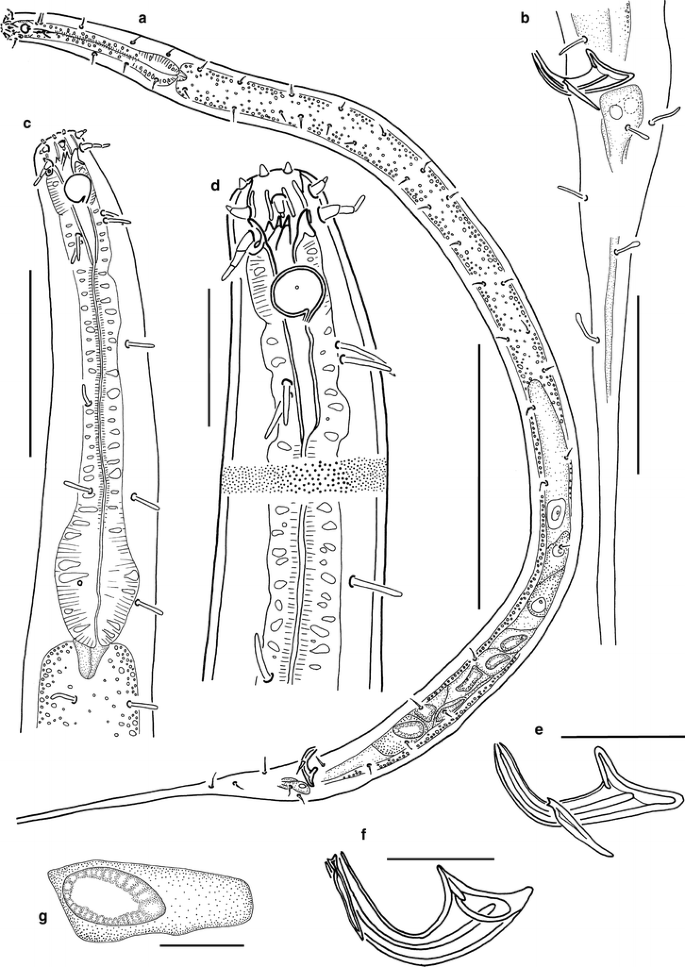
Acantholaimus arthrochaeta sp. n., males. a holotype, total view; b holotype, posterior part; c holotype, anterior end; d holotype, head region; e holotype, spicule; f paratype No. 1, spicule; g holotype, spermatid. Scale bars: a = 200 μm; b, d–g = 20 μm; c = 50 μm
Fig. 7 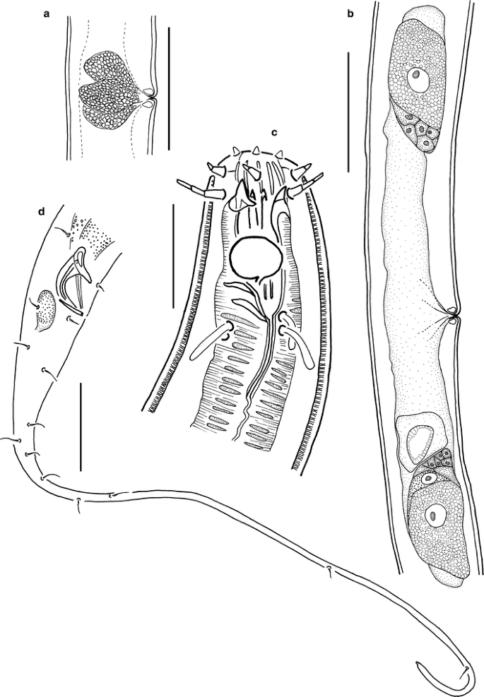
Acantholaimus arthrochaeta sp. n. a female, paratype No. 3, vulvar region; b female, paratype No. 3, female reproductive system; c female, paratype No. 5, head region; d male, paratype No. 1, posterior end. Scale bars: a, b, d = 50 μm; c = 15 μm
Fig. 8 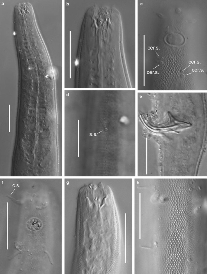
Acantholaimus arthrochaeta sp. n., micrographs. a male, paratype No. 2, anterior end; b, c male, paratype No. 2, head region; d male, paratype No. 2, medio-lateral longitudinal row of small cuticular pores in pharyngeal region; e male, holotype, spicule region; f female, paratype No. 5, head region; g male, holotype, head region; h male, holotype, lateral field with larger irregularly arranged dots in region of mid-body. Scale bars = 20 μm. Abbreviations: cer.s. = cervical seta, c.s. = cephalic seta, s.s. = somatic seta
Table 3 Acantholaimus arthrochaeta sp. n.


Type material
Collection number MNHN-BN498. Holotype: male. Paratypes: 2 males and 4 females (Table 3).
Locality
Etymology
Greek arthrochaeta (= bearing jointed setae).
Description
Main measurements: L = 1,098–1,545 μm; L′ = 839–1,055 μm; a = 20.4–39; a′ = 15.0–31.0; b = 8.5–10.1; b′ = 6.6–7.8; c = 3.2–4.2; c′ = 8.6–13.6; V = 55.0; V′ = 68.1–72.2 (Table 3).
Body cylindrical, with slightly narrowed anterior end and long filiform tail. Somatic setae cylindrical or clavate, 6–9 μm long, numerous, situated along entire body in 4 submedian rows except filiform part of tail where they are irregularly arranged. Numerous pores also visible between somatic setae in these 4 submedian rows. Cuticle densely dotted in lateral fields, beginning posterior to amphideal fovea and extending along entire body except filiform part of tail. Lateral fields with larger dots arranged more sparsely than median dots, which are smaller and more densely arranged. Dots arranged irregularly at level of pharynx (length of body part with irregularly arranged dots varies in different specimens), whereas they are arranged in transverse rows along rest of body. In optical cross-section of cuticle, the dots are discernible as tiny radial struts. Cuticle ca. 1–1.5-μm thick along entire body, except thinner at level of cephalic setae (less than 1 μm), and thicker at level of pharynx and beginning of intestine (ca. 2 μm). Lips not visible. Sensilla of cephalic end arranged in 3 rings: 6 short inner labial papilliform setae 2–3 μm long; 6 short, jointed, bipartite, outer labial setae 4–8 μm long; and 4 longer, jointed, tripartite, cephalic setae 7–8 μm long. In about half of specimens, anterior part of head together with cephalic setae retracted. Amphideal fovea ventrally coiled, single-spiral, round or shaped as transversely oriented oval, 8–9 μm wide in males and 6–8 μm wide in females, with fine but distinct cuticular edging. Very fine concentric striation and more or less defined central spot visible in amphidial fovea. Anterior end of amphideal fovea situated ca. 0.58–0.72 c.b.d. from anterior end. Two pairs (latero-subdorsal and latero-subventral) of cervical setae 5–10 μm long, located posterior to each amphideal fovea. In all individuals, length of cervical setae related to lengths of head setae (they are longer in individuals with longer head setae, and shorter in individuals with shorter head setae). Dorsolateral pair of cervical setae situated at about 0.3 amphidial c.b.d. from amphideal fovea, whereas ventrolateral pair of cervical setae situated farther from amphideal fovea, at about 0.5 amphidial c.b.d. Stoma large, consisting of wide, cup-shaped cheilostoma and narrow, funnel-shaped esophastoma. Cheilostoma ca. 10 μm long, possesses 12 cylindrical rugae. Esophastoma ca. 35 μm long, with thick cuticular walls, containing 5 sclerotized onchia. Basal parts of onchia situated in anterior part of esophastoma, and their apical parts intruding into cheilostoma. One large dorsal or subdorsal onchium ca. 5–6 μm long, one large subventral onchium ca. 5 μm long, one small subventral and two small subdorsal onchia (these latter onchia often appearing as one bifurcated onchium) ca. 1.5–2.0 μm long visible in most specimens. Some small onchia not distinguishable in several specimens. Pharynx narrow in its anterior part, then gradually widening to its posterior end where it forms a poorly developed bulb. Anteriormost part of pharynx ca. 10–15 μm long (evidently consisting of muscles operating onchia) appreciably detached from rest of pharynx by small constriction, possessing another pattern of muscle striation, and almost devoid of cytoplasmic inclusions. In remainder of pharynx, muscle cells alternate with cytoplasmic inclusions along its entire length. Nerve ring not visible. Renetta not visible. Cardia small, triangular. Tail conical in its anterior part and possessing long filiform, cylindrical terminal part constituting 60–80% of entire tail length. Diameter of cylindrical part of tail in its posterior end ca. 2.5–3.0 μm. Cellular bodies of caudal glands visible posterior to anal region. Tails broken in about half of specimens.
Male reproductive system monorchic. Testis directed anteriorly, outstretched, lying to right of intestine, occupying about 0.4–0.5 of pre-anal body length. Curved funnel-shaped paired spicules possessing cuticular sculpture with thickened cuticular areas in shape of longitudinal or bounding ridges. Gubernaculum appearing stick-like, with slightly narrowed proximal end and narrow bifurcated distal end. Supplementary organs not visible.
Female reproductive system consisting of two antidromous, uniformly sized ovaries (anterior ovary lying to right of intestine, and posterior one lying to left of intestine). Each ovary containing one mature oocyte, ca. 40 × 20 μm. One spermatozoon ca. 18 μm in diameter visible in uterus in paratype No. 5. Three pairs of vulvar glands with granular content surrounding short vagina.
Abundance
Acantholaimus arthrochaeta sp. n. was mentioned as Acantholaimus sp. 27 in the description of nematode assemblages inhabiting deep-sea polymetallic nodule fields (Miljutina et al. 2010). Its density varied from 0 to 8.4 inds/10 cm2 (mean 1.8 inds/10 cm2), and its relative abundance within the nematode community was 0–6.2% (mean 1.4%). This species was among the five most abundant species in this area.
Differential diagnosis
Acantholaimus arthrochaeta sp. n. differs from all other Acantholaimus species by the structure of the cephalic setae. There is no other described congener with jointed setae. However, this feature could be difficult to detect in investigations conducted with the help of old or low-power microscopes. Therefore, we will not use this characteristic in comparing species.
The new species shares with A. akvavitus Gerlach et al. 1979, A. barbatus sp. n., A. quintus Gerlach et al. 1979, A. septimus Gerlach et al. 1979, and A. veitkoehlerae sp. n. the mid-sized body length (about 1,000–1,500 μm), the relatively small amphideal fovea (40–65% c.b.d.), and the position of the amphideal fovea relative to the apical part of the body (about 1 c.b.d.).
Acantholaimus arthrochaeta sp. n. differs from A. barbatus sp. n. by the longer cephalic setae (7–8 μm vs. 13–32 μm); the position of the cervical setae (situated 8–10 μm behind the amphideal fovea vs. situated at the level of the amphideal fovea); the shape of the anterior end (truncate vs. conical); and the longer esophastoma (ca. 35 μm vs. 15–20 μm).
Acantholaimus arthrochaeta sp. n. differs from A. veitkoehlerae sp. n. by their body shape (cylindrical with weakly narrowed anterior end vs. spindle-shaped body with strongly narrowed anterior end); the number of cervical setae (4 vs. 2); the shape of the amphideal fovea (oval vs. circular); and the shape of the spicules (thin vs. thick).
Acantholaimus arthrochaeta sp. n. differs from A. quintus by the shape of the anterior end of the body (truncate vs. conical); by the quantity and position of the cervical setae (4 vs. 6, situated far from the amphideal fovea vs. situated at the level of the amphideal fovea); the smaller number of somatic setae; the longer esophastoma (ca. 35 μm vs. 10–15 μm); and the absence of preanal papillae.
Acantholaimus arthrochaeta sp. n. most closely resembles A. akvavitus and A. septimus. It differs from A. akvavitus by the body shape (cylindrical vs. spindle-shaped); the shape of the anterior end (truncate without well developed lips vs. conical with well developed lips); and the number of cervical setae (4 vs. 3).
Acantholaimus arthrochaeta sp. n. differs from A. septimus by the shape and size of the amphideal fovea (transversely oriented oval vs. longitudinally oriented oval, 37–53% vs. 67% of c.b.d.); the position of the cervical setae relative to each other (both pairs of setae are situated very close to one another vs. relatively far); the shape of the spicules (thin vs. thick); and the absence of preanal papillae.
-
Acantholaimus barbatus sp. n. (Figs. 9, 10, 11, 12; Table 4)
Fig. 9 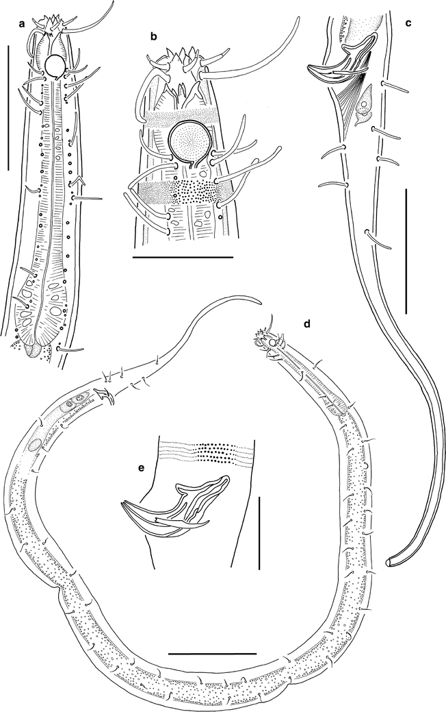
Acantholaimus barbatus sp. n., male, holotype. a anterior end; b head region; c posterior end; d total view; e spicule region. Scale bars: a, c = 50 μm; b, e = 20 μm; d = 100 μm
Fig. 10 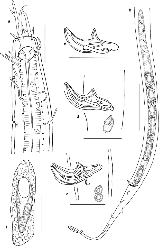
Acantholaimus barbatus sp. n., males. a paratype No. 3, head region with retracted anterior part; b paratype No. 1, posterior end with reproductive system; c paratype No. 3, spicule; d paratype No. 2, spicule region; e paratype No. 5, spicule region; f spermatid. Scale bars: a, c–f = 20 μm; b = 100 μm
Fig. 11 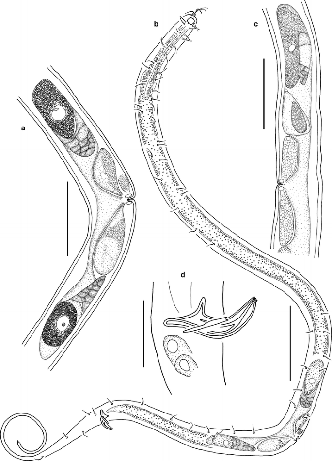
Acantholaimus barbatus sp. n. a paratype No. 6 (intersex), female reproductive system; b paratype No. 6 (intersex), total view; c paratype No. 7 (female), anterior branch of reproductive system with spermatozoon; d paratype No. 6 (intersex), spicule region. Scale bars: a, c = 50 μm; b = 100 μm; d = 20 μm
Fig. 12 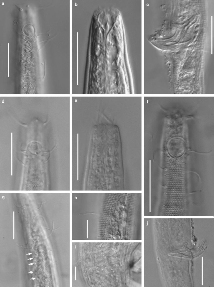
Acantholaimus barbatus sp. n., micrographs. a male, holotype, head region in surface view; b male, holotype, head region; c male, holotype, spicule region; d female, paratype No. 8, head region; e male, paratype No. 3, retracted head; f male, paratype No. 1, head region; g female, paratype No. 10, cervical region; h male, holotype, somatic seta at region of mid-body; i female, paratype No. 10, lateral field with larger, regularly arranged dots in region of mid-body; j intersex, paratype No. 5, spicule region. Scale bars: a–g, j = 20 μm; h, i = 10 μm. Arrows mark location of cuticular pores arranged in medio-lateral rows
Table 4 Acantholaimus barbatus sp. n.


Type material
Collection number MNHN-BN499. Holotype: male. Paratypes: 5 males, 4 females, and 1 intersex [specimen possessing well developed female reproductive system and male copulatory apparatus (spicules with gubernaculum) without testis and vas deferens] (Table 4).
Locality
Etymology
Latin barbatus (= barbate).
Description
Main measurements: L = 1,163–1,340 μm; L′ = 867–1,095 μm; a = 33–40; a′ = 24.5–31.5; b = 8.8–11.5; b′ = 6.8–9.0; c = 3.7–6.1; c′ = 7.1–15.7; V = 55.6–58.9; V′ = 70.3–75.1 (Table 4).
Body slightly spindle-shaped, with narrowed anterior end and filiform posterior end. Somatic setae cylindrical or clavate, 7–11 μm long, numerous, situated along entire body in 4 submedian rows, except filiform part of tail. Numerous pores also visible between somatic setae in these 4 submedian rows. Some of these pores represent distinct outlets of hypodermal glands. Large pores also visible on ventral side at level of pharynx, these pores also connected to hypodermal glands. Cuticle densely dotted, with lateral fields, beginning posterior to amphideal fovea and continuing along entire body length, except filiform part of tail. Dots of lateral fields larger and arranged more sparsely than dots outside lateral fields. Dots arranged irregularly at level of pharynx (length of body part with irregularly situated dots varying in different specimens), whereas they are arranged in transverse rows along remainder of body. In optical cross-section of cuticle, dots discernible as tiny radial struts. Cuticle about 1.0–1.5 μm along entire body length, except thinner at level of cephalic setae (less than 1 μm). Six triangular lips with edged anterior tips present. Three rings of head sensilla visible: very short inner labial setae, 1.5–2.0 μm long; 6 outer labial 2-jointed setae, 5–6 μm long; and 4 longer submedian cephalic setae, 13–32 μm long. In about half of specimens, lips and anterior part of head, together with cephalic setae, retracted. Amphideal fovea situated at level of esophastoma, at 0.7–1.0 c.b.d. from the anterior end, ventrally coiled, single-spiral, round or shaped as transversely oriented oval, 8–10 μm wide in males and 7–8 μm wide in females, with fine but distinct cuticular edging. Very fine concentric striation and more or less distinct central spot visible in amphidial fovea. Two pairs (latero-subdorsal and latero-subventral pair) of cervical setae, 8–12 μm long, located close to posterior part of amphideal fovea at same level, dorsally and ventrally. Also, several somatic setae (usually 2–3 setae in each of 4 latero-median rows) 11–14 μm long, located posterior to cervical setae, these somatic setae longer and set more closely than other somatic setae. Cheilostoma cup-shaped. Esophastoma narrow, funnel-shaped, 15–20 μm long, with thick cuticular walls and 5 sclerotized onchia ca. 2–3 μm long (their arrangement not ascertained clearly). Basal parts of onchia situated in anterior part of esophastoma, and their apical parts intruding into cheilostoma. Some onchia not distinguishable in most specimens. Pharynx narrow, muscular, with few, irregularly located plasmatic inclusions. Pharynx enlarged at level of esophastoma (evidently, this swelling consisting of muscles operating onchia, plasmatic inclusions absent in this part) and gradually widening to its posterior end, where it forms a poorly developed bulb. Nerve ring little visible, lying 52–60 μm from anterior end. Renetta not visible. Cardia small, triangular, surrounded by intestine. Tail consisting of proximal conical part, and long terminal filiform cylindrical part constituting 50–70% of entire tail length. Diameter of cylindrical part of tail in its posterior end ca. 2.5–3.0 μm.
Male reproductive system monorchic. Testis directed anteriorly, outstretched, lying to right of intestine, occupying about 30% of body length. Spermatids oval, large (ca. 40 × 15 μm), with clearly visible, long-oblong nuclei. Curved spicules possessing complex cuticular sculpture with thickened cuticular areas forming longitudinal ridges. In most specimens, gubernaculum in form of slightly curved stick with edged proximal end and bifurcated distal end. In paratype No. 5, gubernaculum curved, hook-like. Supplementary organs not visible.
Female reproductive system possessing two antidromous ovaries (anterior ovary lying to right of intestine, and posterior one lying to left of intestine). Each ovary containing one mature oocyte 42 × 17 μm. One pair of very large vulvar glands and small glands with granular content seen.
Paratype No. 6 is an intersex with a well developed female reproductive system, and an incompletely developed male reproductive system (only spicules are present, and the testis and vas deferens are lacking).
Abundance
Acantholaimus barbatus sp. n. was mentioned as Acantholaimus sp. 19 in the description of nematode assemblages inhabiting deep-sea fields of polymetallic nodules (Miljutina et al. 2010). The density of A. barbatus sp. n. in the samples varied from 0 to 4.1 inds/10 cm2 (mean 1.6 inds/10 cm2). This species ranked fifth in mean relative abundance within the nematode community (mean 1.6%, from 0 to 8.6% in different samples).
Differential diagnosis
The new species shares with A. gigantasetosus Vivier 1985, A. invaginatus Muthumbi and Vincx 1997 and A. maks Gerlach et al. 1979, the relatively small amphideal fovea (40–65% c.b.d.), the position of the amphideal fovea relative to the apical part of the body (about 1 amphideal c.b.d.), and the length of the cephalic setae (about 20–30 μm).
Acantholaimus barbatus sp. n. differs from A. gigantasetosus by the longer body (1,163–1,340 μm long vs. 596–793 μm long); the length of the cephalic setae relative to c.b.d. (1.9 c.b.d. vs. 4.2 c.b.d.); the number of cervical setae (4 vs. 3); and the shape of the spicules.
Acantholaimus barbatus sp. n. differs from A. invaginatus by the longer body (1,163–1,340 μm long vs. 711–1,019 μm long); the length of the cephalic setae (1.9 c.b.d. vs. 1.3–1.4 c.b.d.); the presence of a gubernaculum; and the position of the vulva (V = 55–59 vs. V = 49–52).
Acantholaimus barbatus sp. n. differs from A. maks by the shorter body (1,163–1,340 μm long vs. 1,635–2,057 μm long); the length of the cephalic setae relative to c.b.d. (1.9 c.b.d. vs. 1.2 c.b.d.); the presence of numerous somatic setae at the level of the pharynx; and the shape of the anterior end (gradually narrowing to the tip vs. sharply narrowing).
Acantholaimus barbatus sp. n. shares with A. arthrochaeta sp. n. the middle-sized body, the size and position of the amphideal fovea, and the number of cervical setae. The new species differs from A. arthrochaeta sp. n. by the shorter cephalic setae (7–8 μm long vs. 13–32 μm long); the position of the cervical setae (situated at the level of the amphideal fovea vs. situated far from the amphideal fovea); the shape of the anterior end (conical vs. truncate); and the shorter esophastoma (ca. 20 μm long vs. ca. 35 μm long).
Acantholaimus barbatus sp. n. is closer to A. qintus Gerlach et al. 1979. The new species shares with A. qintus such characteristics as the middle-sized body, the shape of the anterior end, the size and position of the amphideal fovea, the size of the spicules, and the presence of numerous somatic setae at the level of the pharynx. It differs from A. quintus in having longer cephalic and somatic setae (13–32 μm vs. 11 μm and 11–14 μm vs. 5–9 μm, respectively), and by the number of cervical setae (4 vs. 6).
-
Acantholaimus cornutus sp. n. (Figs. 13, 14, 15; Table 5)
Fig. 13 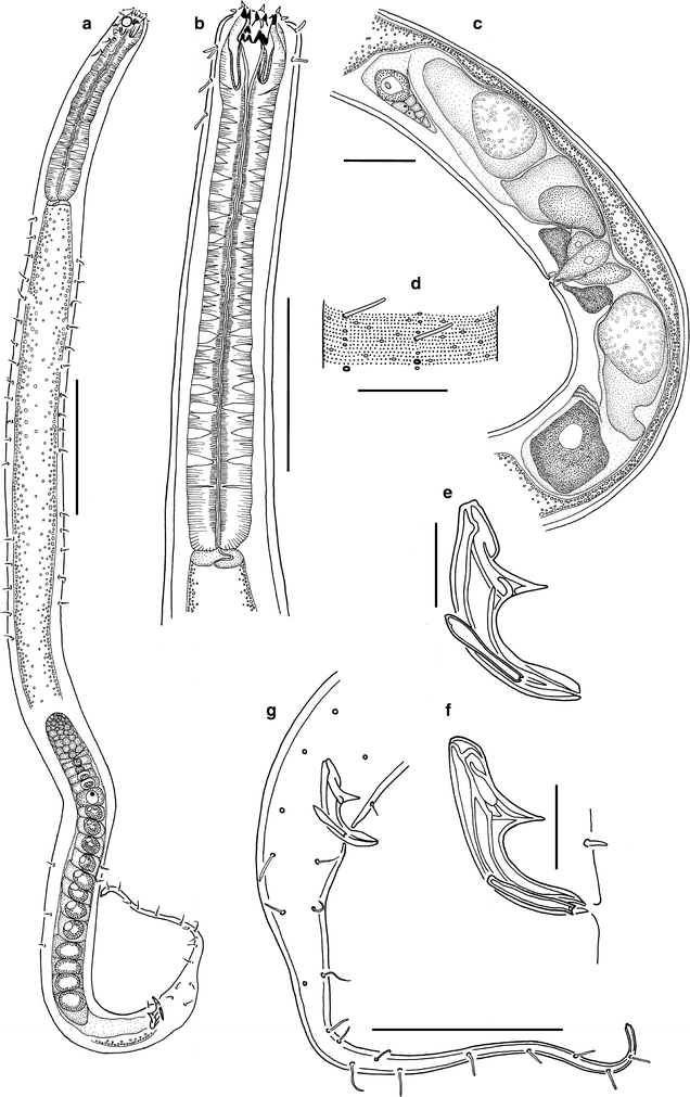
Acantholaimus cornutus sp. n. a male, holotype, total view; b male, holotype, anterior end; c female, paratype No. 3, reproductive system; d holotype, fragment of cuticle surface at mid-body; e male, holotype, spicule; f male, paratype No. 2, spicule; g male, holotype, posterior part. Scale bars: a = 200 μm; b, g = 100 μm; c, d = 50 μm; e, f = 20 μm
Fig. 14 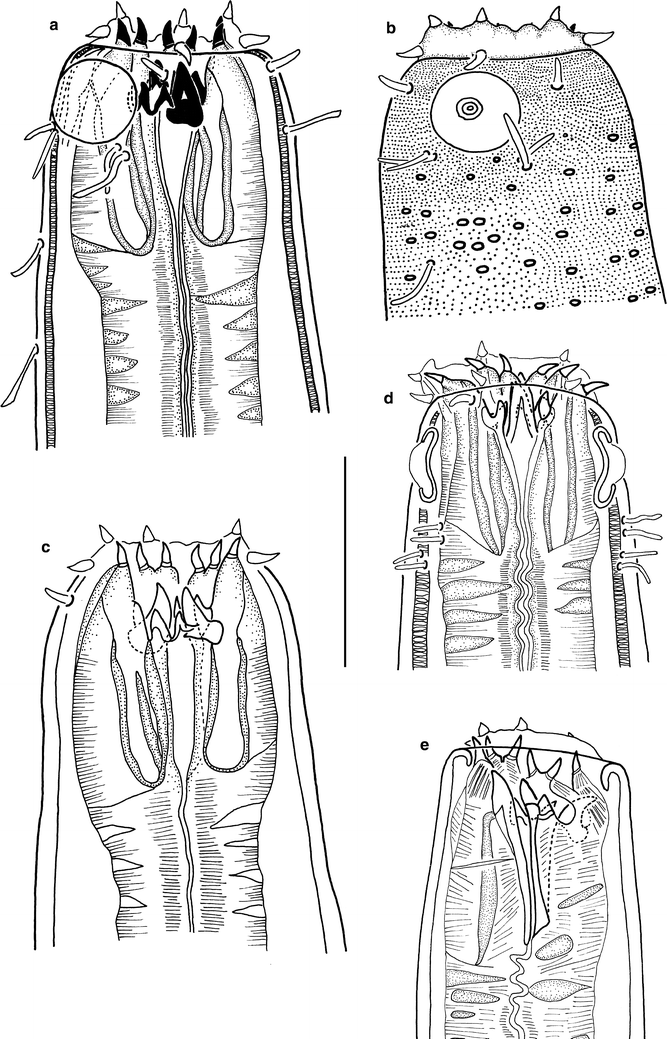
Acantholaimus cornutus sp. n., heads. a, b, c male, holotype, different optical levels; d female, paratype No. 3; e male, paratype No. 1. Scale bars = 40 μm
Fig. 15 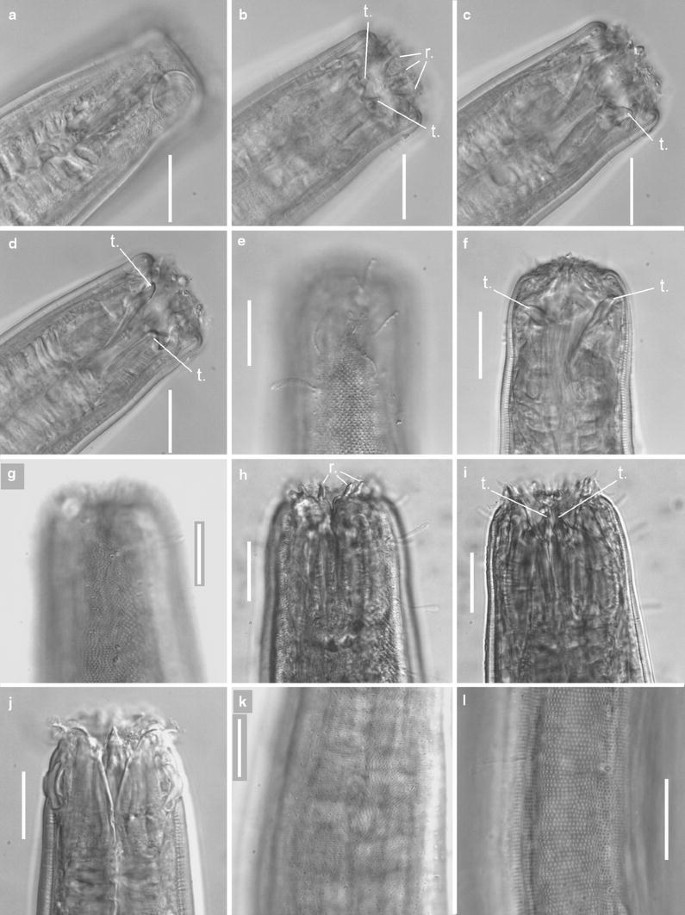
Acantholaimus cornutus sp. n., micrographs. a–d male, paratype No. 1, head region; e, f male, paratype No. 2, head region at different optical levels; g–i male, holotype, head region at different optical levels; j female, paratype No. 4, head region; k female, paratype No. 4, fragment of cuticle in mid-body region, with numerous small, irregularly arranged cuticular pores; l male, paratype No. 2, lateral cuticular field with rows of larger dots. Scale bars = 20 μm. Abbreviations: r. = ruga, t. = onchium
Table 5 Acantholaimus cornutus sp. n.


Type material
Collection number MNHN-BN500. Holotype: male. Paratypes: 2 males and 1 female (Table 5).
Locality
Etymology
Latin cornutus (= horned).
Description
Main measurements: L = 1,932–2,243 μm; L′ = 1,513–1,844 μm; a = 25.5–37.2; a′ = 18.4–30.3; b = 6.4–7.5; b′ = 5.1–5.5; c = 3.5–5.6; c′ = 7.8–14.9; V = 55.3; V′ = 76.5 (Table 5).
Body cylindrical, with truncate anterior end and tail consisting of proximal conical and distal cylindrical filiform parts. Four sublateral rows of round pores ca. 1.5–2.0 μm in diameter with thick cuticular rims beginning posterior to amphideal fovea. Some of these pores bear cylindrical somatic setae 7–15 μm long. Cuticle densely dotted, with short lateral fields beginning posterior to amphideal fovea and ending at posterior third of pharynx. Dots of lateral fields arranged more sparsely and irregularly than dots outside lateral fields, which are arranged in clearly visible, transverse rows and are arranged more densely. In optical cross-section of cuticle, dots are discernible as tiny radial struts. Oval pores with narrow cuticular rims (ca. 2–2.5 μm in diameter at level of anterior third of pharynx, and ca. 1.5 μm along rest of body) distributed irregularly along body. Cuticle ca. 2.5–3 μm thick. Six lips visible. Labial region distinctly set off from remainder of body; its cuticle appreciably thinner (less than 0.5 μm thick). Sensilla of cephalic end arranged in 3 rings: 6 short inner labial conical sensilla, ca. 2–5 μm long; 6 outer labial conical setae, 4–6 μm long; and 4 longer, cylindrical, cephalic setae, 4–7 μm long. Amphideal fovea circular, 14–16 μm in diameter, nearly indiscernible in most specimens because of lack of distinct cuticular rim. Anterior end of amphideal fovea situated ca. 0.2 c.b.d. from anterior end of body. Two pairs (latero-subdorsal and latero-subventral pair) of cervical setae located posterior to amphideal fovea. Latero-subdorsal pair located closer to amphideal fovea than latero-subventral pair; in both pairs, seta situated more distal from amphid being slightly longer than more proximal seta. In holotype and paratype No. 1, pairs of cervical setae located very close to amphideal fovea (ca. 1 μm from posterior end of amphideal fovea); proximal and distal setae in all pairs being 10–11 μm and ca. 13 μm long, respectively. In paratype Nos. 3 and 4, latero-subdorsal and latero-subventral pairs of cervical setae situated ca. 3–4 μm and 5–6 μm posterior to amphideal fovea, respectively; proximal and distal setae in all pairs ca. 5 μm and ca. 7 μm long, respectively. Stoma consisting of wide, cup-shaped cheilostoma and narrow, funnel-shaped esophastoma. Cheilostoma possessing 12 fang-shaped rugae, these protruding ahead of mouth opening in some specimens. Esophastoma 40–45 μm long, in shape of narrow funnel, containing 6 sclerotized onchia: one large dorsal or subdorsal onchium 13–17 μm long, one large subventral onchium 13–17 μm long, and four smaller onchia 5–10 μm long (their exact arrangement unclear). Basal parts of onchia situated in anterior part of esophastoma, and their apical parts intruding into cheilostoma. Longitudinal bands connected with onchia visible in anterior part of pharynx. Pharynx almost cylindrical, with thickened anterior end at level of esophastoma and with slightly widened distal end. Nerve ring and renetta not visible. Cardia round. Tail conical in its anterior part and possessing thin cylindrical terminal part constituting ca. 65–80% of entire tail length.
Male reproductive system monorchic. Testis directed anteriorly, outstretched, lying to right of intestine, occupying ca. 0.4 of pre-anal body length in holotype, and ca. 0.2 in young paratype No. 1. Size of mature spermatids ca. 65 × 40 μm. Curved funnel-shaped paired spicules possessing cuticular sculpture with thickened cuticular ridges. Gubernaculum blade-shaped, with bifurcated distal end. Supplementary organs not found.
Female reproductive system consisting of two antidromous, uniformly sized ovaries (anterior ovary lying to right of intestine, and posterior one lying to left of intestine). Ovaries very short. In single female examined, 3 large spermatozoons ca. 130 × 65 μm present. Vulvar glands with granular content surrounding vulva.
Abundance
The density of A. cornutus sp. n. varied from 0 to 1.7 inds/10 cm2 (0.3 inds/10 cm2 on average), and its relative abundance within the nematode community was 0–1.3% (mean 0.2%).
Differential diagnosis
Acantholaimus cornutus sp. n. differs considerably from most other Acantholaimus species by the shape of the anterior end and the stoma, the position of the amphideal fovea, and the length of the cephalic setae. There is only one similar species in the genus, A. cyathibuca Vivier 1985. The new species is very close to A. cyathibuca and shares with it the shape of the body and anterior end, the position and the size of the amphideal fovea, the arming of the stoma, and the length and position of the outer labial and cephalic setae. A. cornutus sp. n. differs from A. cyathibuca by the body length (1,932–2,243 μm vs. 876 μm); the size and shape of the rugae (fang-shaped vs. rod-shaped); the number of cervical setae (4 vs. 5 plus one pore); the number of longitudinal rows of pores and setae (4 vs. 8); and the length of the somatic setae (7–15 μm vs. 4 μm).
-
Acantholaimus iubilus Gerlach, Schrage, Riemann 1979 (Figs. 16, 17; Table 6)
Fig. 16 
Acantholaimus iubilus, specimen No. 2, female, head region. Scale bar = 20 μm. Abbreviations: d.t. = dorsal onchium, sv.t. = subventral onchium
Fig. 17 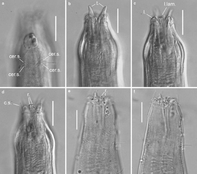
Acantholaimus iubilus, micrographs. a–d female, specimen No. 2, head region at different optical levels; e, f female, specimen No. 1, head region at different optical levels. Scale bars = 20 μm. Abbreviations: cer.s. = cervical seta, c.s. = cephalic seta, l. = lip, l.lam. = lateral cuticular lamina, t. = onchium
Table 6 Acantholaimus iubilus


Material examined
Two females (Table 6).
Locality
Description
Body cylindrical, with narrowed anterior end and very long filiform tail. Somatic setae cylindrical or clavate, 5–9 μm long, arranged in 4 submedian longitudinal rows (except filiform part of tail). Cuticle dotted. Dots arranged in transverse rows; they are significantly larger in tail region than on rest of body. Lateral differentiation of cuticle feebly expressed (dots arranged slightly less regularly and less densely here). Small, irregularly arranged pores along entire body length. Cuticle ca. 3 μm thick at level of mid-body. Lips strongly developed, each lip with pair of large rod-shaped cuticular rugae. Only 4 cephalic setae, 11–14 μm long, visible. These setae taper slightly to their tips, and are straight in all specimens examined. Amphids single-spiral, round (if head not inverted), with fine concentric striation, ca. 14 μm wide. Anterior end of amphideal fovea situated at ca. 1 c.b.d. from the anterior end. Two pairs (latero-subdorsal and latero-subventral) of cervical setae ca. 10 μm long located just posterior to each amphideal fovea. Cervical setae of each pair situated very close to each other. Stoma consisting of wide, cup-shaped cheilostoma and narrow, funnel-shaped esophastoma. Cheilostoma containing 6 pairs of large, well discernible cylindrical rugae. Esophastoma possessing three long, very robust sclerotized onchia: one dorsal and two subventral. Dorsal onchium and right subventral onchium ca. 15–20 μm long, right subventral onchium ca. 8–10 μm long. Basal parts of onchia situated in anterior part of esophastoma, and their apical parts intruding to cheilostoma or even protruding outside mouth opening. In addition, 2 lateral, flat, cuticular laminae with almost right-angled distal ends visible at level of mouth opening. Borders of esophastoma faintly distinguishable, except its anterior part where its thick walls form a prolongation of onchia at a distance of 15–20 μm. Pharynx thin at its anterior part, gradually widening to its posterior end, with large plasmatic interruptions along its entire length. Cardia flattened in direction of main body axis. Nerve ring not visible. Renetta not visible. Tail very long, representing about half of total body length (46–47%), gradually narrowed into filiform, cylindrical terminal part. Cellular bodies of caudal glands visible.
Female reproductive system consisting of two antidromous, uniformly sized ovaries (anterior ovary lying to right of intestine, and posterior one lying to left of intestine), occupying 22–23% of pre-anal body length. Vulvar glands with granular content surrounding short vagina seen.
Abundance
The density of A. iubilus was very low (only 2 specimens were found in the samples).
Remarks
Our individuals closely resemble the type specimens in their appearance (a large strong body with a very long tail); in the size and position of the amphidial fovea; in the number and position of the cervical setae; and in the shape, length, and position of the cephalic setae.
Our individuals differ from the type specimens by their larger body (body length is 2,703–3,366 μm vs. 1,950–2,320 μm in the type specimens, and the maximum body diameter is 66–95 μm vs. 57–60 μm); and by the number of onchia (3 vs. 5). Differences in body shape can be explained by interpopulation variability.
Various authors have described different numbers of onchia in this species. Gerlach et al. (1979) found, presumably, 5 onchia. Gourbault and Vincx (1985) described 3 onchia and 2 lateral right-angled cuticular plates. This structure was also found in our specimens. Possibly, Gerlach et al. (1979) described these cuticular plates as 2 additional onchia.
Distribution
South-eastern Pacific, Peru Basin, 3,086–6,313 m, fine silt (Gerlach et al. 1979); North-eastern Atlantic, Bay of Biscay, 4,725 m (Vivier 1985); South-eastern Atlantic, 2,063–4,308 m (Gourbault and Vincx 1985); North Atlantic, Norway Sea, 970–3,294 m (Jensen 1988); Central western Atlantic, Hatteras plain, 5,411 m, Puerto Rico Trench, 7,460–8,380 m (Tietjen 1989); North-eastern tropical Pacific, Clarion–Clipperton Fracture Zone, nodule fields and area without nodules, 5,000–5,035 m (present report) (Fig. 5).
-
Acantholaimus maks Gerlach, Schrage, Riemann 1979 (Figs. 18, 19; Table 7)
Fig. 18 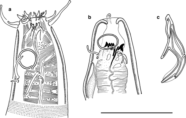
Acantholaimus maks. a juvenile, head region; b male, head with retracted anterior part; c male, spicule. Scale bar = 50 μm
Fig. 19 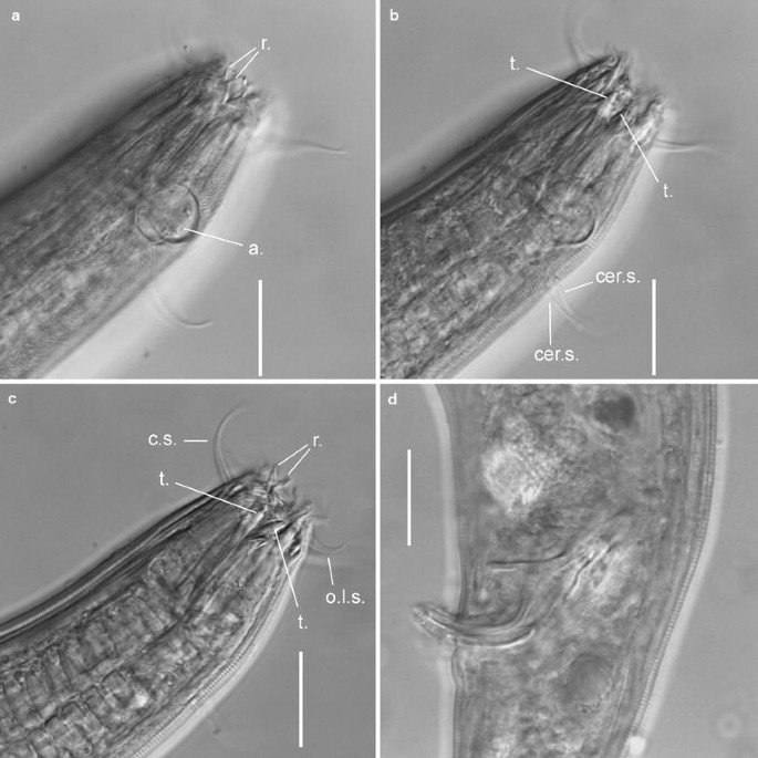
Acantholaimus maks, micrographs. a–c juvenile, head region at different optical levels; d male, spicule region. Scale bars = 20 μm. Abbreviations: cer.s. = cervical seta, c.s. = cephalic seta, i.l.s. = inner labial seta, o.l.s. = outer labial seta, r. = ruga, t. = onchium
Table 7 Acantholaimus maks


Material examined
One male, one female, one juvenile (Table 7).
Locality
Description
Body cylindrical, with slightly narrowed anterior end and long filiform tail. Somatic setae few, cylindrical or clavate, 9–12 μm long, arranged in 4 submedian longitudinal rows (except filiform part of tail). Cuticle dotted. Dots arranged in transverse rows. Lateral fields not seen. Small, irregularly situated pores present at level of pharynx. Cuticle ca. 2–2.5 μm thick at level of mid-body. Lips not visible. 3 rings of sensilla of cephalic end visible: 6 short inner labial setae ca. 5 μm long; 6 short outer labial setae ca. 8 μm long (some of them appearing 2-jointed); and 4 longer cephalic setae ca. 23 μm long. Amphids single-spiral, round (if head not inverted), ca. 16 μm wide. Anterior end of amphideal fovea situated ca. 1.0 c.b.d. from the anterior end. Two pairs (latero-subdorsal and latero-subventral) of cervical setae 14–15 μm long, located just posterior to each amphideal fovea. Stoma consisting of wide, cup-shaped cheilostoma and narrow, funnel-shaped esophastoma. Cheilostoma containing 12 large, well discernible cylindrical rugae. Esophastoma possessing 5 (presumably) sclerotized onchia: one large subventral onchium ca. 6 μm long, and four other onchia 3–4 μm long, one of them appearing bifurcated. Arrangement of small onchia not clear. Basal parts of onchia situated in anterior part of esophastoma, and their apical parts intruding into cheilostoma. Borders of esophastoma feebly distinguishable, except its anterior part where its thick walls form a prolongation of onchia at a distance of ca. 20 μm. Pharynx thin at its anterior part, gradually widening to its posterior end, with numerous plasmatic interruptions. Cardia oblate. Nerve ring not visible. Renetta not visible. Tail conical in its anterior part and possessing long filiform, cylindrical terminal part constituting ca. 60–70% of entire tail length. Three cellular bodies of caudal glands visible in conical part of tail.
Male reproductive system monorchic. Testis directed anteriorly, outstretched, lying to right of intestine, occupying 40% of pre-anal body length. Curved funnel-shaped paired spicules possessing cuticular sculpture with thickened cuticular areas, forming longitudinal or bounding ridges. Gubernaculum wide, with slightly narrowed proximal end and narrow bifurcated distal end. Size of mature spermatids ca. 70 × 40 μm. Supplementary organs not visible.
Female reproductive system consisting of two antidromous, uniformly sized ovaries (anterior ovary lying to right of intestine, and posterior one lying to left of intestine), occupying ca. 25% of pre-anal body length. Large vulvar glands with granular content surrounding short vagina.
Abundance
The density of A. maks was very low: only 3 specimens were found in the samples.
Remarks
Our individuals closely resemble the type specimens in most parameters, such as body size, position and shape of amphideal fovea, number of cervical setae, and shape of spicules.
Thiel (2003) suggested that this species inhabits only crevices inside polymetallic nodules. In our investigation, A. maks was found in the 26-year-old track after the experimental nodule dredging, which was therefore devoid of nodules (Miljutin et al. 2011).
Distribution
South-eastern Pacific, Peru Basin, 3,364–6,313 m, silt (Gerlach et al. 1979); North-eastern Atlantic, Bay of Biscay, 2,690 m (Vivier 1985); South-eastern Atlantic, 2,992–4,308 m (Gourbault and Vincx 1985); South-eastern Pacific, Peru Basin, nodule field, internal cavity of manganese nodules, 4,142–4,150 m (Bussau 1993); North-eastern tropical Pacific, Clarion–Clipperton Fracture Zone, track remaining after experimental nodule dredging (present report) (Fig. 5).
-
Acantholaimus occultus Bussau 1993 (Figs. 20, 21; Table 8)
Fig. 20 
Acantholaimus occultus, males. a specimen No. 1, anterior end; b specimen No. 1, head region; c specimen No. 2, head region; d specimen No. 2, total view; e specimen No. 1, cloacal region with spicule; f specimen No. 1, spermatid. Scale bars: a = 50 μm; b, c = 20 μm; d = 200 μm; e, f = 10 μm
Fig. 21 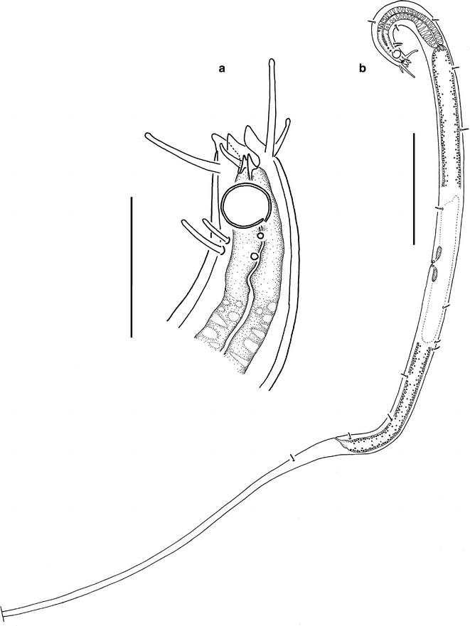
Acantholaimus occultus, female. a head region; b total view. Scale bars: a = 20 μm; b = 100 μm
Table 8 Acantholaimus occultus


Material examined
Two males, one female (Table 8).
Locality
Description
Body slender (in comparison with many other Acantholaimus species), slightly spindle-shaped, with slightly narrowing anterior end and very long filiform tail (length of tail more than half of total body length). Somatic setae cylindrical, 6–7 μm long, situated along entire body. Cuticle punctuated; punctuation feeble in males. Dots arranged in transverse rows. Lateral differentiation of cuticle present, lateral field quite narrow. Dots of lateral fields larger. Cuticle ca. 1 μm thick along entire body, except thinner at level of cephalic setae (less than 1 μm). Lips large and well discernible. Two rings of sensilla of cephalic end discernible: 6 shorter clavate outer labial setae, 5–6 μm long; and 4 longer, thicker, conical with slightly clavate tips, cephalic setae, 14–17 μm long. Amphideal fovea ventrally coiled, single-spiral; in shape of longitudinally oriented oval and ca. 9 × 15 μm in males; ca. 8 μm in diameter and round in female. Anterior end of amphideal fovea situated ca. 1 amphidial length from anterior end of body. Two pairs (latero-subdorsal and latero-subventral) of cervical clavate or cylindrical setae located just posterior to each amphideal fovea. Anterior cervical seta of each pair ca. 5 μm long; posterior seta ca. 7 μm long. Cheilostoma containing 3 sclerotized onchia 3–4 μm long: 1 dorsal and 2 subventral. Basal parts of onchia situated in anterior part of esophastoma, and their apical parts intruding into cheilostoma. Esophastoma long (20–32 μm long, one-third of total length of pharynx), its internal lumen with thicker cuticular wall. Muscle envelope of esophastoma appreciably detached from rest of pharynx by small constriction, and differing from other part of pharyngeal muscle envelope by lack of plasmatic inclusions and more distinct muscle striation. In its posterior two-thirds, pharynx containing large and numerous, regularly altering plasmatic interruptions. Cardia cordate. Nerve ring not visible. Renetta not visible. Tail very long, conical in its anterior part and possessing long filiform, cylindrical terminal part constituting ca. 85–90% of total tail length.
Male reproductive system monorchic. Testis directed anteriorly, outstretched, lying to right of intestine, occupying about 0.5 of pre-anal body length. Spicule thick, shaped as curved funnel, with thick cuticular walls. Gubernaculum blade-shaped, with narrowed proximal end and bifurcated distal end. Mature spermatids ca. 30 × 13 μm. Supplementary organs not found.
Female reproductive system consisting of two antidromous, uniformly sized ovaries (anterior one lying to right of intestine, and posterior one lying to left of intestine).
Abundance
The relative abundance of A. occultus within the nematode community was 0–2.3% (mean 0.4%). The density varied from 0 to 0.7 inds/10 cm2 (0.1 inds/10 cm2 on average).
Remarks
The new individuals closely resemble the type specimens in their appearance and many body parameters. The shape and construction of the spicule are also very similar.
The new individuals have a smaller and differently shaped amphideal fovea (9 × 15 μm and oval in the males vs. 18 μm in diameter and round in the type males). The amphids of the new specimens are situated closer to the anterior end (at 10–11 μm vs. 16 μm in the type specimens).
Distribution
South-eastern Pacific, Peru Basin, nodule field, 4,147 m, sediment from nodule field (Bussau 1993); North-eastern tropical Pacific, Clarion–Clipperton Fracture Zone, nodule field, 4950, ooze (present report) (Fig. 5).
-
Acantholaimus robustus sp. n. (Figs. 22, 23, 24; Table 9)
Fig. 22 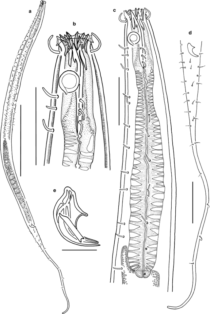
Acantholaimus robustus sp. n., males. a holotype, total view; b holotype, head region; c holotype, anterior end; d holotype, tail region; e paratype No. 1, spicule. Scale bars: a = 500 μm; b, c = 50 μm; d = 100 μm; e = 20 μm
Fig. 23 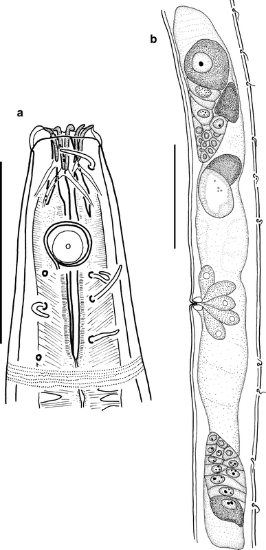
Acantholaimus robustus sp. n., paratype No. 3, female. a: head region; b: reproductive system. Scale bars: a = 50 μm; b = 100 μm
Fig. 24 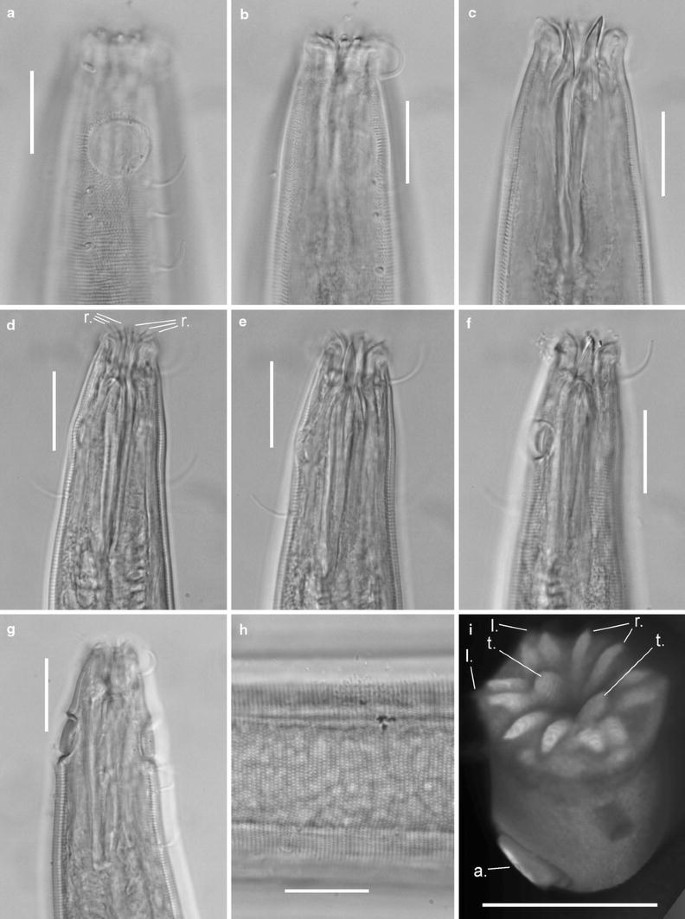
Acantholaimus robustus sp. n. a–c male, paratype, head region at different optical levels; d–f juvenile, paratype No. 5, head region at different optical levels; g male, paratype No. 2, head region; h female, paratype No. 3, head; i male, paratype, head region, reconstruction based on micrographs made using a confocal microscope. Scale bars = 20 μm. Abbreviations: a. = amphid, cer.s. = cervical seta, l. = lip, r. = ruga, t. = onchium
Table 9 Acantholaimus robustus sp. n.


Type material
Collection number MNHN-BN501. Holotype: male. Paratypes: 2 males, 2 females, and 1 juvenile (Table 9).
Locality
Etymology
Latin robustus (= robust, stout).
Description
Main measurements: L = 1,500–2,487 μm; L′ = 1,226–1,928 μm; a = 30.6–40.2; a′ = 20.0–28.7; b = 6.6–10.6; b′ = 5.3–7.6; c = 3.2–5.5; c′ = 8.8–19.6; V = 45.6–53.8; V′ = 65.4–69.4 (Table 9).
Body cylindrical, with narrowed anterior end and long filiform tail. Somatic setae numerous, cylindrical, 8–12 μm long, situated in 4 submedian longitudinal rows (except filiform part of tail). Cuticle dotted. Dots arranged in transverse rows. No lateral differentiation of cuticle observed. Small cuticular pores arranged irregularly along entire body length. Cuticle ca. 3.0–3.5 μm thick. Six lips visible. Each lip possessing a pair of large rod-shaped cuticular rugae connecting in pairs at their distal ends. Labial sensilla papilloid; 4 cephalic setae cylindrical, 12–17 μm long. Amphideal fovea ventrally coiled, single-spiral, round, 11–14 μm wide. Anterior end of amphideal fovea situated ca. 1 c.b.d. from the anterior end. Two rows (latero-subdorsal and latero-subventral) of 3 cervical setae, 7–12 μm long, located posterior to each amphideal fovea. Stoma consisting of cup-shaped cheilostoma and narrow esophastoma with thick cuticular walls. Cheilostoma containing 6 pairs of large, well discernible cylindrical rugae. Esophastoma 42–48 μm long, possessing 2 very large sclerotized onchia, 1 dorsal and 1 presumably subventral. Basal parts of onchia situated in anterior part of esophastoma, and their apical parts intruding into cheilostoma. Dorsal onchium 17–19 μm long, subventral one 22–24 μm long. Pharynx cylindrical, narrowing in the first third. Numerous large plasmatic interruptions visible posterior to esophastomal region. Cardia small, cordate. Nerve ring not visible. Renetta not visible. A small intestinal cecum directed anteriorly and situated at level of posterior part of pharynx, visible in all specimens examined. Tail gradually narrowed into filiform, cylindrical terminal part constituting ca. 60% of entire tail length. Cellular bodies of caudal glands visible.
Male reproductive system monorchic. Testis directed anteriorly, outstretched, lying to right of intestine, occupying about 30% of pre-anal body length. Size of mature spermatids ca. 50–60 to 30–40 μm. Curved funnel-shaped paired spicules, quite thick and possessing complicated cuticular sculpture with thickened cuticular areas in shape of longitudinal or transverse ridges. Gubernaculum blade-shaped, with narrow proximal end and wide bifurcated distal end. Supplementary organs not found.
Female reproductive system consisting of two antidromous, uniformly sized ovaries (anterior ovary lying to right of intestine, and posterior one lying to left of intestine), occupying ca. 20% of pre-anal body length. Numerous, well developed vulvar glands with granular content surrounding short vagina seen.
Abundance
The density of A. robustus sp. n. varied from 0 to 3.6 inds/10 cm2 (mean 0.8 inds/10 cm2), and its relative abundance within the nematode community was 0 to 2.1% (mean 0.5%).
Differential diagnosis
Acantholaimus robustus sp. n. is characterized by its very large body size, two very large onchia, and thick, strongly developed rugae. The cervical setae of the new species are grouped in threes.
There are only four equally large Acantholaimus species known: A. arminius Gerlach et al. 1979; A. iubilus Gerlach et al. 1979; A. maks Gerlach et al. 1979; and A. quadridentatus Jensen 1985.
Acantholaimus robustus sp. n. differs from A. arminius by its thicker body (a = 30–40 vs. 53); the absence of lateral differentiation of the cuticle; the shorter cephalic setae (13–17 μm vs. 21 μm); the arrangement and number of cervical setae (2 groups of 3 setae posterior to each amphideal fovea vs. 2 single setae in A. arminius); and the longer esophastoma (ca. 47–50 μm vs. ca. 20 μm).
Acantholaimus robustus sp. n. differs from A. quadridentatus in many parameters, of which the most significant are: the amphid is farther from the anterior end (located ca. 1–1.5 amphidial c.b.d. from the anterior end vs. 0.2 c.b.d. in A. quadridentatus); the cephalic setae are shorter (12–17 μm vs. 25 μm); the different shape of the outer labial sensilla (short and papilliform vs. long and setiform); and the much thicker spicules.
Acantholaimus robustus sp. n. is close to A. maks in the location of the amphids and in the length of the setae of the cephalic end. The new species differs from A. maks by the number of onchia (2 vs. 5–6 in A. maks); the much longer esophastoma (47–50 μm vs. ca. 15 μm); the much thicker cuticular wall of the internal lumen of the esophastoma (this thick wall is easily distinguishable from the much thinner wall of the other, posterior part of the pharynx in A. robustus, whereas it does not differ from the other part of the pharynx in A. maks); the arrangement of the cervical setae (2 groups of 3 sparsely located setae posterior to each amphideal fovea in A. robustus vs. 2 groups of 2 closely grouped setae in A. maks); and the shape of the spicules (thick vs. thin).
The new species shows the most resemblance to A. iubilus sp. n. in the size and location of the amphids, the size of the onchia, its very long esophastoma, and the very well developed rugae. The new species differs from A. iubilus by the length of the esophastoma (ca. 47–50 μm vs. ca. 25 μm in A.iubilus); the arrangement of the cervical setae (2 groups of 3 widely set setae posterior to each amphideal fovea in A. robustus sp. n. vs. 2 groups of 2 closely set setae in A.iubilus); the number of onchia (2 onchia vs. 3 onchia supplemented by a pair of lateral cuticular laminae); and the shorter tail (22–31% of the entire body length vs. ca. 50%).
-
Acantholaimus sieglerae sp. n. (Figs. 25, 26; Table 10)
Fig. 25 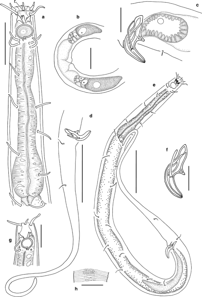
Acantholaimus sieglerae sp. n. a male, holotype, anterior end; b female, paratype No. 4, reproductive system; c male, paratype No. 2, spicule region; d holotype, tail region; e holotype, total view; f holotype, spicule; g paratype No. 4, head region; h paratype No. 4, lateral differentiation of cuticular punctation. Scale bars: a, b = 20 μm; c, f, g, h = 10 μm; d, e = 50 μm. Abbreviation: r. = a pair of rugae
Fig. 26 
Acantholaimus sieglerae sp. n., male, holotype, micrographs. a, b head region at different optical levels; c spicule region. Scale bars = 20 μm. Abbreviations: cer.s. = cervical seta, t. = onchium
Table 10 Acantholaimus sieglerae sp. n.


Type material
Collection number MNHN-BN502. Holotype: male. Paratypes: 2 males and 2 females (Table 10).
Locality
Etymology
In honor of Viola Siegler (German Centre for Marine Biodiversity Research, Senckenberg am Meer, Wilhelmshaven, Germany).
Description
Main measurements: L = 457–668 μm; L′ = 360–443 μm; a = 19.9–32.7; a′ = 15.7–25.2; b = 5.6–6.1; b′ = 3.9–4.7; c = 2.8–4.7; c′ = 7.5–13.9; V = 52.8–54.3; V′ = 67.8–68.9 (Table 10).
Body cylindrical, with slightly tapered anterior end and gradually narrowed tail. Maximum body width at posterior end of pharynx. Somatic setae numerous, arranged in 4 submedian longitudinal rows (except on filiform part of tail), 8–10 μm long. These setae cylindrical, sometimes with slightly widened tips. Cuticle densely dotted. Dots arranged in regular transverse rows. Lateral field narrow; dots appearing larger there but also arranged in transverse rows. Numerous irregularly arranged pores visible at level of cervical region. Cuticle ca. 1.5–1.7 μm thick. Lips not visible. Sensilla of cephalic end arranged in 3 rings: 6 short inner labial papilloid sensilla ca. 0.7 μm long; 6 short, outer labial setae 1.5–3.5 μm long; and 4 longer, cylindrical cephalic setae 7–10 μm long. In several specimens, outer labial setae appearing jointed, consisting of two segments. Amphideal fovea ventrally coiled, single-spiral, in shape of transversely oriented oval, 5–9 μm wide, with distinct cuticular edging and internal fine concentric striation. In females, amphideal fovea smaller in diameter than in males. Anterior end of amphideal fovea situated ca. 0.7–1 c.b.d. from the anterior end. Two pairs (latero-subdorsal and latero-subventral) of cervical setae 7–8 μm long, situated just behind each amphideal fovea. Stoma large. Cheilostoma barrel-shaped, bearing 12 indistinct cuticular rugae. Esophastoma narrow, ca. 8 μm long, with thick cuticular walls, possessing six sclerotized onchia. Right subventral and dorsal onchia are larger and more robust (ca. 2–3 μm long) and easily distinguishable. Basal parts of onchia situated in anterior part of esophastoma, and their apical parts intruding into cheilostoma. Pharynx cylindrical, with slight bulb-like widening at its distal end. Numerous cytoplasmic inclusions visible along entire length of pharynx. Nerve ring not visible. Renetta not visible. Cardia triangular or oval. Tail elongated-conical in its anterior part and gradually narrowing into thin cylindrical terminal part constituting ca. 50–70% of entire tail length.
Male reproductive system monorchic. Testis directed anteriorly, outstretched, lying to right of intestine, occupying 37–51% of pre-anal body length. Size of mature spermatids ca. 30 × 11 μm. Curved funnel-shaped paired spicules possessing cuticular sculpture with thickened cuticular areas in shape of longitudinal or transverse ridges. Gubernaculum a thin, slightly curved rod, with bifurcated distal end. Supplementary organs not found.
Female reproductive system consisting of two antidromous, uniformly sized ovaries (anterior ovary lying to right of intestine, and posterior one lying to left of intestine), occupying 33–38% of pre-anal body length.
Abundance
The density of A. sieglerae sp. n. was 0–2.0 inds/10 cm2 (0.1 inds/10 cm2 on average), and its relative abundance within the nematode community was 0–2.5% (mean 0.2%).
Differential diagnosis
Acantholaimus sieglerae sp. n. is a comparatively small species, and at the same time it possesses very large onchia. The latter feature is not characteristic for small Acantholaimus species.
The new species shows the most resemblance to A. gathumai Muthumbi and Vincx 1997 and to A. incomptus Vivier 1985 in the body length and maximum diameter, the length of the cephalic setae, and the location of the amphids (ca. 1 amphidial c.b.d. from the anterior end).
Acantholaimus sieglerae sp. n. differs from A. gathumai by the shape of the cephalic end (cylindrical, with a sharply truncated end, vs. triangular, with a gradually narrowing end in A. gathumai), and by its larger onchia.
Acantholaimus sieglerae sp. n. differs from A. incomptus by its larger amphids (diameter of amphids ca. 50–69% of amphidial c.b.d. vs. 45–51% in A. incomptus); the shape of the tail (gradually narrowing tail vs. tail with sharply distinguishable conical and flagelliform parts in A. incomptus); and its larger onchia.
-
Acantholaimus tchesinovi sp. n. (Figs. 27, 28, 29; Table 11)
Fig. 27 
Acantholaimus tchesunovi sp. n., holotype. a total view; b head region; c anterior end; d spicule; e spermatid. Scale bars: a = 50 μm; b, c, e = 20 μm; d = 10 μm
Fig. 28 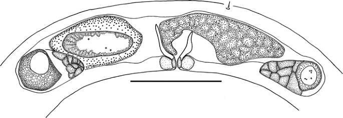
Acantholaimus tchesunovi sp. n., paratype No. 2, female reproductive system. Scale bar = 40 μm
Fig. 29 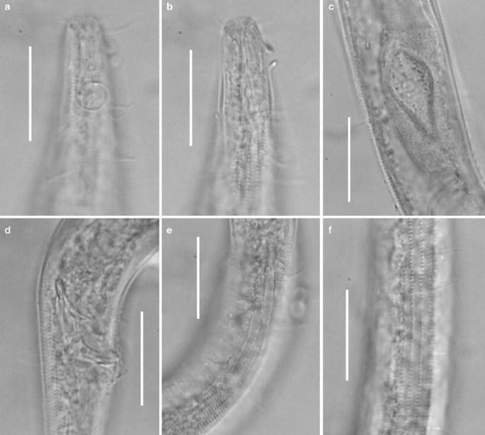
Acantholaimus tchesunovi sp. n., micrographs. a, b male, holotype, head region at different optical levels; c holotype, spermatid; d holotype, spicule region; e holotype, lateral differentiation of cuticle at mid-body; f male, paratype No. 1, lateral differentiation of cuticle at mid-body. Scale bars = 20 μm
Table 11 Acantholaimus tchesunovi sp. n.


Type material
Collection number MNHN-BN503. Holotype: male. Paratypes: 1 male and 1 female (Table 11).
Locality
Etymology
In honor of Prof. Dr. A. V. Tchesunov (Lomonosov’s Moscow State University, Moscow, Russia).
Description
Main measurements: L = 707–750 μm; L′ = 474–536 μm; a = 21.4–26.2; a′ = 15.3–19.2; b = 7.1–7.4; b′ = 5.0–5.1; c = 3.0–3.5; c′ = 10.7–12.9; V = 52.0; V′ = 72.8 (Table 11).
Body cylindrical, with slightly and gradually narrowed anterior end, and tail consisting of proximal conical and long, thin cylindrical parts. Somatic setae numerous, arranged in 4 submedian longitudinal rows (except filiform part of tail where they are arranged irregularly), 6–8 μm long at level of pharynx, 4–6 μm long at mid-body, and 6–8 μm long at caudal part. These setae cylindrical, sometimes with slightly widened tips, on entire pre-anal part of body, but clavate on caudal part. Somatic setae thicker at level of pharynx than on remainder of body. Numerous cuticular pores alternating irregularly with somatic setae in 4 longitudinal rows. Cuticle densely dotted, with lateral fields, beginning posterior to amphideal fovea and extending along entire body except filiform part of tail. Dots of lateral fields appearing larger and distributed more sparsely than median ones; lateral dots arranged in clearly visible, longitudinal rows. Four such rows located closely posterior to amphideal fovea; one additional, slightly posterior row; and 1 or 2 additional rows at level of cardia, for a total of 6–7 longitudinal lateral rows extending from beginning of intestine to approximately middle of conical part of tail. Median dots smaller, arranged more densely and in transverse rows. In optical cross-section of cuticle, dots discernible as tiny radial struts. Cuticle ca. 2–3 μm thick at mid-body level and 1–1.5 μm thick at pharyngeal and caudal levels. Lips not visible. Sensilla of cephalic end arranged in 3 rings: 6 short inner labial papilloid sensilla ca. 1 μm long; 6 short, 2-jointed, outer labial setae 3–4 μm long; and 4 longer, clavate, cephalic setae ca. 12 μm long. Amphideal fovea ventrally coiled, single-spiral, round, 6–8 μm wide, with distinct cuticular edging, situated ca. 1.2 c.b.d. from anterior end. Two pairs (latero-subdorsal and latero-subventral) of cervical setae 6–7 μm long, located at level of posterior part of amphideal fovea. Cheilostoma barrel-shaped, containing 12 cylindrical rugae; esophastoma narrow, 5–8 μm long, with thickened cuticular walls of internal pharyngeal lumen, and possessing at least 4 sclerotized onchia. Basal parts of onchia situated in anterior part of esophastoma, and their apical parts intruding into cheilostoma. One larger dorsal and one larger subventral onchia ca. 1.5 μm long, more distinguishable than remaining onchia. Pharynx gradually widening to its posterior end, with numerous cytoplasmic inclusions along its entire length. Nerve ring not visible. Renetta not visible. Cardia round. Tail conical in its anterior part, and with thin cylindrical terminal part constituting ca. 70–75% of total tail length.
Male reproductive system monorchic. Testis directed anteriorly, outstretched, lying to right of intestine, occupying about 0.4–0.55 of pre-anal body length. Size of mature spermatids ca. 38 × 16 μm. Curved funnel-shaped paired spicules possessing cuticular sculpture with thickened cuticular areas forming longitudinal or transverse ridges. Gubernaculum blade-shaped, with narrow proximal end and wide bifurcated distal end. Supplementary organs not found.
Female reproductive system consisting of two antidromous, uniformly sized ovaries (anterior ovary lying to right of intestine, and posterior ovary lying to left of intestine). In single female examined, one large spermatozoon ca. 40 × 25 μm in anterior part of uterus, and presumably outgoing fertilized egg at early stage of division in posterior part of uterus present. Vulvar glands surrounding vulva.
Abundance
The density of A. tchesunovi sp. n. was 0–2.5 inds/10 cm2 (0.3 inds/10 cm2 on average), and its relative abundance in the nematode community was 0–2.3% (mean 0.3%).
Differential diagnosis
The new species shares with A. calathus Gerlach et al. 1979; A. ewensis Platt and Zhang 1982; A. polydentatus Gerlach 1951; and A. pilosus Kreis 1963 the lateral differentiation of the cuticle in the form of regular longitudinal rows of larger punctuations. However, all these species possess 2 rows of larger dots vs. 4–6 rows in A. tchesunovi sp. n. In addition, the lateral differentiation begins at the level of the bulb in A. calathus, A. ewensis, and A. polydentatus. In A. pilosus the rows of dots begin just behind the cephalic setae. In contrast, in the new species the lateral differentiation begins behind the amphideal fovea.
Besides these differences, A. tchesunovi sp. n. differs from A. polydentatus and A. pilosus by the position of the amphideal fovea (about 1.8 head diameter from the anterior end vs. 0.5 head diameter from the anterior end, just behind the cephalic setae); the presence of outer labial setae (vs. papillae); and the length of the somatic setae (5–7 μm vs. 9–13.5 μm in A. pilosus and up to 30 μm in A. polydentatus).
Acantholaimus tchesunovi sp. n. differs from A. ewensis by the position of the amphideal fovea (about 1.8 head diameter from the anterior end vs. less than 0.5 head diameter from the anterior end, just behind the cephalic setae); by the shorter cephalic setae (12 μm vs. 30–33 μm); and the shorter somatic setae (5–7 μm vs. 15–40 μm).
The new species is closer to A. calathus in the body size and shape, the tail shape, the position and size of the amphideal fovea, and the length of the cephalic setae. A. tchesunovi sp. n. differs from A. calathus by the thicker body (a = 21–26 vs. a = 35), the longer pharynx (b = 7–7.4 vs. b = 8–9), and the shorter somatic setae (5–7 μm vs. 4–27 μm).
-
Acantholaimus veitkoehlerae sp. n. (Figs. 30, 31, 32, 33; Table 12)
Fig. 30 
Acantholaimus veitkoehlerae sp. n. a male, holotype, total view; b male, holotype, anterior end; c female, paratype No. 5, reproductive system. Scale bars: a = 100 μm; b, c = 50 μm
Fig. 31 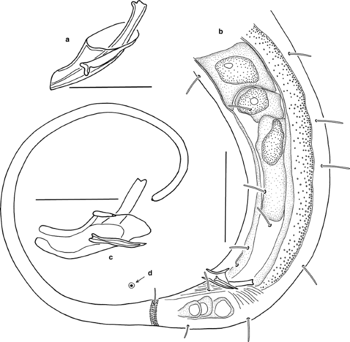
Acantholaimus veitkoehlerae sp. n., males. a holotype, spicule; b paratype No. 1, posterior end; c paratype No. 1, spicule; d paratype No. 1, cuticle punctation pattern at postcloacal region. Scale bars: a, c = 20 μm; b = 50 μm
Fig. 32 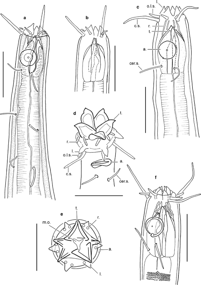
Acantholaimus veitkoehlerae sp. n., heads. a, b male, holotype; c male, paratype No. 3; d male, paratype No. 2 (everted stoma); e female, paratype No. 8 (everted stoma, front view); f female, paratype No. 6 (retracted anterior part). Scale bars = 20 μm. Abbreviations: a = amphid, c.s. = cephalic seta, cer.s. = cervical seta, l. = labium, m.o. = mouth opening, o.l.s. = outer labial seta, r. = ruga, t. = onchium
Fig. 33 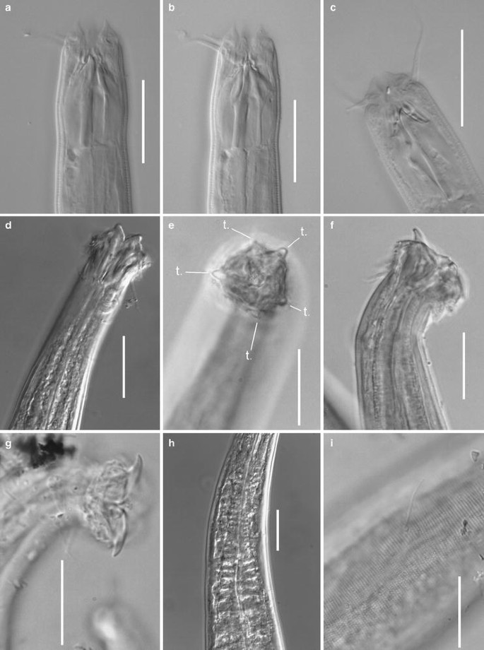
Acantholaimus veitkoehlerae sp. n., micrographs. a, b female, paratype No. 7, head region at different optical levels; c female, paratype No. 6, head region; d male, paratype No. 2, head region with everted stoma; e female, paratype No. 8, head with everted stoma, apical view; f male, paratype No. 3, head region with everted stoma; g female, paratype No. 11, head region with everted head; h male, paratype No. 2, pharyngeal region; i female, paratype No. 6, lateral view of cuticle surface (lateral differentiation of cuticular punctation not discernible, numerous small pores seen). Scale bars = 20 μm. Abbreviation: t. = onchium
Table 12 Acantholaimus veitkoehlerae sp. n.


Type material
Collection number MNHN-BN504. Holotype: male. Paratypes: 4 males and 7 females (Table 12).
Locality
Etymology
In honor of Dr. Gritta Veit-Köhler [Senckenberg am Meer, German Centre for Marine Biodiversity Research (DZMB), Wilhelmshaven, Germany].
Description
Main measurements: L = 1,069–1,541 μm; L′ = 746–1,252 μm; a = 20.7–31.4; a′ = 13.3–24.1; b = 4.6–6.6; b′ = 3.5–5.4; c = 2.3–5.3; c′ = 7.1–19.0; V = 39.6–50.2; V′ = 60.1–69.9 (Table 12).
Body spindle-shaped, with sharply narrowed anterior end and filiform posterior end. Somatic setae cylindrical, 6–13 μm long, present along entire body except filiform part of tail; these setae arranged in 4 submedian rows. Cuticle densely dotted, with indistinct lateral differentiation. Dots in lateral fields larger and spaced slightly farther apart than dots in median fields. Dots of cuticle outside lateral fields smaller, except in postanal part, where they are larger than dots of lateral fields in midbody region and appear toroidal. Dots usually arranged more or less in transverse rows. In optical cross-section of cuticle, dots discernible as tiny radial struts. Small cuticular pores irregularly situated along entire body length. Cuticle thickness ca. 1.5 μm along entire body length except cervical part where it is thinner (ca. 0.8 μm). There are 6 triangular lips with edged anterior tips. Inner labial sensilla not visible. 6 outer labial setae 3–6 μm long and 4 submedian cephalic setae 9–17 μm long, appearing confluent and lying at same level. Amphideal fovea ventrally coiled, single-spiral, round or shaped as transversely oriented oval, with fine but distinct cuticular edging, situated at level of middle of esophastoma, at 0.5–0.7 c.b.d. posterior to the anterior end. Very fine concentric striation and more or less distinct central spot visible in amphidial fovea. One pair of cervical setae 7–12 μm long (latero-subdorsal and latero-subventral) located at a short distance posterior to each amphideal fovea. Stoma consisting of cup-shaped cheilostoma and narrow, funnel-shaped esophastoma. Cheilostoma possessing 6 pairs of long, thick, slightly undulating, cuticular rugae (each pair located posterior to lip), esophastoma with 5 sclerotized onchia: 1 dorsal, 2 subventral, and 2 small lateral (1 on each side), Basal parts of onchia situated in anterior part of esophastoma, and their apical parts intruding into cheilostoma. Dorsal and one subventral onchia 7–8 μm long, three other smaller onchia 4–5 μm long. Esophastoma 15–20 μm in length, with thick cuticular walls, its tissues distinctly separated from tissues of pharynx. Pharynx regularly muscular, narrow in its anterior half and gradually widening to its posterior half, without formation of distinct bulb. Posterior part of pharynx containing numerous large plasmatic interruptions. Nerve ring not visible, but numerous neuron bodies present at level of middle of pharynx. Renetta not visible. Cardia large, triangular, surrounded by intestine. In about half of specimens, anterior part of pharynx everted and protruding outside, including onchia. Some specimens with partly retracted lips. Tail consisting of proximal conical part and long terminal filiform cylindrical part constituting 50–75% of entire tail length. Diameter of cylindrical part of tail at its posterior end 3–5 μm. Bodies of three caudal glands seen posterior to anus.
Male reproductive system. Single testis directed anteriorly, outstretched, lying to right of intestine. Moderately curved spicules consisting of two (wide distal and narrow proximal) parts. Gubernaculum in shape of slightly curved stick with edged proximal end and bifurcated distal end. Supplementary organs not found.
Female reproductive system consisting of two antidromous ovaries (anterior ovary lying to right of intestine, and posterior one lying to left of intestine), and short oviducts. Uterus not defined. Each ovary containing one mature ovocyte. Three pairs of vulvar glands with granular content surrounding short vagina.
Abundance
Acantholaimus veitkoehlerae sp. n. was mentioned as Acantholaimus sp. 6 in the description of nematode assemblages inhabiting deep-sea polymetallic nodule fields (Miljutina et al. 2010). The density of A. veitkoehlerae sp. n. varied from 0 to 8.4 inds/10 cm2 (mean 2.6 inds/10 cm2). This species ranked third in relative abundance within the nematode community (from 0 to 4.8% in different samples, mean 1.9%).
Differential diagnosis
The new species shares with A. arthrochaeta sp. n., A. akvavitus Gerlach et al. 1979, A. quintus Gerlach et al. 1979, and A. septimus Gerlach et al. 1979 the middle-sized body length (about 1,000–1,500 μm), relatively small amphideal fovea (40–65% c.b.d.), the position of the amphideal fovea relative to the apical part of the body (about 1 amphideal c.b.d.) and the length of the cephalic setae (about 10–15 μm). Acantholaimus veitkoehlerae sp. n. shares with A. elegans Jensen 1988 the spindle-shaped body; the long narrow eversible anterior end; and the length and shape of the pharynx (b = 4–6, consisting of two parts, a long narrow anterior muscular part and a thick posterior part with numerous plasmatic interruptions).
Acantholaimus veitkoehlerae sp. n. differs from A. elegans in the body length (L = 1,069–1,541 μm vs. L = 500–700 μm); the size (35–53% c.b.d. vs 75%); the position of the amphideal fovea (0.5–0.7 c.b.d. posterior to the anterior end vs. 1.3–1.7 c.b.d.); and the presence of lateral differentiation.
Acantholaimus veitkoehlerae sp. n. distinctly differs from A. quintus by their body shape (spindle-shaped body with strongly narrowed anterior end vs. cylindrical body with slightly narrowed anterior end); the absence of preanal papillae; the number of cervical setae (2 vs. 6); and the absence of the numerous somatic setae at the level of the pharynx that are present in A. quintus.
Acantholaimus veitkoehlerae sp. n. differs from A. arthrochaeta by the spindle-shaped body with strongly narrowed anterior end vs. the cylindrical body with weakly narrowed anterior end; the absence of jointed outer labial and cephalic setae; the shape of the amphideal fovea (circular vs oval); the number of cervical setae (2 vs. 4); and the shape of the spicules (thick vs. thin).
Acantholaimus veitkoehlerae sp. n. is closer to A. septimus and A. akvavitus. The new species differs from A. septimus by the shape and thickness of the body (spindle-shaped vs. cylindrical; a = 21–31 vs. a = 31–60); the absence of preanal papillae; the number of cervical setae (2 vs. 4); the shape of the amphideal fovea (circular vs. circular-oval); the length of the outer labial setae (3–6 μm vs. 8 μm); and the length of the inflated anterior part of the pharynx (1.2 vs. 3 cephalic diameters).
Acantholaimus veitkoehlerae sp. n. differs from A. akvavitus by the number of cervical setae (2 vs. 3); and by the length of the outer labial and cervical setae: the outer labial setae are three times longer than the cephalic setae in the new species, 3–6 μm and 9–17 μm, respectively, vs. the corresponding setae of almost identical length, 7 μm and 11 μm, respectively, in A. akvavitus.
References
Allgén CA (1933) Freilebende Nematoden aus dem Trondhjemsfjord. Capita Zool 4(2):1–162
Bussau C (1993) Taxonomische und ökologische Untersuchungen an Nematoden des Peru-Beckens. Dissertation, Christian-Albrechts-Universität, Kiel
Bussau C (1995) New deep-sea nematoda (Enoplida, Thoracostomopsidae, Oncholaimidae, Enchelidiidae) from a manganese nodule area of the eastern South Pacific. Zool Scr 24(1):1–12
Bussau C, Vopel K (1999) New nematode species and genera (Chromadorida, Microlaimidae) from the deep sea of the eastern tropical South Pacific (Peru Basin). Ann Naturhist Mus Wien 101B:405–421
De Mesel ID, Lee HJ, Vanhove S, Vincx M, Vanreusel A (2006) Species diversity and distribution within the deep-sea nematode genus Acantholaimus on the continental shelf and slope in Antarctica. Polar Biol 29:860–871
Fonseca G, Decraemer W, Vanreusel A (2006) Taxonomy and species distribution of the genus Manganonema Bussau, 1993 (Nematoda: Monhysterida). Cah Biol Mar 47:189–203
Gerlach SA (1951) Drei bemerkenswerte neue Nematoden aus der Kieler Bucht. Zool Anz 147(1/2):37–43
Gerlach SA, Schrage M, Riemann F (1979) Die Gattung Acantholaimus (Nematoda, Chromaodoridae), und Beobachtungen über einen mutmaßlichen Transportmechanismus für Spermien bei A. calathus sp.n. Veröff Inst Meeresforsch Bremerh 18:35–67
Gourbault N, Vincx M (1985) Nématodes abyssaux (Campagne Walda du N/O „Jean Charcot“). VI. Le genre Acantholaimus: espèces nouvelles et étude de l’appareil reproducteur à spermatozoïdes géants. Bull Mus Natl Hist Nat, Paris, 4-e série 7:611–632
Jensen P (1985) The nematode fauna in the sulphide-rich brine seep and adjacent bottoms of the East Flower Garden, NW Gulf of Mexico. I. Chromadorida. Zoologica Scripta 14(4):247–263
Jensen P (1988) Four new nematode species, abundant in the deep-sea benthos of the Norwegian Sea. Sarsia 73:149–155
Khripounoff A, Caprais JC, Crassous P (2006) Geochemical and biological recovery of the disturbed seafloor in polymetallic nodule fields of the Clipperton–Clarion Fracture Zone (CCFZ) at 5, 000-m depth. Limnol Oceanogr 51(5):2033–2041
Kreis HA (1963) Marine nematoda. In: Friđriksson A, Tuxen SL (eds) The zoology of Iceland 2(14). Ejnar Munksgaard Press, Copenhagen and Reykjavík, pp 1–68
Lambshead PJD, Brown BJ, Ferrero T, Hawkins LE, Smith CR, Mitchell NJ (2003) Biodiversity of nematode assemblages from the region of the Clarion–Clipperton Fracture Zone, an area of commercial mining interest. BMC Ecol 3(1). doi:10.1186/1472-6785-3-1
Miljutin DM, Miljutina MA (2009a) Description of Bathynema nodinautensis gen. n., sp. n. and four new Trophomera species (Nematoda: Benthimermithidae) from the Clarion–Clipperton Fracture Zone (Eastern Tropic Pacific), supplemented with the keys to genera and species. In: Brökeland W, George KH (eds) Deep-Sea taxonomy—a contribution to our knowledge of biodiversity. Zootaxa 2096:173–196
Miljutin DM, Miljutina MA (2009b) Deep-sea nematode species of family Microlaimidae from the Clarion–Clipperton Fracture Zone (North-Eastern Tropic Pacific), with the descriptions of three new species. In: Brökeland W, George KH (eds) Deep-Sea taxonomy—a contribution to our knowledge of biodiversity. Zootaxa 2096:137–172
Miljutin DM, Miljutina MA, Martínez Arbizu P, Galéron J (2011) Deep-sea nematode assemblage has not recovered 26 years after experimental mining of polymetallic nodules (Clarion–Clipperton Fracture Zone, Tropical Eastern Pacific). Deep-Sea Res I 58:885–897
Miljutina MA, Miljutin DM, Mahatma R, Galéron J (2010) Deep-sea nematode assemblages of the Clarion–Clipperton Nodule Province (Tropical North-Eastern Pacific). Mar Biodivers 40:1–15
Muthumbi AW, Vincx M (1997) Acantholaimus (chromadoridae: nematoda) from the Indian Ocean: description of seven species. Hydrobiologia 346:59–76
Platt HM, Zhang ZN (1982) New species of marine nematodes from Loch Ewe, Scotland. Bulletin of the British Museum (Natural History). Zoology 42(4):227–246
Roggen DR (1970) Functional aspects of the lower size-limit of nematodes. Nematologica 16(4):532–536
Sebastian S, Raes M, De Mesel I, Vanreusel A (2007) Comparison of the nematode fauna from the Weddell Sea Abyssal Plain with two North Atlantic abyssal sites. Deep Sea Res II 54:1727–1736
Seinhorst JW (1959) A rapid method for the transfer of nematodes from fixative to anhydrous glycerin. Nematologica 4:67–69
Thiel H (2001) Use and protection of the deep sea—an introduction. Deep Sea Res II 48:3427–3431
Thiel H (2003) Anthropogenic impact on the deep-sea. In: Tyler PA (ed) Ecosystems of the deep oceans. Elsevier, Amsterdam, pp 427–472
Tietjen JH (1984) Distribution and species diversity of deep-sea nematodes in the Venezuela Basin. Deep-Sea Res I 31:119–132
Tietjen JH (1989) Ecology of deep-sea nematodes from the Puerto Rico Trench area and Hatteras Abyssal Plain. Deep Sea Res 36(10):1579–1594
Vanhove S, Arntz W, Vincx M (1999) Comparative study of the nematode communities on the southeastern Weddell Sea shelf and slope (Antarctica). Mar Ecol Prog Ser 181:237–256
Vanreusel A, Van den Bossche I, Thiermann F (1997) Free-living marine nematodes from hydrothermal sediments: similarities with communities from diverse reduced habitats. Mar Ecol Prog Ser 157:207–219
Vanreusel A, Clough L, Jacobsen K, Ambrose W, Jivaluk J, Ryheul V, Herman R, Vincx M (2000) Meiobenthos of the central Arctic Ocean with special emphasis on the nematode community structure. Deep Sea Res I 47:1855–1879
Vivier MH (1985) Espèces du genre Acantholaimus (Nematoda, Chromadoridae, Spilipherinae). In: Laubier L, Monniot C (eds) Peuplements profonds du Golfe de Gascogne. IFREMER, Brest, pp 331–349
Acknowledgments
The authors thank Prof. Dr. Pedro Martínez Arbizu (Senckenberg am Meer, the German Centre for Marine Biodiversity Research, Wilhelmshaven, Germany), the chief scientist of the NODINAUT cruise Dr. Joelle Galéron (IFREMER, Brest, France), the captain and crew of RV l’Atalante, and the Nautile team for collecting the material during the NODINAUT cruise. We are also grateful to Dr. Gritta Veit-Koehler (Senckenberg am Meer, the German Centre for Marine Biodiversity Research, Wilhelmshaven, Germany) for placing her laboratory at our disposal for making the descriptions of several species, and to Viola Siegler (Senckenberg am Meer, the German Centre for Marine Biodiversity Research, Wilhelmshaven, Germany) for help and support in many ways. We also express our gratitude to two anonymous reviewers for valuable remarks and Dr. Janet W. Reid (JWR Associates, USA) for her critical revision of the English text. This study was financially supported by CeDAMar and CoML.
Author information
Authors and Affiliations
Corresponding author
Additional information
Communicated by H.-D. Franke.
Rights and permissions
About this article
Cite this article
Miljutina, M.A., Miljutin, D.M. Seven new and four known species of the genus Acantholaimus (Nematoda: Chromadoridae) from the abyssal manganese nodule field (Clarion-Clipperton Fracture Zone, North-Eastern Tropical Pacific). Helgol Mar Res 66, 413–462 (2012). https://doi.org/10.1007/s10152-011-0282-z
Received:
Revised:
Accepted:
Published:
Issue Date:
DOI: https://doi.org/10.1007/s10152-011-0282-z