Abstract
Infectious bursal disease (IBD) is a viral immunosuppressive disease of chickens attacking mainly an important lymphoid organ in birds [the bursa of Fabricius (BF)]. The emergence of new variant strains of the causative agent [infectious bursal disease virus (IBDV)] has made it more urgent to develop new vaccination strategies against IBD. One of these strategies is the use of recombinant vaccines (DNA and viral-vectored vaccines). Several studies have investigated the host immune response towards IBDV. This review will present a detailed background on the disease and its causative agent, accompanied by a summary of the most recent findings regarding the host immune response to IBDV infection and the use of recombinant vaccines against IBD.
IBDV
Infectious bursal disease virus (IBDV) has a selective tropism for bursal B cells. Infectious bursal disease (IBD) involves massive destruction of B cells in lymphoid organs, resulting in lymphopenia (immunosuppression) and secondary infection of the infected birds [52]. Immunosuppression is a state of immune system dysfunction that leads to increased disease susceptibility [73]. Immunosuppression is considered to be one of the major problems threatening the poultry industry. In general, any infectious disease can cause immunosuppression.
There are two distinct types of IBDV, designated as serotypes I and II [52, 61]. While serotype I viruses are pathogenic to chickens, serotype II viruses are isolated from turkeys and are avirulent for chickens [59]. IBDV serotype I isolates have different levels of virulence and different replication efficiency in bursal cells [82]. Beginning in 1990, variant strains of serotype I virus emerged in the United States, Western Europe, and parts of Southeast Asia that were more virulent than classical strains and caused mortality rates of over 50 % [52, 61]. These variant strains were isolated from flocks that had been vaccinated with classical-strain vaccines, and they are antigenically different from the classical strains and resistant to current commercial vaccines. Comparing the disease outcome of one of the classical virulent IBDV (vIBDV) strains (F52/70) to the variant/very virulent IBDV (vvIBDV) strains, there was an increase in the mortality rate, from 50 % with vIBDV to 90 % with vvIBDV, which was accompanied by more-severe immunosuppression across the lymphoid organs.
IBDV is a member of the genus Avibirnavirus, family Birnaviridae [52, 61]. Its genome consists of two segments of linear double-stranded RNA, designated A and B, 6 kb in length in total. Segment A is 3.2 kb in length and contains two partly overlapping open reading frames (ORF). The largest ORF encodes a polyprotein that is autocatalytically cleaved into two structural proteins, VP2 and VP3, and a serine protease, VP4 [6, 45].
VP2 is considered to be the major host-protective antigen and contains the major antigenic site responsible for eliciting neutralizing antibodies (Abs) [26]. At least two neutralizing epitopes are located on this polypeptide. VP2 induces virus-neutralizing Abs that protect susceptible chickens from vIBDV. It is responsible for antigenic variation, tissue-culture adaptation and viral virulence [12].
VP2 is folded into three main domains (the base, shell and projection domains) [8, 20, 46, 53, 65]. The base and shell domains are formed by the conserved N- and C-termini of VP2. The projection domain is formed by the hypervariable region of VP2 [amino acids (AAs) 206 to 350] (Fig. 1) [3]. Within the VP2 region, two hydrophilic regions (A and B) were identified (Fig. 1). Region A spans AAs 212 to 224, and region B spans AAs 314 to 325 [2]. These regions constitute two loops, PBC and PHI (neutralising Ab-binding domains), which represent the outmost part of the projection domain (Fig. 1) [46]. Two additional loops were identified in the projection domain, PDE and PFG (Fig. 1) [20]. Moreover, the putative AAs responsible for virulence and cellular tropism were identified to be glutamine at AA position 253, aspartic acid at AA position 279, and alanine at AA position 284 [12] (Fig. 1).
Deduced AA sequence of VP2, from AA positions 1 to 441 of vIBDV strain F52/70 and vvIBDV strain UK661. Sequences were taken from Brown and Skinner [13]. Yellow highlighted residues are AAs that differ the two strains. Green highlighted residues are the AAs responsible for virulence and cellular tropism. The two underlined segments represent hydrophilic regions A and B, respectively. Blue segment, PBC loop; green segment, PDE loop; brown segment, PFG loop; red segment, PHI loop (color figure online)
Segment A also encodes a 17-kD non-structural protein, VP5, from the small ORF [60]. VP5 is a class II membrane protein with a cytoplasmic N-terminus and an extracellular C-terminal domain [50]. It is highly basic, cysteine-rich, and semi-conserved among all serotype I IBDV strains [60], and it has been incriminated in the induced bursal pathology [92]. Moreover, it has a role in virus dissemination from infected cells [50]. VP5 accumulates within the cell membrane, resulting in its disruption and decreasing cellular viability. VP2 and VP5 have been shown to induce apoptosis in in vitro culture [70, 92].
Segment B is 2.8 kb in length and encodes VP1, a 97-kD protein with polymerase activity [61]. VP1 exists as a genome-linked protein, circularizing segments A and B. DNA vector-based RNA interference, directed towards VP1, prevents IBDV replication in Vero cells [29].
Receptors
The first report investigating the IBDV receptor was made by Ogawa et al. [62]. They described the protein as an N-glycosylated protein associated with the expression of B cell surface immunoglobulin (Ig) M+. In a later study by Lin et al. [49], chicken heat shock protein 90 was suggested to be part of the IBDV-receptor complex. A more recent report demonstrated the ability of α4β1 integrin to act as an IBDV receptor [22].
Pathogenesis, clinical signs, and pathology of infected bursae
Following oral infection, the virus replicates in gut-associated macrophages and lymphoid cells and enters the portal circulation, leading to primary viraemia [61]. The viral antigen is detectable in the macrophages and lymphoid cells of the caecum as early as 4 h postinfection (hpi) and reaches the liver by 5 hpi, and following primary viraemia, the virus reaches the BF by 11 hpi [52, 61]. Following IBDV replication in the BF, the virus enters the blood stream to cause secondary viraemia, which results in virus spread to other tissues.
Apoptosis is an individual and active type of cell death that is characterised by nuclear fragmentation and breakdown into apoptotic vesicles without extracellular release of the cellular contents, and consequently without eliciting an inflammatory reaction [19]. Apoptosis has an important role in IBDV pathogenesis and immunosuppression. A high level of apoptosis is evident in chicken peripheral blood lymphocytes infected with serotype I IBDV strain L [85], and peripheral blood lymphocytes showed a high apoptotic index (nuclear fragmentation and cellular breakdown into apoptotic vesicles).
Morbidity in IBDV-infected flocks can reach up to 100 %, and mortality rates can be as high as 90 % [61]. The most severe clinical manifestations are seen in chicks of 3 to 6 weeks of age, when the BF approaches its maximal stage of development. Birds of 1 to 14 days of age are less susceptible; they are usually protected by maternal Abs. Infected birds more than 6 weeks old rarely develop clinical signs of disease. However, they produce Abs to the virus. The incubation period is usually 2 to 3 days, after which birds show distress, depression, ruffled feathers, anorexia, diarrhoea, trembling and dehydration. The clinical disease lasts for 3 to 4 days, followed by rapid recovery in surviving birds.
Macroscopically, the infected birds are dehydrated, with haemorrhages frequently seen in the thigh and pectoral muscles [52]. The mucous content of the intestine increases. A yellowish transudate starts to cover the BF at 2 or 3 dpi. The BF begins to increase in size and weight at 3 dpi. It reaches double its normal weight by 4 dpi, begins to decrease in size, and returns to its normal weight by 5 dpi. The transudate disappears as the BF returns to its normal size. The BF starts to atrophy at 8 dpi.
Microscopically, degeneration and necrosis of lymphocytes start in the medullary area of the bursal follicles as early as 1 dpi [52]. Lymphocytes become replaced by heterophils. By day 3 or 4 postinfection, all of the lymphoid follicles are affected. Severe oedema, hyperaemia, and marked accumulation of heterophils are evident, which cause the increased bursal weight. Cystic cavities develop in the follicular medulla. These cystic cavities are caused by necrosis and phagocytosis by heterophils and plasma cells. During the stage of bursal atrophy, fibroplasia of the bursal tissue becomes evident. In addition, the bursal epithelium becomes proliferative, forming a glandular-like structure, which consists of the bursal columnar epithelium containing globules of mucin. In the late stages, scattered lymphocyte foci appear without the ability to form functional follicles.
Immune response towards IBDV
The role of the BF in IBDV infection was revealed when bursectomized birds survived infection with lethal doses of IBDV without showing any clinical illness [61]. The stage of B cell differentiation in the BF is important for viral replication, as stem cells and peripheral B cells do not support replication of the virus. The acute phase of the disease lasts for about 7-10 days [76]. Within this phase, bursal follicles are depleted of B cells and the BF becomes atrophic, which results in a diminished Ab response and increased susceptibility to secondary infection in recovered birds [61, 76]. The resulting immunosuppression leads to diminished Ab production following vaccination against other viral diseases, leading to subsequent outbreaks. Recovered birds show high anti-IBDV Ab titres.
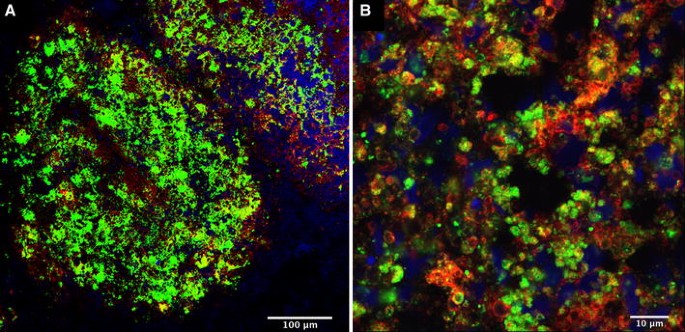
Following IBDV infection, rapid progressive loss of B cells occurs in the bursal cortex and medulla (Fig. 2), peripheral blood and thymic medulla [66]. Bursal epithelial cells are also affected through the loss of some surface antigens, and it has been reported that T cells are not susceptible to infection. Kim et al. [39] revealed the infiltration of IBDV-infected BF with CD4+ and CD8+ T cells starting from 4 dpi and increasing until T cells represented 65 % of the bursal cell population at 7 dpi. In a more recent report by Withers et al. [90], the numbers of CD4+ cells, CD8+ cells, and macrophages increased in the BF following IBDV infection as early as 1 dpi. Moreover, the bursal cells and the splenocytes up-regulated IFN-γ transcription. IBDV-infected CD4+ and CD8+ T cells inhibited the mitogenic response of splenocytes to ConA treatment [38]. Chemical induction of T cell depletion caused an increase in the bursal viral load [39]. In addition, a combined surgical and chemical induction of T cell depletion produced the same effect and decreased the transcriptional level of interferon (IFN)-γ and interleukin (IL)-2 [67]. It also delayed the recovery of IBDV-infected BF.
The interaction between B cells and vIBDV strain F52/70 in an IBDV-infected BF at 4 dpi [54]. Rhode Island red (RIR) birds (3 weeks of age) were infected intranasally with vIBDV strain F52/70 at egg infectious dose (EID)50 101.6. (A) A section of an infected BF showing the interaction between B cells (red) and IBDV (green). (B) A higher magnification showing the same features. Some of the B cells have lost their cellular integrity. IBDV particles can be seen co-localising with and inside most of the B cells. Areas of co-localisation are seen in yellow. The B cell marker (IgG1 isotype) was used with a secondary conjugate (Alexafluor 568). The IBDV marker (IgG2a isotype) was used with a secondary conjugate (Alexafluor 488) (color figure online)
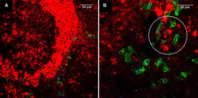
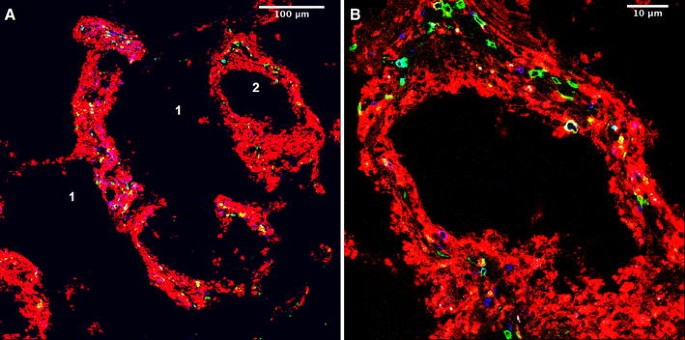
The protection against IBDV infection does not depend solely on the induction of virus-neutralising Abs [68]; T cell involvement is critical. IgM+ cells serve as targets for IBDV [76]. Viral particles are detected in the BF and other peripheral lymphoid organs, such as the caecal tonsils, spleen, and thymus [69, 76, 89]. In these reports, CD4+ and CD8+ T cells accumulated at areas of virus replication. After recovery, the BF becomes repopulated with IgM+ B cells. Depletion of Bu-1+, IgM+, and IgY+ (the avian equivalent of IgG) cells from the BF, spleen, and thymus of chickens infected with vvIBDV strain UK611 was evident [89]. The BF was repopulated with small numbers of Bu-1+ cells 14 days postinfection. Few of these cells expressed IgM or IgY. Bursal macrophages increased for the first 3 to 5 days after infection, and this was followed by an influx of CD4+ and CD8+ T cells. Depletion of cortical thymocytes was evident during the acute phase of infection. Withers et al. [90] reported a bursal recovery as early as one week after infection with vIBDV strain F52/70. Such recovery was associated with the development of two types of follicles, differentiated (resembling the uninfected control BF) and undifferentiated (lacking a recognisable cortex and medulla). The abundance of undifferentiated follicles in the BF structure was associated with an inability to mount an Ab response against IBDV. IBDV infection caused reduction in the number of chicken splenic macrophages at 3 and 5 dpi [63]. Such cells contained IBDV particles. IBDV infection was associated with the up-regulation of CD8αα+ TCR2−cells, CD4− CD8αα− TCR2+ cells, CD8αα+ TCR2+ cells, CD4+ TCR2− cells, and CD4+ TCR2+ cells in the BF and the CT [54]. In addition, IBDV particles were seen co-localised with and contained by CD8αα+ TCR2- cells, CD4+ TCR2− cells, CD4− CD8αα− TCR2+ cells, CD8αα+ TCR2+ cells, and CD4+ TCR2+ (Figs. 3, 4) in the BF and co-localised with CD8αα+ TCR2− cells, CD4− CD8αα− TCR2+ cells, and CD4+ TCR2− cells in the CT.
The interaction between CD8αα+ TCR2+ cells and IBDV in a BF of infected with vvIBDV strain UK661 at 4 dpi [54]. RIR birds (3 weeks of age) were infected intranasally with vvIBDV (strain UK661) at a dose of 101.3 EID50. (A) A section of an infected BF showing CD8αα+ cells (green) and TCR2+ cells (blue) surrounding IBDV-infected areas (red). (B) A higher magnification showing areas of co-localisation between CD8αα and TCR2 markers (cyan). Areas showing the localisation of IBDV particles in CD8αα+ TCR2+ cells are circled. The TCR2 marker (IgG1 isotype) was used with a secondary conjugate (Alexafluor 633). The CD8αα marker (IgG2b isotype) was used with a secondary conjugate (Alexafluor 488). The IBDV marker (IgG2a isotype) was used with a secondary conjugate (Alexafluor 568) (color figure online)
The interaction of CD4+ and TCR2+ cells with vvIBDV strain UK661 in the BF at 4 dpi [54]. RIR birds (3 weeks of age) were infected intranasally with vvIBDV (strain UK661) at a dose of 101.3 EID50. (A) A section of an IBDV-infected BF showing CD4+ (green) and TCR2+ cells (blue) in IBDV-infected areas (red). The image was taken using ×10 magnification. (B) A higher magnification showing the same features. The TCR2 marker (IgG1 isotype) was used with a secondary conjugate (Alexafluor 633). The CD4 marker (IgG2b isotype) was used with a secondary conjugate (Alexafluor 488). The IBDV marker (IgG2a isotype) was used with a secondary conjugate (Alexafluor 568) 1 = destroyed bursal tissue. 2 = cystic cavity at the centre of an infected lymphoid follicle (color figure online)
The role of cell-mediated immunity in IBDV infection was made clear by the increased bursal mRNA transcription of the pro-inflammatory cytokines IL-1β, IL-6, CXCLi2, and IFN-γ following in vivo infection of chickens with vIBDV strain F52/70 and vvIBDV strain UK661, together with down-regulation of transforming growth factor-β4 [24]. The up-regulation of IFN-γ was correlated with an up-regulation of IL-12α but not IL-12β or IL-18. In a different study, an increased level of IL-18 mRNA in splenic macrophages was detected five days after infection with IBDV strain Irwin Moulthrop [63]. The levels of mRNA transcripts of other pro-inflammatory mediators, including IL-1β, IL-6, and inducible nitric oxide synthase, were also increased. In addition, in vivo challenge with vvIBDV UK661 strain up-regulated the transcriptional levels of type I, II, and III IFN as well as IL-18, IL-4, and IL-13 [54].
Detection and control of IBDV in commercial flocks
The differentiation between field and vaccine strains of IBDV is crucial and can be achieved by a number of techniques, including enzyme-linked immunosorbent assay [7], reverse transcription polymerase chain reaction (RT-PCR) directed towards single nucleotide polymorphisms in the VP2 region [36], and restriction fragment length polymorphism (where viral-genotype-specific restriction enzyme sites are analysed) combined with the use of RT-PCR [5]. Avian viruses [avian influenza virus, infectious bronchitis virus (IBV), Newcastle disease virus (NDV), and IBDV] have also been distinguished using microarrays coupled with RT-PCR [81]. In addition, pan-viral detection microarrays have been developed and tested successfully for sero-/genotyping of key veterinary viruses with a potential use in the field or as a surveillance tool [30].
IBDV vaccines include live attenuated, inactivated/killed, and immune complex vaccines (a mixture of hyperimmune sera from specific-pathogen-infected chickens and embryo-adapted live pathogens [74]. The application route varies from in ovo administration to addition to drinking water or intramuscular injection. Breeder chickens are vaccinated by adding IBDV vaccine to drinking water to prevent infection of newly hatched chicks or by oral live-virus vaccination of breeding stock at 18 weeks of age, together with injection of inactivated vaccine in oil adjuvant just before laying. This is repeated a year later. This results in a well-maintained high level of neutralizing Ab throughout the laying life of the birds. Maternal Abs provide effective protection for chicks for 4 to 7 weeks after hatching. If chicks have low or inconsistent levels of maternal Abs, an attenuated virus vaccine is given at 1-2 weeks of age. Maternal Abs are transferred from the mother to the chick via the egg yolk. IgY begins to be absorbed from the yolk in the late stages of embryonic development until shortly after hatching [58].
Recombinant DNA-IBDV vaccines
There is growing concern that intensive use of live attenuated vaccines against IBDV could be driving this pathogen to increasing virulence due to potential mutation. IBDV is immunosuppressive, and the response of the poultry industry to disease outbreaks in vaccinated flocks has been through the use of more-virulent (hot) vaccines. With immunosuppressive pathogens, there is a risk that such hot vaccines could be harmful to susceptible genotypes or chicks that are poorly protected by maternal Abs. Therefore, these problems have raised increasing interest in the development of subunit vaccines, where one or more genes encoding specific pathogen antigens are expressed by a recombinant DNA vaccine.
DNA vaccination offers several advantages for delivering protective antigens; DNA vaccines mimic a natural viral infection in that the antigens they encode are produced in their native structure and are presented in the context of MHC class I and II, evoking a balanced immune response. In addition, it is possible to administer multicomponent vaccines in a single dose. There is no evidence of injection-site reactions and no risks resulting from reversion to the wild-type. And finally, neonates can be immunized with minimal interference from maternal Abs. However, it still remains to be determined whether DNA vaccines can always overcome maternal Abs. DNA vaccines are stable at high ambient temperatures, removing the need for maintaining a cold chain. Finally, constructing and purifying plasmid DNA is relatively quick and easy compared to conventional vaccines.
New approaches to immunization have sprung from the understanding of DNA and the ability to construct expression plasmids, recombinant viruses and recombinant bacteria. Recently, genetically engineered vaccines have been developed to elicit cell-mediated as well as humoral immunity. DNA vaccines expressing either VP2 or the VP4-2-3 polyprotein (segment A) of IBDV have been used as plasmid DNA vaccines for IBDV [17, 18, 27, 41, 47, 48, 55, 56, 71, 72, 88]. The resulting vaccines produced variable levels of protection, ranging from partial to complete protection, against IBDV challenge. Complete protection, against clinical disease, mortality and damage to the BF was only observed when the DNA vaccine was applied in a prime-boost regimen [17, 18, 47]. Of the several administration routes tested (the intramuscular, intraperitoneal, oral and eyedrop routes), the intramuscular route was the only one that provided protection and an anti-viral Ab response [47]. In contrast, topical administration of DNA vaccines has been reported to induce IgA and IgM anti-viral Abs [34]. When compared, DNA vaccines containing the polyprotein gene are generally more protective than DNA vaccines containing the single VP2 gene [27, 47]. However, Chang et al. [18] reported that a VP2-containing DNA plasmid induced complete protection.
In ovo injection of a 40-μg DNA vaccine (pCI-VP2) based on the plasmid vector pCI-neo carrying the coding sequence for VP2 from the vIBDV strain F52/70, in the amniotic fluid at 18 days of incubation, accompanied by a boost with fpIBD1 post-hatch, produced complete protection against IBDV-induced mortality and bursal pathology [31]. This protection was not evident with the use of either vaccine on its own. An Ab response against IBDV was not detected after the prime-boost vaccination, even after chicks were challenged with IBDV. Moreover, the in vitro incubation of empty vector (pCI-neo) with HD11 cells, a chicken macrophage cell line, and monocyte-derived macrophages stimulated the release of nitric oxide and IL-6 from both, proving the immunostimulatory effect of unmethylated CpG motifs (DNA motifs, prevalent in bacteria and viral DNA but heavily suppressed and methylated in vertebrate genomes, are also present in plasmid DNA) in the form of plasmid DNA [32]. This immunostimulatory effect was inhibited when the plasmid was digested with DNase and methylated with cytosine, confirming the immunomodulatory role of CpG motifs in the plasmid DNA and suggesting the potential role of CpG motifs in plasmid DNA vaccines as vaccine adjuvants.
Particularly powerful among T cell vaccines have been combinations of DNA and live vectors in which a live vector vaccine is used to boost the response to a DNA prime, or in which one recombinant viral vector is used for priming and a second viral vector for boosting [75]. These heterologous prime-boost immunizations elicit stronger immune responses than can be achieved by priming and boosting with the same vector. The first vaccination initiates memory cells; the second vaccination expands the memory response. Outside of the immune response to the common vaccine insert, which undergoes a tremendous boost, the two agents do not raise responses against each other and thus do not interfere with each other’s activity. DNA vaccines used alone induce T cell responses in animals, as can antigen with many adjuvants. Various strategies have been considered to improve DNA vaccines, such as cytokine augmentation.
Cytokines as vaccine adjuvants with recombinant DNA-IBDV vaccines
The successful elimination of pathogens following prophylactic immunization depends to a large extent on the ability of the host’s immune system to recognize when it is necessary to become activated and how to respond most effectively, preferably with minimal injury to healthy tissue. In the design of effective, non-replicating vaccines, immunological adjuvants serve as critical components that instruct and control the selective induction of the appropriate type of antigen-specific immune response. Hence, vaccine adjuvants are essential to stimulate the host’s immune response to antigens that lack immunogenicity. Theoretically, adjuvants can be divided into either facilitators of signal 1 (enhancing the duration or magnitude of either whole antigen or its peptide fragments presented by MHC molecules on APC in lymphoid organs) and/or inducers of endogenous signal 2 molecules (cytokines, membrane-bound co-stimulatory molecules or other host-derived natural adjuvants).
Effective vaccine adjuvants, like Freund’s adjuvant, have limited clinical use due to their potential toxicity, as they mediate their action through non-specific induction of several cytokines; e.g., Freund’s adjuvant induces T cell proliferation and the production of IL-2 and IFN-γ [37]. Therefore, the use of specific cytokines as vaccine adjuvants should augment immunogenicity without the side effects exerted by nonspecific cytokine induction. A number of such host-derived immunostimulatory factors have been described as immunopotentiators of vaccines when co-administered either as a heterologous expression product or following delivery by a viral vector [33, 40, 87]. They likely act by providing the second signal directly to adaptive effector T or B cells or by regulating indirectly the generation of other essential signal 2 molecules. Based on their biological activity, mammalian and avian cytokines have been used as adjuvants to enhance antigen presentation (granulocyte macrophage-colony stimulation factor (GM-CSF), macrophage-CSF, IL-1α/β, and IFN-γ), enhance T cell immune responses (IL-2, IL-12, IL-6, IFN-γ), and enhance B cell humoral immune responses (GM-CSF, IL-1α/β, IL-2, IL-6, IFN-γ, IFN-α, and IFN-β) [23, 33, 42, 48, 51, 80].
Co-administration of recombinant chicken IFN-γ with antigen resulted in enhanced secondary Ab responses that persisted at higher levels and for longer periods compared to antigen injected in the absence of IFN-γ [51]. In addition, IFN-γ treatment allowed lower doses of antigen to be more effective. Recombinant IL-18 was used as a vaccine adjuvant with inactivated NDV, IBV, and Clostridium perfringens α-toxoid [21]. Raised Ab titres were observed for NDV and C. perfringens α-toxoid but not for IBV. Protection against challenge with virulent IBDV was enhanced when a DNA plasmid expressing chicken IL-2 was co-administered with a DNA vaccine encoding VP2 [35, 48]. Plasmid vectors carrying chicken IL-6 and IBDV VP2-4-3 were used by Sun et al. [80] to immunize chickens against vvIBDV strain SH95. Immunization with a mixture of both genes conferred protection on 90 % of chickens. In addition, partial protection and raised Ab titre were observed when chicken IL-2 was used as a vaccine adjuvant with VP2 [42, 48] when compared to the effect of VP2 alone. The combined in ovo administration of IL-12 or IL-18 with or without pCI-VP2 protected bursal cells and splenocytes from in vitro infection with vvIBDV strain UK661 [54].
Viral-vectored IBDV vaccines
Fowlpox IBD1 (fpIBD1)
Various viruses have been used as vectors for IBDV, e.g., Marek’s disease virus [83, 84], Semliki Forest virus [64], baculoviruses [57], avian adenovirus [28, 78], and recently, herpesvirus of turkey [14]. The use of Fowlpox virus (FPV) as a recombinant vector for use in poultry started in the 1980s. Recombinant FPV vectors have been successful in protection against a wide variety of diseases, e.g., NDV [9], IBV, avian influenza virus, and Marek’s disease virus [10]. Moreover, recombinant FPV vaccines have been used for non-avian species [10, 86]. FP9 is the best characterized of the FPV strains used for recombinant vaccine purposes. FP9 was isolated in the late 1980s at the Houghton Poultry Research Station, U.K. [79].
The FPV genome is 288 kb in length and contains 260 ORFs [1]. The genome consists of a central coding region bounded by two identical inverted terminal repeat (ITR) regions of approximately 9.5 kb each. The boundaries between the ITRs and the central coding region are marked by the 3′ 148 codons of ORF FPV010 and FPV251. Poxviruses have immunomodulatory strategies; one of them is the ability to bind IL-18 [1, 44, 91]. Eldaghayes [25] proved that FPV-IL-18 binding protein is encoded by ORF 214.
Bayliss et al. [4] constructed a recombinant FPV vaccine, fpIBD1 (FPV strain FP9, a tissue-culture adapted strain of the parent FPV strain HP-1, encoding VP2 from vIBDV strain F52/70 as a β-galactosidase fusion protein, which was inserted in the ITR of the FPV genome). fpIBD1 (in a prime-boost vaccination regimen) protected outbred Rhode Island red (RIR) chickens from mortality induced by vIBDV strain F52/70 or the highly virulent strain CS89, although not vIBDV-induced bursal damage. Successful vaccination with fpIBD1 was revealed to be dependent on the titre of the challenge virus, as high titres of challenge virus were able to overcome protection induced by fpIBD1, whereas challenge with a low titre of virus did not [77]. Protection by fpIBD1 was induced in the absence of detectable serum Abs [4, 16, 77], suggesting a significant role for cell-mediated immunity in protection from IBDV challenge. In contrast, Ab responses to influenza virus and FPV were detected in chickens vaccinated with a recombinant FPV expressing VP2 and influenza virus haemagglutinin [11]. fpIBD1 also protected against IBDV-induced immunosuppression, as shown by measuring the chicken’s Ab response [16].
Eldaghayes [25] manipulated fpIBD1 to express chicken IL-18 (fpIBD1::IL-18) and investigated the potential of chicken IL-18 to act as a vaccine adjuvant against vIBDV (F52/70). Chicken IL-18 was inserted into the ORF FPV030 locus (FPV homologue of human PC-1, plasma cell membrane glycoprotein, which has alkaline phosphodiesterase and nucleotide pyrophosphatase activities), which was found to be non-essential for FPV replication in tissue culture [1, 15, 43]. fpIBD1::IL-18 provided complete protection against challenge with vIBDV; no viral RNA or bursal damage was detected in vaccinated challenged birds. In a different study, both fpIBD1 and fpIBD1::IL-18 protected RIR from challenge with the vvIBDV UK661 strain [54]. Both vaccines raised the humoral antibody response. In addition, they both raised transcriptional levels of type I, II, and III IFN as well as T helper 1 and T helper 2 cytokines, IL-18 and IL-4 and IL-13, respectively.
Conclusion
Various vaccination strategies have been tested experimentally against IBD. In comparison with the traditional live attenuated and inactivated vaccines, recombinant vaccines have proved a success in providing protection against IBDV infection, depending on the route of administration, the amount or titre of the vaccine, and the challenge dose of the virus, without the risk of reverting to virulence or inducing bursal pathology. While vaccination regimens using the traditional live attenuated vaccine starts at day 14 of bird’s life (in the case of the presence of maternal Abs), DNA vaccines can be applied at an earlier age without any interference from maternal Abs. In addition, recombinant vaccines can provide protection against multiple infectious agents (through the insertion of their specific immunogenic genes in the vaccine construct in a single carrier construct), saving labour costs, vaccination costs, and stress to the vaccinated flock. However, it is still in question if these vaccines are cost competitive.
References
Afonso CL, Tulman ER, Lu Z, Zsak L, Kutish GF, Rock DL (2000) The genome of fowlpox virus. J Virol 74:3815–3831
Azad AA, Jagadish MN, Brown MA, Hudson PJ (1987) Deletion mapping and expression in Escherichia coli of the large genomic segment of a birnavirus. Virology 161:145–152
Bayliss CD, Spies U, Shaw K, Peters RW, Papageorgiou A, Muller H, Boursnell ME (1990) A comparison of the sequences of segment A of four infectious bursal disease virus strains and identification of a variable region in VP2. J Gen Virol 71(6):1303–1312
Bayliss CD, Peters RW, Cook JK, Reece RL, Howes K, Binns MM, Boursnell ME (1991) A recombinant fowlpox virus that expresses the VP2 antigen of infectious bursal disease virus induces protection against mortality caused by the virus. Arch Virol 120:193–205
Biet F (2002) Results of recent IBD field survey in France using bursal imaging analysis, RT-PCR-RFLP and serology. In: Annual Report and Proceedings of immunosuppressive viral disease in poultry, COST Action 839
Birghan C, Mundt E, Gorbalenya AE (2000) A non-canonical lon proteinase lacking the ATPase domain employs the ser-Lys catalytic dyad to exercise broad control over the life cycle of a double-stranded RNA virus. Embo J 19:114–123
Boot HJ, ter Huurne AA, Hoekman AJ, Peeters BP, Gielkens AL (2000) Rescue of very virulent and mosaic infectious bursal disease virus from cloned cDNA: VP2 is not the sole determinant of the very virulent phenotype. J Virol 74:6701–6711
Bottcher B, Kiselev NA, Stel’Mashchuk VY, Perevozchikova NA, Borisov AV, Crowther RA (1997) Three-dimensional structure of infectious bursal disease virus determined by electron cryomicroscopy. J Virol 71:325–330
Boursnell ME, Green PF, Campbell JI, Deuter A, Peters RW, Tomley FM, Samson AC, Chambers P, Emmerson PT, Binns MM (1990) Insertion of the fusion gene from Newcastle disease virus into a non-essential region in the terminal repeats of fowlpox virus and demonstration of protective immunity induced by the recombinant. J Gen Virol 71(3):621–628
Boyle DB, Heine HG (1993) Recombinant fowlpox virus-vaccines for poultry. Immunol Cell Biol 71:391–397
Boyle DB, Heine HG (1994) Influence of dose and route of inoculation on responses of chickens to recombinant fowlpox virus vaccines. Vet Microbiol 41:173–181
Brandt M, Yao K, Liu M, Heckert RA, Vakharia VN (2001) Molecular determinants of virulence, cell tropism, and pathogenic phenotype of infectious bursal disease virus. J Virol 75:11974–11982
Brown MD, Skinner MA (1996) Coding sequences of both genome segments of a European ‘very virulent’ infectious bursal disease virus. Virus Res 40:1–15
Bublot M, Pritchard N, Le Gros FX, Goutebroze S (2007) Use of a vectored vaccine against infectious bursal disease of chickens in the face of high-titred maternally derived antibody. J Comp Pathol 137(Suppl 1):S81–S84
Buckley MF, Loveland KA, Mckinstry WJ, Garson OM, Goding JW (1990) Plasma-cell membrane glycoprotein Pc-1-Cdna cloning of the human molecule, amino-acid-sequence, and chromosomal location. J Biol Chem 265:17506–17511
Butter C, Sturman TD, Baaten BJ, Davison TF (2003) Protection from infectious bursal disease virus (IBDV)-induced immunosuppression by immunization with a fowlpox recombinant containing IBDV-VP2. Avian Pathol 32:597–604
Chang HC, Lin TL, Wu CC (2001) DNA-mediated vaccination against infectious bursal disease in chickens. Vaccine 20:328–335
Chang HC, Lin TL, Wu CC (2003) DNA vaccination with plasmids containing various fragments of large segment genome of infectious bursal disease virus. Vaccine 21:507–513
Cohen JJ (1991) Programmed cell death in the immune system. Adv Immunol 50:55–85
Coulibaly F, Chevalier C, Gutsche I, Pous J, Navaza J, Bressanelli S, Delmas B, Rey FA (2005) The birnavirus crystal structure reveals structural relationships among icosahedral viruses. Cell 120:761–772
Degen WG, van Zuilekom HI, Scholtes NC, van Daal N, Schijns VE (2005) Potentiation of humoral immune responses to vaccine antigens by recombinant chicken IL-18 (rChIL-18). Vaccine 23:4212–4218
Delgui L, Ona A, Gutierrez S, Luque D, Navarro A, Caston JR, Rodriguez JF (2009) The capsid protein of infectious bursal disease virus contains a functional alpha 4 beta 1 integrin ligand motif. Virology 386:360–372
Dong P, Brunn C, Ho RJY (1995) Cytokines as vaccine adjuvants. Current status and potential applications. In: Powell MF, Newman MJ, Burdman JR (eds) Vaccine design: the subunit and adjuvant approach. Pharmaceutical Biotechnology; Plenum Press, New York, pp 625–643
Eldaghayes I, Rothwell L, Williams A, Withers D, Balu S, Davison F, Kaiser P (2006) Infectious bursal disease virus: strains that differ in virulence differentially modulate the innate immune response to infection in the chicken bursa. Viral Immunol 19:83–91
Eldaghayes IM (2005) Use of chicken interleukin-18 as a vaccine adjuvant with a recombinant fowlpox virus fpIBD1, a subunit vaccine giving partial protection against IBDV. University of Bristol, Bristol
Fahey KJ, Erny K, Crooks J (1989) A conformational immunogen on VP-2 of infectious bursal disease virus that induces virus-neutralizing antibodies that passively protect chickens. J Gen Virol 70(6):1473–1481
Fodor I, Horvath E, Fodor N, Nagy E, Rencendorsh A, Vakharia VN, Dube SK (1999) Induction of protective immunity in chickens immunised with plasmid DNA encoding infectious bursal disease virus antigens. Acta Vet Hung 47:481–492
Francois A, Chevalier C, Delmas B, Eterradossi N, Toquin D, Rivallan G, Langlois P (2004) Avian adenovirus CELO recombinants expressing VP2 of infectious bursal disease virus induce protection against bursal disease in chickens. Vaccine 22:2351–2360
Gao Y, Liu W, Gao H, Qi X, Lin H, Wang X, Shen R (2008) Effective inhibition of infectious bursal disease virus replication in vitro by DNA vector-based RNA interference. Antiviral Res 79:87–94
Gurrala R, Dastjerdi A, Johnson N, Nunez-Garcia J, Grierson S, Steinbach F, Banks M (2009) Development of a DNA microarray for simultaneous detection and genotyping of lyssaviruses. Virus Res 144:202–208
Haygreen EA, Kaiser P, Burgess SC, Davison TF (2006) In ovo DNA immunisation followed by a recombinant fowlpox boost is fully protective to challenge with virulent IBDV. Vaccine 24:4951–4961
Haygreen L, Davison F, Kaiser P (2005) DNA vaccines for poultry: the jump from theory to practice. Expert Rev Vaccines 4:51–62
Heath AW (1995) Cytokines as immunological adjuvants. Pharm Biotechnol 6:645–658
Heckert RA, Elankumaran S, Oshop GL, Vakharia VN (2002) A novel transcutaneous plasmid-dimethylsulfoxide delivery technique for avian nucleic acid immunization. Vet Immunol Immunopathol 89:67–81
Hulse DJ, Romero CH (2002) Fate of plasmid DNA encoding infectious bursal disease virus VP2 capsid protein gene after injection into the pectoralis muscle of the chicken. Poult Sci 81:213–216
Jackwood DJ (2002) Molecular diagnosis of infectious bursal disease viruses using conventional and real-time RT-PCR. In: Annual Report and Proceedings of immunosuppressive viral disease in poultry, COST Action 839, pp 136–143
Jensen FC, Savary JR, Diveley JP, Chang JC (1998) Adjuvant activity of incomplete Freund’s adjuvant. Adv Drug Deliv Rev 32:173–186
Kim I-J, Sharma JM (2000) IBDV-induced bursal T lymphocytes inhibit mitogenic response of normal splenocytes. Vet Immunol Immunop 74:47–57
Kim IJ, You SK, Kim H, Yeh HY, Sharma JM (2000) Characteristics of bursal T lymphocytes induced by infectious bursal disease virus. J Virol 74:8884–8892
Kim JJ, Yang JS, VanCott TC, Lee DJ, Manson KH, Wyand MS, Boyer JD, Ugen KE, Weiner DB (2000) Modulation of antigen-specific humoral responses in rhesus macaques by using cytokine cDNAs as DNA vaccine adjuvants. J Virol 74:3427–3429
Kim SJ, Sung HW, Han JH, Jackwood D, Kwon HM (2004) Protection against very virulent infectious bursal disease virus in chickens immunized with DNA vaccines. Vet Microbiol 101:39–51
Kumar S, Ahi YS, Salunkhe SS, Koul M, Tiwari AK, Gupta PK, Rai A (2009) Effective protection by high efficiency bicistronic DNA vaccine against infectious bursal disease virus expressing VP2 protein and chicken IL-2. Vaccine 27:864–869
Laidlaw SM, Anwar MA, Thomas W, Green P, Shaw K, Skinner MA (1998) Fowlpox virus encodes nonessential homologs of cellular alpha-SNAP, PC-1, and an orphan human homolog of a secreted nematode protein. J Virol 72:6742–6751
Laidlaw SM, Skinner MA (2004) Comparison of the genome sequence of FP9, an attenuated, tissue culture-adapted European strain of Fowlpox virus, with those of virulent American and European viruses. J Gen Virol 85:305–322
Lejal N, Da Costa B, Huet JC, Delmas B (2000) Role of Ser-652 and Lys-692 in the protease activity of infectious bursal disease virus VP4 and identification of its substrate cleavage sites. J Gen Virol 81:983–992
Letzel T, Coulibaly F, Rey FA, Delmas B, Jagt E, van Loon AA, Mundt E (2007) Molecular and structural bases for the antigenicity of VP2 of infectious bursal disease virus. J Virol 81:12827–12835
Li J, Huang Y, Liang X, Lu M, Li L, Yu L, Deng R (2003) Plasmid DNA encoding antigens of infectious bursal disease viruses induce protective immune responses in chickens: factors influencing efficacy. Virus Res 98:63–74
Li J, Liang X, Huang Y, Meng S, Xie R, Deng R, Yu L (2004) Enhancement of the immunogenicity of DNA vaccine against infectious bursal disease virus by co-delivery with plasmid encoding chicken interleukin 2. Virology 329:89–100
Lin TW, Lo CW, Lai SY, Fan RJ, Lo CJ, Chou YM, Thiruvengadam R, Wang AH, Wang MY (2007) Chicken heat shock protein 90 is a component of the putative cellular receptor complex of infectious bursal disease virus. J Virol 81:8730–8741
Lombardo E, Maraver A, Espinosa I, Fernandez-Arias A, Rodriguez JF (2000) VP5, the nonstructural polypeptide of infectious bursal disease virus, accumulates within the host plasma membrane and induces cell lysis. Virology 277:345–357
Lowenthal JW, O’Neil TE, Broadway M, Strom AD, Digby MR, Andrew M, York JJ (1998) Coadministration of IFN-gamma enhances antibody responses in chickens. J Interferon Cytokine Res 18:617–622
Lukert PD, Saif YM (1997) Infectious bursal disease. In: Calnek BW, Barnes HJ, Beard CW, Mcdougald LR, Saif YM (eds) Diseases of poultry. Iowa State University Press, Iowa, pp 721–738
Luque D, Saugar I, Rodriguez JF, Verdaguer N, Garriga D, Martin CS, Velazquez-Muriel JA, Trus BL, Carrascosa JL, Caston JR (2007) Infectious bursal disease virus capsid assembly and maturation by structural rearrangements of a transient molecular switch. J Virol 81:6869–6878
Mahgoub HA (2010) Improved vaccination strategies for IBDV: cytokines as vaccine adjuvants. University of Bristol, Bristol
Mahmood MS, Siddique M, Hussain I, Khan A, Mansoor MK (2006) Protection capability of recombinant plasmid DNA vaccine containing VP2 gene of very virulent infectious bursal disease virus in chickens adjuvanted with CpG oligodeoxynucleotide. Vaccine 24:4838–4846
Mahmood MS, Hussain I, Siddique M, Akhtar M, Ali S (2007) DNA vaccination with VP2 gene of very virulent infectious bursal disease virus (vvIBDV) delivered by transgenic E-coli DH5 alpha given orally confers protective immune responses in chickens. Vaccine 25:7629–7635
Martinez-Torrecuadrada JL, Saubi N, Pages-Mante A, Caston JR, Espuna E, Casal JI (2003) Structure-dependent efficacy of infectious bursal disease virus (IBDV) recombinant vaccines. Vaccine 21:3342–3350
Mast J, Gilson D, Morales D, Muelemans G, TP vdB (2001) Transfer of maternal antibodies from the yolk sac to the chicken. In: Annual Report and Proceedings of immunosuppressive viral disease in poultry, COST Action 839
McFerran JB, McNulty MS, McKillop ER, Connor TJ, McCracken RM, Collins DS, Allan GM (1980) Isolation and serological studies with infectious bursal disease viruses from fowl, turkeys and ducks: demonstration of a second serotype. Avian Pathol 9:395–404
Mundt E, Beyer J, Muller H (1995) Identification of a novel viral protein in infectious bursal disease virus-infected cells. J Gen Virol 76(2):437–443
Murphy FA, Gibbs EPJ, Horzinek MC, Studdert MJ (1999) Veterinary virology, 3rd edn. Academic Press, London, pp 405–409
Ogawa M, Yamaguchi T, Setiyono A, Ho T, Matsuda H, Furusawa S, Fukushi H, Hirai K (1998) Some characteristics of a cellular receptor for virulent infectious bursal disease virus by using flow cytometry. Arch Virol 143:2327–2341
Palmquist JM, Khatri M, Cha RM, Goddeeris BM, Walcheck B, Sharma JM (2006) In vivo activation of chicken macrophages by infectious bursal disease virus. Viral Immunol 19:305–315
Phenix KV, Wark K, Luke CJ, Skinner MA, Smyth JA, Mawhinney KA, Todd D (2001) Recombinant Semliki Forest virus vector exhibits potential for avian virus vaccine development. Vaccine 19:3116–3123
Pous J, Chevalier C, Ouldali M, Navaza J, Delmas B, Lepault J (2005) Structure of birnavirus-like particles determined by combined electron cryomicroscopy and X-ray crystallography. J Gen Virol 86:2339–2346
Ramm HC, Wilson TJ, Boyd RL, Ward HA, Mitrangas K, Fahey KJ (1991) The effect of infectious bursal disease virus on B lymphocytes and bursal stromal components in specific pathogen-free (SPF) white leghorn chickens. Dev Comp Immunol 15:369–381
Rautenschlein S, Yeh HY, Njenga MK, Sharma JM (2002) Role of intrabursal T cells in infectious bursal disease virus (IBDV) infection: T cells promote viral clearance but delay follicular recovery. Arch Virol 147:285–304
Rautenschlein S, Yeh HY, Sharma JM (2002) The role of T cells in protection by an inactivated infectious bursal disease virus vaccine. Vet Immunol Immunopathol 89:159–167
Rautenschlein S, von Samson-Himmelstjerna G, Haase C (2007) A comparison of immune responses to infection with virulent infectious bursal disease virus (IBDV) between specific-pathogen-free chickens infected at 12 and 28 days of age. Vet Immunol Immunop 115:251–260
Rodriguez-Lecompte JC, Nino-Fong R, Lopez A, Frederick Markham RJ, Kibenge FS (2005) Infectious bursal disease virus (IBDV) induces apoptosis in chicken B cells. Comp Immunol Microbiol Infect Dis 28:321–337
Rong J, Cheng T, Liu X, Jiang T, Gu H, Zou G (2005) Development of recombinant VP2 vaccine for the prevention of infectious bursal disease of chickens. Vaccine 23:4844–4851
Rong J, Jiang TZ, Cheng TP, Shen MH, Du YZ, Li SL, Wang SM, Xu BJ, Fan GC (2007) Large-scale manufacture and use of recombinant VP2 vaccine against infectious bursal disease in chickens. Vaccine 25:7900–7908
Schat KA, Skinner MA (2008) Avian immunosuppressive diseases and immune evasion. In: Davison TF, Kaspers B, Schat KA (eds) Avian immunology. Academic press and Elsevier Ltd, Amsterdam; London, pp 299–322
Schijns VEJC, Sharma J, Tarpey I (2008) Practical aspects of poultry vaccination. In: Davison F, Kaspers B, Schat KA (eds) Avian immunology. Academic Press and Elsevier Ltd, Amsterdam; London, pp 373–393
Schneider J, Gilbert SC, Hannan CM, Degano P, Prieur E, Sheu EG, Plebanski M, Hill AV (1999) Induction of CD8+ T cells using heterologous prime-boost immunisation strategies. Immunol Rev 170:29–38
Sharma JM, Kim IJ, Rautenschlein S, Yeh HY (2000) Infectious bursal disease virus of chickens: pathogenesis and immunosuppression. Dev Comp Immunol 24:223–235
Shaw I, Davison TF (2000) Protection from IBDV-induced bursal damage by a recombinant fowlpox vaccine, fpIBD1, is dependent on the titre of challenge virus and chicken genotype. Vaccine 18:3230–3241
Sheppard M, Fahey KJ (1989) Herpesviruses and adenoviruses as potential vectors for vaccines in poultry. Aust Vet J 66:421–423
Skinner MA, Laidlaw SM, Eldaghayes I, Kaiser P, Cottingham MG (2005) Fowlpox virus as a recombinant vaccine vector for use in mammals and poultry. Expert Rev Vaccines 4:63–76
Sun JH, Yan YX, Jiang J, Lu P (2005) DNA immunization against very virulent infectious bursal disease virus with VP2-4-3 gene and chicken IL-6 gene. J Vet Med B Infect Dis Vet Public Health 52:1–7
Tao Q, Wang X, Bao H, Wu J, Shi L, Li Y, Qiao C, Yakovlevich SA, Mikhaylovna PN, Chen H (2009) Detection and differentiation of four poultry diseases using asymmetric reverse transcription polymerase chain reaction in combination with oligonucleotide microarrays. J Vet Diagn Invest 21:623–632
Tsukamoto K, Tanimura N, Mase M, Imai K (1995) Comparison of virus replication efficiency in lymphoid tissues among three infectious bursal disease virus strains. Avian Dis 39:844–852
Tsukamoto K, Sato T, Saito S, Tanimura N, Hamazaki N, Mase M, Yamaguchi S (2000) Dual-viral vector approach induced strong and long-lasting protective immunity against very virulent infectious bursal disease virus. Virology 269:257–267
Tsukamoto K, Saito S, Saeki S, Sato T, Tanimura N, Isobe T, Mase M, Imada T, Yuasa N, Yamaguchi S (2002) Complete, long-lasting protection against lethal infectious bursal disease virus challenge by a single vaccination with an avian herpesvirus vector expressing VP2 antigens. J Virol 76:5637–5645
Vasconcelos AC, Lam KM (1994) Apoptosis induced by infectious bursal disease virus. J Gen Virol 75(7):1803–1806
Vazquez Blomquist D, Green P, Laidlaw SM, Skinner MA, Borrow P, Duarte CA (2002) Induction of a strong HIV-specific CD8+ T cell response in mice using a fowlpox virus vector expressing an HIV-1 multi-CTL-epitope polypeptide. Viral Immunol 15:337–356
Villinger F (2003) Cytokines as clinical adjuvants: how far are we? Expert Rev Vaccines 2:317–326
Williams AE (2002) The pathogenesis of very virulent Infectious Bursal Disease virus and its modulation by DNA vaccination. Royal Veterinary College, London
Williams AE, Davison TF (2005) Enhanced immunopathology induced by very virulent infectious bursal disease virus. Avian Pathol 34:4–14
Withers DR, Young JR, Davison TF (2005) Infectious bursal disease virus-induced immunosuppression in the chick is associated with the presence of undifferentiated follicles in the recovering bursa. Viral Immunol 18:127–137
Xiang Y, Moss B (1999) IL-18 binding and inhibition of interferon gamma induction by human poxvirus-encoded proteins. Proc Natl Acad Sci USA 96:11537–11542
Yao K, Goodwin MA, Vakharia VN (1998) Generation of a mutant infectious bursal disease virus that does not cause bursal lesions. J Virol 72:2647–2654
Author information
Authors and Affiliations
Corresponding author
Rights and permissions
About this article
Cite this article
Mahgoub, H.A. An overview of infectious bursal disease. Arch Virol 157, 2047–2057 (2012). https://doi.org/10.1007/s00705-012-1377-9
Received:
Accepted:
Published:
Issue Date:
DOI: https://doi.org/10.1007/s00705-012-1377-9


