
Abstract
Neuroscience has become one of the most exciting contemporary research areas with major breakthroughs expected in the coming decades. Modern imaging techniques have enabled scientific understanding of the neural system by revealing anatomical, functional, metabolic, and molecular information about the brain. Among these techniques, photoacoustic tomography (PAT), drawing more and more attention, is playing an increasingly important role in brain studies, thanks to its rich optical absorption contrast, high spatiotemporal resolution, and deep penetration. More importantly, PAT’s unique scalability empowers neuroscientists to examine the brain at multiple spatial scales using the same contrast mechanism, bridging microscopic insights to macroscopic observations of the brain. In this chapter, we review the principles of PAT, present the major implementations, and summarize the representative neuroscience applications. We also discuss challenges in translating PAT to human brain imaging and envision its potential promise.
Access this chapter
Tax calculation will be finalised at checkout
Purchases are for personal use only
References
A.G. Bell, ART. XXXIV.--On the production and reproduction of sound by light. Am. J. Sci. (1880–1910) 20(118), 305 (1880)
L. Li et al., Label-free photoacoustic tomography of whole mouse brain structures ex vivo. NEUROW 3(3), 035001 (2016)
T.T. Wong et al., Use of a single xenon flash lamp for photoacoustic computed tomography of multiple-centimeter-thick biological tissue ex vivo and a whole mouse body in vivo. J. Biomed. Opt. 22(4), 041003 (2016)
T. Imai et al., High-throughput ultraviolet photoacoustic microscopy with multifocal excitation. J. Biomed. Opt. 23(3), 036007 (2018)
Y. Qu et al., Dichroism-sensitive photoacoustic computed tomography. Optica 5(4), 495–501 (2018)
J. Yao et al., In vivo photoacoustic imaging of transverse blood flow by using Doppler broadening of bandwidth. Opt. Lett. 35(9), 1419–1421 (2010)
D.-K. Yao et al., Optimal ultraviolet wavelength for in vivo photoacoustic imaging of cell nuclei. J. Biomed. Opt. 17(5), 056004 (2012)
D.-K. Yao et al., In vivo label-free photoacoustic microscopy of cell nuclei by excitation of DNA and RNA. Opt. Lett. 35(24), 4139–4141 (2010)
L. Lin et al., In vivo photoacoustic tomography of myoglobin oxygen saturation. J. Biomed. Opt. 21(6), 061002 (2015)
X.L. Deán-Ben, D. Razansky, Functional optoacoustic human angiography with handheld video rate three dimensional scanner. Photo-Dermatology 1(3–4), 68–73 (2013)
S. Gottschalk et al., Noninvasive real-time visualization of multiple cerebral hemodynamic parameters in whole mouse brains using five-dimensional optoacoustic tomography. J. Cereb. Blood Flow Metab. 35(4), 531–535 (2015)
D. Razansky, A. Buehler, V. Ntziachristos, Volumetric real-time multispectral optoacoustic tomography of biomarkers. Nat. Protoc. 6(8), 1121–1129 (2011)
Y. Li et al., Snapshot photoacoustic topography through an ergodic relay for high-throughput imaging of optical absorption. Nat. Photonics 14(3), 1–7 (2020)
V. Ntziachristos, D. Razansky, Molecular imaging by means of multispectral optoacoustic tomography (MSOT). Chem. Rev. 110(5), 2783–2794 (2010)
D. Razansky, C. Vinegoni, V. Ntziachristos, Multispectral photoacoustic imaging of fluorochromes in small animals. Opt. Lett. 32(19), 2891–2893 (2007)
A. Taruttis et al., Real-time imaging of cardiovascular dynamics and circulating gold nanorods with multispectral optoacoustic tomography. Opt. Express 18(19), 19592–19602 (2010)
E. Herzog et al., Optical imaging of cancer heterogeneity with multispectral optoacoustic tomography. Radiology 263(2), 461–468 (2012)
N.C. Burton et al., Multispectral Opto-Acoustic Tomography (MSOT) of the brain and glioblastoma characterization. NeuroImage 65, 522–528 (2013)
J. Yao et al., Reversibly switchable photoacoustic tomography using a genetically encoded near-infrared phytochrome, in Photons Plus Ultrasound: Imaging and Sensing 2016 97082U (2016)
L. Li et al., In vivo photoacoustic multi-contrast imaging and detection of protein interactions using a small near-infrared photochromic protein, in Photons Plus Ultrasound: Imaging and Sensing 2019 1087818 (2019)
N.C. Deliolanis et al., Deep-tissue reporter-gene imaging with fluorescence and optoacoustic tomography: A performance overview. Mol. Imaging Biol. 16(5), 652–660 (2014)
C. Vinegoni et al., Transillumination fluorescence imaging in mice using biocompatible upconverting nanoparticles. Opt. Lett. 34(17), 2566–2568 (2009)
A.C. Stiel et al., High-contrast imaging of reversibly switchable fluorescent proteins via temporally unmixed multispectral optoacoustic tomography. Opt. Lett. 40(3), 367–370 (2015)
G.S. Filonov et al., Deep-tissue photoacoustic tomography of a genetically encoded near-infrared fluorescent probe. Angew. Chem. Int. Ed. 51(6), 1448–1451 (2012)
C. Kim et al., In vivo photoacoustic mapping of lymphatic systems with plasmon-resonant nanostars. J. Mater. Chem. 21(9), 2841–2844 (2011)
J. Yao et al., Evans blue dye-enhanced capillary-resolution photoacoustic microscopy in vivo. J. Biomed. Opt. 14(5). 054049 (2009)
M. Baker, Whole-animal imaging: The whole picture. Nature 463(7283), 977–980 (2010)
L.H.V. Wang, S. Hu, Photoacoustic tomography: In vivo imaging from organelles to organs. Science 335(6075), 1458–1462 (2012)
L. Li et al., Single-impulse panoramic photoacoustic computed tomography of small-animal whole-body dynamics at high spatiotemporal resolution. Nat. Biomed. Eng. 1, 0071 (2017)
V.E. Gusev, A.A. Karabutov, Laser optoacoustics. NASA STI/Recon Technical Report A 93 (1991)
M. Xu, L.V. Wang, Universal back-projection algorithm for photoacoustic computed tomography. Phys. Rev. E 71(1), 016706 (2005)
Y. Xu et al., Reconstructions in limited-view thermoacoustic tomography. Med. Phys. 31(4), 724–733 (2004)
Y. Xu, L.V. Wang, Effects of acoustic heterogeneity in breast thermoacoustic tomography. IEEE Trans. Ultrason. Ferroelectr. Freq. Control 50(9), 1134–1146 (2003)
J. Xia et al., Enhancement of photoacoustic tomography by ultrasonic computed tomography based on optical excitation of elements of a full-ring transducer array. Opt. Lett. 38(16), 3140–3143 (2013)
K. Wang et al., Investigation of iterative image reconstruction in three-dimensional optoacoustic tomography. Phys. Med. Biol. 57(17), 5399–5423 (2012)
C. Huang et al., Full-wave iterative image reconstruction in photoacoustic tomography with acoustically inhomogeneous media. IEEE Trans. Med. Imaging 32(6), 1097–1110 (2013)
Q. Sheng et al., A constrained variable projection reconstruction method for photoacoustic computed tomography without accurate knowledge of transducer responses. IEEE Trans. Med. Imaging 34(12), 2443–2458 (2015)
J. Poudel et al., Mitigation of artifacts due to isolated acoustic heterogeneities in photoacoustic computed tomography using a variable data truncation-based reconstruction method. J. Biomed. Opt. 22(4), 041018 (2017)
T.P. Matthews et al., Parameterized joint reconstruction of the initial pressure and sound speed distributions for photoacoustic computed tomography. SIAM J. Imag. Sci. 11(2), 1560–1588 (2018)
B.E. Treeby, B.T. Cox, k-Wave: MATLAB toolbox for the simulation and reconstruction of photoacoustic wave fields, SPIE (2010)
B.E. Treeby, E.Z. Zhang, B.T. Cox, Photoacoustic tomography in absorbing acoustic media using time reversal. Inverse Probl. 26(11), 115003 (2010)
B.E. Treeby et al., Modeling nonlinear ultrasound propagation in heterogeneous media with power law absorption using a k-space pseudospectral method. J. Acoust. Soc. Am. 131(6), 4324–4336 (2012)
X.L. Dean-Ben et al., Accurate model-based reconstruction algorithm for three-dimensional optoacoustic tomography. IEEE Trans. Med. Imaging 31(10), 1922–1928 (2012)
A. Rosenthal, V. Ntziachristos, D. Razansky, Model-based optoacoustic inversion with arbitrary-shape detectors. Med. Phys. 38(7), 4285–4295 (2011)
A. Rosenthal, V. Ntziachristos, D. Razansky, Acoustic inversion in optoacoustic tomography: A review. Curr. Med. Imaging Rev. 9(4), 318–336 (2013)
T. Jetzfellner et al., Performance of iterative optoacoustic tomography with experimental data. Appl. Phys. Lett. 95(1), 013703 (2009)
J. Yao, L.V. Wang, Sensitivity of photoacoustic microscopy. Photo-Dermatology 2(2), 87–101 (2014)
X. Wang et al., Noninvasive laser-induced photoacoustic tomography for structural and functional in vivo imaging of the brain. Nat. Biotechnol. 21(7), 803 (2003)
K. Maslov, G. Stoica, L.V. Wang, In vivo dark-field reflection-mode photoacoustic microscopy. Opt. Lett. 30(6), 625–627 (2005)
H.F. Zhang et al., Functional photoacoustic microscopy for high-resolution and noninvasive in vivo imaging. Nat. Biotechnol. 24(7), 848 (2006)
J. Gamelin et al., A real-time photoacoustic tomography system for small animals. Opt. Express 17(13), 10489–10498 (2009)
L. Song, K. Maslov, L.V. Wang, Multifocal optical-resolution photoacoustic microscopy in vivo. Opt. Lett. 36(7), 1236–1238 (2011)
H.-P.F. Brecht et al., Whole-body three-dimensional optoacoustic tomography system for small animals. J. Biomed. Opt. 14(6), 064007 (2009)
E. Z. Zhang, J. Laufer, P. Beard, Three-dimensional photoacoustic imaging of vascular anatomy in small animals using an optical detection system, in Photons Plus Ultrasound: Imaging and Sensing 2007: The Eighth Conference on Biomedical Thermoacoustics, Optoacoustics, and Acousto-optics 64370S (2007)
Z. Wu et al., A microrobotic system guided by photoacoustic computed tomography for targeted navigation in intestines in vivo. Sci. Robot. 4(32), eaax0613 (2019)
J. Yao et al., Label-free oxygen-metabolic photoacoustic microscopy <i>in vivo</i>, SPIE (2011)
J. Yao et al., Noninvasive photoacoustic computed tomography of mouse brain metabolism in vivo. NeuroImage 64, 257–266 (2013)
R. Cao et al., Functional and oxygen-metabolic photoacoustic microscopy of the awake mouse brain. NeuroImage 150, 77–87 (2017)
L. Li et al., Photoacoustic imaging of lacZ gene expression in vivo. J. Biomed. Opt. 12(2), 020504 (2007)
X. Cai et al., Multi-scale molecular photoacoustic tomography of gene expression. PloS one 7(8), e43999 (2012)
J. Aguirre et al., Precision assessment of label-free psoriasis biomarkers with ultra-broadband optoacoustic mesoscopy. Nat. Biomed. Eng. 1(5), 0068 (2017)
T.T.W. Wong et al., Fast label-free multilayered histology-like imaging of human breast cancer by photoacoustic microscopy. Sci. Adv. 3(5), e1602168 (2017)
T.T.W. Wong et al., Label-free automated three-dimensional imaging of whole organs by microtomy-assisted photoacoustic microscopy. Nat. Commun. 8(1), 1386 (2017)
L. Lin et al., In vivo deep brain imaging of rats using oral-cavity illuminated photoacoustic computed tomography. J. Biomed. Opt. 20(1), 016019–016019 (2015)
P. Zhang et al., In vivo superresolution photoacoustic computed tomography by localization of single dyed droplets. Light-Sci. Appl. 8(1), 1–9 (2019)
F. Knieling et al., Multispectral optoacoustic tomography for assessment of Crohn’s disease activity. N. Engl. J. Med. 376(13), 1292–1294 (2017)
J.-M. Yang et al., Photoacoustic endoscopy. Opt. Lett. 34(10), 1591–1593 (2009)
J.-M. Yang et al., Simultaneous functional photoacoustic and ultrasonic endoscopy of internal organs in vivo. Nat. Med. 18(8), 1297 (2012)
B. Dong et al., Photoacoustic probe using a microring resonator ultrasonic sensor for endoscopic applications. Opt. Lett. 39(15), 4372–4375 (2014)
K. Jansen et al., Intravascular photoacoustic imaging of human coronary atherosclerosis. Opt. Lett. 36(5), 597–599 (2011)
S. Sethuraman et al., Spectroscopic intravascular photoacoustic imaging to differentiate atherosclerotic plaques. Opt. Express 16(5), 3362–3367 (2008)
L. Lin et al., Handheld optical-resolution photoacoustic microscopy. J. Biomed. Opt. 22(4), 041002 (2016)
J.-T. Oh et al., Three-dimensional imaging of skin melanoma in vivo by dual-wavelength photoacoustic microscopy. J. Biomed. Opt. 11(3), 034032 (2006)
L. Li et al., Fully motorized optical-resolution photoacoustic microscopy. Opt. Lett. 39(7), 2117–2120 (2014)
X. Luís Deán-Ben, D. Razansky, Adding fifth dimension to optoacoustic imaging: Volumetric time-resolved spectrally enriched tomography. Light-Sci Appl 3, e137 (2014)
H. Ke et al., Performance characterization of an integrated ultrasound, photoacoustic, and thermoacoustic imaging system. J. Biomed. Opt. 17(5), 056010 (2012)
A. Danielli et al., Label-free photoacoustic nanoscopy. J. Biomed. Opt. 19(8), 086006 (2014)
V. Ermolayev et al., Simultaneous visualization of tumour oxygenation, neovascularization and contrast agent perfusion by real-time three-dimensional optoacoustic tomography. Eur. Radiol. 26(6), 1843–1851 (2016)
C. Lutzweiler et al., Real-time optoacoustic tomography of indocyanine green perfusion and oxygenation parameters in human finger vasculature. Opt. Lett. 39(14), 4061–4064 (2014)
S. Gottschalk et al., Correlation between volumetric oxygenation responses and electrophysiology identifies deep thalamocortical activity during epileptic seizures, SPIE (2016)
L. Wang et al., Ultrasonically encoded photoacoustic flowgraphy in biological tissue. Phys. Rev. Lett. 111(20), 204301 (2013)
J. Xia et al., Calibration-free quantification of absolute oxygen saturation based on the dynamics of photoacoustic signals. Opt. Lett. 38(15), 2800–2803 (2013)
S. Hu, K. Maslov, L.V. Wang, Second-generation optical-resolution photoacoustic microscopy with improved sensitivity and speed. Opt. Lett. 36(7), 1134–1136 (2011)
S. Hu et al., Functional transcranial brain imaging by optical-resolution photoacoustic microscopy, SPIE (2009)
S. Hu, K. Maslov, L.V. Wang, Noninvasive label-free imaging of microhemodynamics by optical-resolution photoacoustic microscopy. Opt. Express 17(9), 7688–7693 (2009)
Z. Guo, S. Hu, L.V. Wang, Calibration-free absolute quantification of optical absorption coefficients using acoustic spectra in 3D photoacoustic microscopy of biological tissue. Opt. Lett. 35(12), 2067–2069 (2010)
Y. Zhou et al., Calibration-free in vivo transverse blood flowmetry based on cross correlation of slow time profiles from photoacoustic microscopy. Opt. Lett. 38(19), 3882–3885 (2013)
S.N. Hennen et al., Photoacoustic tomography imaging and estimation of oxygen saturation of hemoglobin in ocular tissue of rabbits. Exp. Eye Res. 138, 153–158 (2015)
X. Wang et al., Noninvasive imaging of hemoglobin concentration and oxygenation in the rat brain using high-resolution photoacoustic tomography, SPIE (2006)
L. Li, J. Yao, L.V. Wang, Photoacoustic tomography enhanced by nanoparticles, in Wiley Encyclopedia of Electrical and Electronics Engineering, (2016), pp. 1–14
G.P. Luke, D. Yeager, S.Y. Emelianov, Biomedical applications of photoacoustic imaging with exogenous contrast agents. Ann. Biomed. Eng. 40(2), 422–437 (2012)
L.V. Wang, J. Yao, A practical guide to photoacoustic tomography in the life sciences. Nat. Methods 13, 627 (2016)
Y. Zhou et al., Handheld photoacoustic probe to detect both melanoma depth and volume at high speed in vivo. J. Biophotonics 8(11–12), 961–967 (2015)
C.H. Lee et al., Chemical imaging in vivo: Photoacoustic-based 4-dimensional chemical analysis. Anal. Chem. 91(4), 2561–2569 (2019)
J. Jo et al., Detecting joint inflammation by an LED-based photoacoustic imaging system: A feasibility study, SPIE (2018)
J. Jo et al., Photoacoustic tomography for human musculoskeletal imaging and inflammatory arthritis detection. Photoacoustics 12, 82–89 (2018)
Y. Zhu et al., Identifying intestinal fibrosis and inflammation by spectroscopic photoacoustic imaging: An animal study in vivo. Biomed. Opt. Express 9(4), 1590–1600 (2018)
A. Garcia-Uribe et al., Dual-modality photoacoustic and ultrasound imaging system for noninvasive sentinel lymph node detection in patients with breast cancer. Sci. Rep. 5, 15748 (2015)
B. Huang et al., Improving limited-view photoacoustic tomography with an acoustic reflector. J. Biomed. Opt. 18(11), 110505 (2013)
R. Ellwood et al., Photoacoustic imaging using acoustic reflectors to enhance planar arrays. J. Biomed. Opt. 19(12), 126012 (2014)
G. Li et al., Tripling the detection view of high-frequency linear-array-based photoacoustic computed tomography by using two planar acoustic reflectors. Quant. Imaging Med. Surg. 5(1), 57 (2015)
P. Zhang et al., High-resolution deep functional imaging of the whole mouse brain by photoacoustic computed tomography in vivo. J. Biophotonics 11(1), e201700024 (2018)
G. Li et al., Multiview Hilbert transformation for full-view photoacoustic computed tomography using a linear array, SPIE (2015)
J. Xia et al., Whole-body ring-shaped confocal photoacoustic computed tomography of small animals in vivo, SPIE (2012)
C. Li et al., Real-time photoacoustic tomography of cortical hemodynamics in small animals, SPIE (2010)
J. Xia et al., Three-dimensional photoacoustic tomography based on the focal-line concept, SPIE (2011)
M.R. Chatni et al., Tumor glucose metabolism imaged <i>in vivo</i> in small animals with whole-body photoacoustic computed tomography, SPIE (2012)
J. Xia et al., Retrospective respiration-gated whole-body photoacoustic computed tomography of mice, SPIE (2014)
L. Li et al., Imaging small animal whole-body dynamics by single-impulse panoramic photoacoustic computed tomography, in Photons Plus Ultrasound: Imaging and Sensing 2017 100640M (2017)
L. Lin et al., Single-breath-hold photoacoustic computed tomography of the breast. Nat. Commun. 9(1), 2352 (2018)
X.L. Deán-Ben et al., Spiral volumetric optoacoustic tomography visualizes multi-scale dynamics in mice. Light-Sci Appl 6(4), e16247 (2017)
H.-P.F. Brecht et al., Whole-body three-dimensional optoacoustic tomography system for small animals, SPIE (2009)
K. Nagae et al., Real-time 3D photoacoustic visualization system with a wide field of view for imaging human limbs [version 2; referees: 2 approved]. F1000Research 7(1813) (2019)
R.A. Kruger et al., Dedicated 3D photoacoustic breast imaging. Med. Phys. 40(11), 113301 (2013)
R.A. Kruger et al., Photoacoustic angiography of the breast. Med. Phys. 37(11), 6096–6100 (2010)
I. Yamaga et al., Vascular branching point counts using photoacoustic imaging in the superficial layer of the breast: A potential biomarker for breast cancer. Photoacoustics 11, 6–13 (2018)
T. Shiina, M. Toi, T. Yagi, Development and clinical translation of photoacoustic mammography. Biomed. Eng. Lett. 8(2), 157–165 (2018)
Y. Matsumoto et al., Visualising peripheral arterioles and venules through high-resolution and large-area photoacoustic imaging. Sci. Rep. 8(1), 14930 (2018)
K. Nagae et al., Real-time 3D photoacoustic visualization system with a wide field of view for imaging human limbs. F1000Research 7, 1813–1813 (2019)
R. Su et al., Small animal optoacoustic tomography system for molecular imaging of contrast agents, SPIE (2016)
O. Ogunlade et al., In vivo three-dimensional photoacoustic imaging of the renal vasculature in preclinical rodent models. Am. J. Physiol. Renal Physiol. 314(6), F1145–F1153 (2018)
J. Buchmann et al., Evaluation of Fabry-Perot polymer film sensors made using hard dielectric mirror deposition, in Photons Plus Ultrasound: Imaging and Sensing 2016 970856 (2016)
E. Zhang, J. Laufer, P. Beard, Backward-mode multiwavelength photoacoustic scanner using a planar Fabry-Perot polymer film ultrasound sensor for high-resolution three-dimensional imaging of biological tissues. Appl. Opt. 47(4), 561–577 (2008)
E.Z.Y. Zhang, P.C. Beard, Ultrahigh-sensitivity wideband Fabry-Perot ultrasound sensors as an alternative to piezoelectric PVDF transducers for biomedical photoacoustic detection, SPIE (2004)
E.Z. Zhang et al., In vivo high-resolution 3D photoacoustic imaging of superficial vascular anatomy. Phys. Med. Biol. 54(4), 1035–1046 (2009)
J.G. Laufer et al., <i>In vivo</i> preclinical photoacoustic imaging of tumor vasculature development and therapy, SPIE (2012)
A.P. Jathoul et al., Deep in vivo photoacoustic imaging of mammalian tissues using a tyrosinase-based genetic reporter. Nat. Photonics 9, 239 (2015)
R. Ellwood et al., Photoacoustic tomography using orthogonal Fabry–Pérot sensors, SPIE (2016)
A.A. Plumb et al., Rapid volumetric photoacoustic tomographic imaging with a Fabry-Perot ultrasound sensor depicts peripheral arteries and microvascular vasomotor responses to thermal stimuli. Eur. Radiol. 28(3), 1037–1045 (2018)
J. Laufer et al., Three-dimensional noninvasive imaging of the vasculature in the mouse brain using a high resolution photoacoustic scanner. Appl. Opt. 48(10), D299–D306 (2009)
M. Xu, L.V. Wang, Analytic explanation of spatial resolution related to bandwidth and detector aperture size in thermoacoustic or photoacoustic reconstruction. Phys. Rev. E 67(5), 056605 (2003)
K. Maslov et al., Optical-resolution photoacoustic microscopy for in vivo imaging of single capillaries. Opt. Lett. 33(9), 929–931 (2008)
J. Yao, L.V. Wang, Photoacoustic microscopy. Laser Photonics Rev. 7(5), 758–778 (2013)
L.V. Wang, Multiscale photoacoustic microscopy and computed tomography. Nat. Photonics 3(9), 503 (2009)
L.V. Wang, Tutorial on photoacoustic microscopy and computed tomography. IEEE J. Sel. Top. Quantum Electron. 14(1), 171–179 (2008)
E.W. Stein, K.I. Maslov, L.V. Wang, Noninvasive, in vivo imaging of blood-oxygenation dynamics within the mouse brain using photoacoustic microscopy. J. Biomed. Opt. 14(2), 020502 (2009)
C.P. Favazza et al., In vivo photoacoustic microscopy of human cutaneous microvasculature and a nevus. J. Biomed. Opt. 16(1), 016015 (2011)
L. Wang et al., Fast voice-coil scanning optical-resolution photoacoustic microscopy. Opt. Lett. 36(2), 139–141 (2011)
J. Yao et al., High-speed label-free functional photoacoustic microscopy of mouse brain in action. Nat. Methods 12, 407 (2015)
Y.S. Zhang et al., Optical-resolution photoacoustic microscopy for volumetric and spectral analysis of histological and immunochemical samples. Angew. Chem. 126(31), 8237–8241 (2014)
C. Zhang et al., In vivo photoacoustic microscopy with 7.6-μm axial resolution using a commercial 125-MHz ultrasonic transducer. J. Biomed. Opt. 17(11), 116016–116016 (2012)
H.-C. Hsu et al., Dual-axis illumination for virtually augmenting the detection view of optical-resolution photoacoustic microscopy. J. Biomed. Opt. 23(7), 076001 (2018)
B. Ning et al., Ultrasound-aided multi-parametric photoacoustic microscopy of the mouse brain. Sci. Rep. 5, 18775 (2015)
T.P. Matthews et al., Label-free photoacoustic microscopy of peripheral nerves, SPIE (2014)
J. Shi et al., High-resolution, high-contrast mid-infrared imaging of fresh biological samples with ultraviolet-localized photoacoustic microscopy. Nat. Photonics 13(9), 609–615 (2019)
S.V. Ovsepian et al., Pushing the boundaries of neuroimaging with optoacoustics. Neuron 96(5), 966–988 (2017)
S. Hu et al., Intravital imaging of amyloid plaques in a transgenic mouse model using optical-resolution photoacoustic microscopy. Opt. Lett. 34(24), 3899–3901 (2009)
J. Yao, L.V. Wang, Photoacoustic brain imaging: From microscopic to macroscopic scales. NEUROW 1(1), 011003 (2014)
M.-L. Li et al., Simultaneous molecular and hypoxia imaging of brain tumors in vivo using spectroscopic photoacoustic tomography. Proc. IEEE 96(3), 481–489 (2008)
L. Li et al., Small near-infrared photochromic protein for photoacoustic multi-contrast imaging and detection of protein interactions in vivo. Nat. Commun. 9(1), 2734 (2018)
J. Yao et al., Multiscale photoacoustic tomography using reversibly switchable bacterial phytochrome as a near-infrared photochromic probe. Nat. Methods 13(1), 67 (2016)
J. Yang et al., Focusing light inside live tissue using reversibly switchable bacterial phytochrome as a genetically encoded photochromic guide star. Sci. Adv. 5(12), eaay1211 (2019)
L. Wang, K. Maslov, L.V. Wang, Single-cell label-free photoacoustic flowoxigraphy in vivo. Proc. Natl. Acad. Sci. 110(15), 5759–5764 (2013)
M. Nasiriavanaki et al., High-resolution photoacoustic tomography of resting-state functional connectivity in the mouse brain. Proc. Natl. Acad. Sci. 111(1), 21–26 (2014)
C. Rui et al., Comprehensive characterization of cerebrovascular dysfunction in blast traumatic brain injury using photoacoustic microscopy. J. Neurotrauma 0(0), null
M. Kneipp et al., Functional real-time optoacoustic imaging of middle cerebral artery occlusion in mice. PloS one 9(4), e96118 (2014)
S. Hu et al., Optical-resolution photoacoustic microscopy of ischemic stroke, in Photons Plus Ultrasound: Imaging and Sensing 2011 789906 (2011)
R. Cao et al., Photoacoustic microscopy reveals the hemodynamic basis of sphingosine 1-phosphate-induced neuroprotection against ischemic stroke. Theranostics 8(22), 6111 (2018)
S. Gottschalk et al., Correlation between volumetric oxygenation responses and electrophysiology identifies deep thalamocortical activity during epileptic seizures. NEUROW 4(1), 011007 (2016)
V. Tsytsarev et al., In vivo imaging of epileptic activity using 2-NBDG, a fluorescent deoxyglucose analog. J. Neurosci. Methods 203(1), 136–140 (2012)
Q. Zhang et al., Non-invasive imaging of epileptic seizures in vivo using photoacoustic tomography. Phys. Med. Biol. 53(7), 1921–1931 (2008)
B. Rao et al., Photoacoustic imaging of voltage responses beyond the optical diffusion limit. Sci. Rep. 7(1), 2560 (2017)
X.L. Deán-Ben et al., Functional optoacoustic neuro-tomography for scalable whole-brain monitoring of calcium indicators. Light-Sci. Appl. 5, e16201 (2016)
T.-W. Chen et al., Ultrasensitive fluorescent proteins for imaging neuronal activity. Nature 499, 295 (2013)
Y. Gong et al., High-speed recording of neural spikes in awake mice and flies with a fluorescent voltage sensor. Science 350(6266), 1361–1366 (2015)
G. Ku, L.V. Wang, Scanning microwave-induced thermoacoustic tomography: Signal, resolution, and contrast. Med. Phys. 28(1), 4–10 (2001)
R.A. Kruger, D.R. Reinecke, G.A. Kruger, Thermoacoustic computed tomography–technical considerations. Med. Phys. 26(9), 1832–1837 (1999)
M. Xu, L.V. Wang, Time-domain reconstruction for thermoacoustic tomography in a spherical geometry. IEEE Trans. Med. Imaging 21(7), 814–822 (2002)
R.A. Kruger et al., Thermoacoustic CT with radio waves: A medical imaging paradigm. Radiology 211(1), 275–278 (1999)
G. Ku et al., Thermoacoustic and photoacoustic tomography of thick biological tissues toward breast imaging. Technol. Cancer Res. Treat. 4(5), 559–565 (2005)
Y. Xu, L.V. Wang, Rhesus monkey brain imaging through intact skull with thermoacoustic tomography. IEEE Trans. Ultrason. Ferroelectr. Freq. Control 53(3), 542–548 (2006)
Author information
Authors and Affiliations
Corresponding author
Editor information
Editors and Affiliations
Homework
Homework
-
1.
Show that the units of the pressure and the energy density are the same.
-
2.
Estimate the temperature and the initial pressure rises upon short-pulsed laser excitation of whole arterial blood at the body temperature, with an optical fluence of 10 mJ cm−2 at 532 nm.
-
3.
Estimate the local initial pressure rise per one-degree local temperature rise at the body temperature.
-
4.
In water, estimate the fractional PA amplitude change upon one-degree local temperature rise with the baseline temperature of (a) 20 °C and (b) 37 °C.
-
5.
Given d c = 1 mm or 0.01 mm, compute τ th and τ s in muscles.
-
6.
Derive the photoacoustic equation shown in Eq. (12.10).
-
7.
Show that the time reversal of the temporal function is equivalent to the complex conjugation of the temporal spectrum.
-
8.
Use Eq. (12.17) to derive and plot the PA pressure wave as a function of time observed outside a sphere excited by (a) a delta pulse and (b) a Gaussian pulse.
-
9.
Use Eq. (12.17) to derive and plot the pressure wave as a function of time observed outside a line object excited by (a) a delta pulse and (b) a Gaussian pulse.
-
10.
The line in Question 9 has a finite length; please simulate the PA pressure wave detected by (a) a linear transducer array and (b) a ring array (see the geometry below). Please reconstruct the PA image using the forward data from the linear array and the ring array. Hint: please use the MATLAB k-wave toolbox for both the forward and reconstruction simulations. Please download the k-wave toolbox from http://www.k-wave.org/.
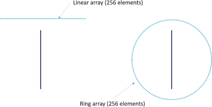
Fig. 21.33
-
11.
For the circular geometry, if the designed imaging FOV is 25 mm in diameter, to satisfy the spatial Nyquist sampling requirement, what is the minimum number of sampling channels for detection at a cutoff frequency of (a) 2.25 MHz and (b) 15 MHz?
-
12.
Under the same conditions in Q.10, please calculate the minimum number of sampling channels for the full spherical geometry.
-
13.
Derive Eq. (12.31), assuming the frequency response of the detector has a Gaussian profile.
-
14.
Assuming you are engineering a “perfect” PA contrast agent for molecular imaging, please list all the desired key characteristics and explain the reasons.

Rights and permissions
Copyright information
© 2020 Springer Nature Switzerland AG
About this chapter

Cite this chapter
Li, L., Yao, J., Wang, L.V. (2020). Photoacoustic Tomography of Neural Systems. In: He, B. (eds) Neural Engineering. Springer, Cham. https://doi.org/10.1007/978-3-030-43395-6_12
Download citation
DOI: https://doi.org/10.1007/978-3-030-43395-6_12
Published:
Publisher Name: Springer, Cham
Print ISBN: 978-3-030-43394-9
Online ISBN: 978-3-030-43395-6
eBook Packages: Biomedical and Life SciencesBiomedical and Life Sciences (R0)