
Abstract
Point-of-care transthoracic echocardiography (TTE) is a rapid, noninvasive diagnostic tool that can be indispensable for the management of symptomatic or hemodynamically unstable patients in any clinical environment. Image acquisition and interpretation can be challenging for some patients, but many important goal-directed clinical questions can be answered with a modest amount of training. Often, not every piece of information is available from every view, but a structure inaccessible from one view may be visible from another, allowing clinicians to guide their management. A basic exam may consist of a series of four views, including the parasternal long-axis, parasternal short-axis, apical four-chamber, and subxiphoid views. Additional views can augment this exam and may provide additional information. Point-of-care TTE findings may prompt a subsequent comprehensive exam, however, can display critical patient data in the interim providing a basis for initial decision making.
Access this chapter
Tax calculation will be finalised at checkout
Purchases are for personal use only
Abbreviations
- TTE:
-
Transthoracic echocardiography
- ASE:
-
American Society of Echocardiography
- ACEP:
-
American College of Emergency Physicians
- LLD:
-
Left lateral decubitus
- LAX:
-
Long-axis
- LA:
-
Left atrium
- LV:
-
Left ventricle
- LVOT:
-
Left ventricular outflow tract
- RVOT:
-
Right ventricular outflow tract
- TEE:
-
Transesophageal echocardiography
- ME:
-
Midesophageal
- RV:
-
Right ventricle
- SAX:
-
Short-axis
- TG:
-
Transgastric
- PMI:
-
Point of maximal impulse
- TAPSE:
-
Tricuspid annular plane systolic excursion
- PEEP:
-
Positive end-expiratory pressure
- IVC:
-
Inferior vena cava
References
Labovitz AJ, Noble VE, Bierig M, et al. Focused cardiac ultrasound in the emergent setting: a consensus statement of the American Society of Echocardiography and American College of Emergency Physicians. J Am Soc Echocardiogr. 2010;23:1225–30.
Bahner DP, Blickendorf JM, Bockbrader M, et al. Language of transducer manipulation: codifying terms for effective teaching. J Ultrasound Med. 2016;35:183–8.
Author information
Authors and Affiliations
Editor information
Editors and Affiliations
Electronic Supplementary Material
Parasternal LAX view (MP4 25880 kb)
Parasternal right ventricular inflow view (MP4 53440 kb)
Parasternal SAX view of the AV (MP4 26689 kb)
Parasternal SAX basal view of the LV (MP4 26689 kb)
Parasternal SAX mid-papillary view of the LV (MP4 27497 kb)
Parasternal SAX apical view of the LV (MP4 26689 kb)
Apical four-chamber view (MP4 127973 kb)
Apical five-chamber view (MP4 137817 kb)
Apical two-chamber view (MP4 133598 kb)
Apical long-axis (three-chamber) view (MP4 75941 kb)
Subcostal four-chamber view (MP4 147661 kb)
Suprasternal view of the aortic arch (MP4 1005 kb)
Questions
Questions
-
1.
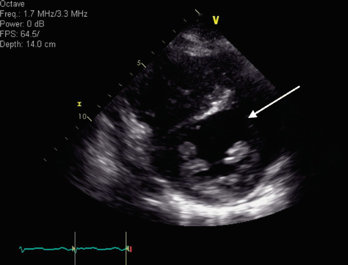
Which anatomical structure is indicated by the arrow in the image below?
-
a.
Left ventricular mid-anterior wall
-
b.
Left ventricular mid-inferior wall
-
c.
Left ventricular mid-anterolateral wall
-
d.
Left ventricular mid-inferolateral wall
-
a.
-
2.
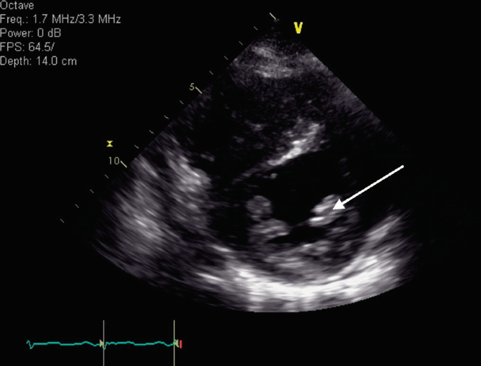
Which adjustment to the probe was made between the first and second image below?


-
a.
Tilting medially
-
b.
Fanning inferiorly
-
c.
Twisting clockwise
-
d.
Twisting counterclockwise
-
a.
-
3.
In which of the following views is the aortic valve visible?
-
a.
Apical long-axis view
-
b.
Apical four-chamber view
-
c.
Subcostal four-chamber view
-
d.
None of the above
-
a.
-
4.
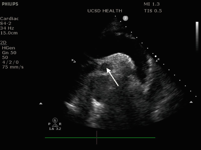
Which anatomical structure is indicated by the arrow in the image below?
-
a.
LV thrombus
-
b.
Posteromedial papillary muscle
-
c.
Inferolateral papillary muscle
-
d.
Anterolateral papillary muscle
-
a.
-
5.
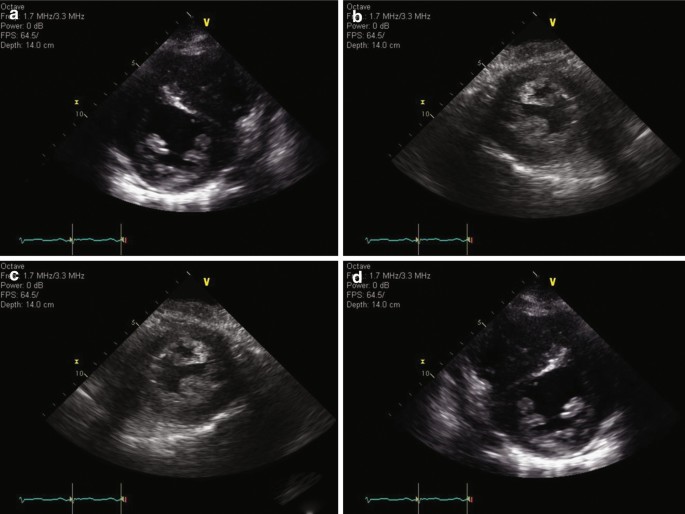
Which of the following images shows the correct orientation for a parasternal short-axis view?
-
a.
Image A
-
b.
Image B
-
c.
Image C
-
d.
Image D
-
a.
-
6.
Which of the following most accurately describes the approximate position for the probe footprint to obtain a parasternal long-axis view?
-
a.
2nd or 3rd intercostal space, to the left of the sternum, with probe marker oriented toward the patient’s right shoulder
-
b.
2nd or 3rd intercostal space, to the right of the sternum, with probe marker oriented toward the patient’s right shoulder
-
c.
2nd or 3rd intercostal space, to the left of the sternum, with probe marker oriented toward the patient’s left shoulder
-
d.
2nd or 3rd intercostal space, to the left of the sternum, with probe marker oriented toward the patient’s left shoulder
-
a.
-
7.
From the following image, which structure is indicated by the arrow?
-
a.
Main pulmonary artery
-
b.
Left pulmonary artery
-
c.
Right pulmonary artery
-
d.
Superior vena cava
-
a.
-
8.
Which of the following statement is most true regarding the differences between transesophageal and transthoracic echocardiography?
-
a.
Transthoracic echocardiography provides more accurate Doppler evaluation of the aortic valve
-
b.
Transthoracic echocardiography provides more accurate Doppler evaluation of the mitral valve
-
c.
Transthoracic echocardiography is not possible in mechanically ventilated patients
-
d.
Transthoracic echocardiography is unable to visualize the aortic arch
-
a.
-
9.
Which of the following is most likely to improve visualization of an apical four-chamber view?
-
a.
Having the patient hold in their breath
-
b.
Laying the patient supine
-
c.
Turning the patient to the left lateral decubitus position
-
d.
Raising the head of the bed
-
a.
-
10.
Which adjustment to the probe was made between the first and second image below?


-
a.
The probe footprint was moved anterior one rib space (swept)
-
b.
The probe was angled anteriorly (fanned)
-
c.
The probe was rotated clockwise
-
d.
The probe was rotated counterclockwise
-
a.


Rights and permissions
Copyright information
© 2022 The Author(s), under exclusive license to Springer Nature Switzerland AG
About this chapter

Cite this chapter
Tainter, C.R., Nhieu, S. (2022). Point-of-Care Transthoracic Echocardiography: Probe Manipulation, Positioning, and Essential Views. In: Maus, T.M., Tainter, C.R. (eds) Essential Echocardiography. Springer, Cham. https://doi.org/10.1007/978-3-030-84349-6_3
Download citation
DOI: https://doi.org/10.1007/978-3-030-84349-6_3
Published:
Publisher Name: Springer, Cham
Print ISBN: 978-3-030-84348-9
Online ISBN: 978-3-030-84349-6
eBook Packages: MedicineMedicine (R0)