
Abstract
Background
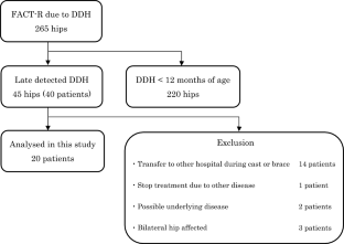
The relationship between hip morphological changes and joint concentricity in infants with late-detected developmental dysplasia of the hip (DDH) treated with gradual reduction remains unclear. Therefore, we investigated hip morphological changes and concentricity in infants with late-detected unilateral DDH using magnetic resonance imaging (MRI) during gradual reduction.
Methods
We enrolled 20 infants aged ≥ 12 months with unilateral DDH. Treatment comprised continuous traction, a hip-spica cast, and an abduction brace. MRI was performed before treatment, immediately after hip-spica cast placement, after cast removal, and at the end of the brace. We evaluated the acetabulum and femoral head morphology and joint concentricity.
Results
The mean age was 25 months, and female sex and the left side were predominant. Before treatment, the acetabulum was small and shallow and the femoral head was spherically flat on the affected side. Immediately after the continuous traction, the affected acetabulum and femoral head were still smaller than the healthy/contralateral one. However, they improved to a deeper acetabulum and round femoral head. Intra-articular soft tissue (IAST) and femoral–acetabular distance (FAD) continuously decreased, indicating gradual improvement in joint concentricity. Deeper formation of the acetabulum and round shaping of the femoral head had occurred even in non-concentric reduction.
Conclusion
The shape and concentricity of the hip joint improved after treatment; however, the acetabulum and femoral head remained small. The deeper acetabulum and round femoral head were observed the non-concentric reduction before the concentric reduction was achieved. The continuous decrease in IAST and FAD indicates effective post-traction therapy.


Data Availability
The datasets analyzed in this study are available from the corresponding author on reasonable request.
Code Availability
Not applicable.
References
Vaquero-Picado, A., González-Morán, G., Garay, E. G., & Moraleda, L. (2019). Developmental dysplasia of the hip: Update of management. EFORT Open Reviews, 2019(4), 548–556.
Studer, K., Williams, N., Studer, P., Baker, M., Glynn, A., Foster, B. K., & Cundy, P. J. (2017). Obstacles to reduction in infantile developmental dysplasia of the hip. Journal of Children’s Orthopaedics, 2017(11), 358–366.
Glorion, C. (2018). Surgical reduction of congenital hip dislocation. Orthopaedics and Traumatology, Surgery and Research, 2018(104), S147–S157.
Meng, X., Yang, J., & Wang, Z. (2021). Magnetic resonance imaging follow-up can screen for soft tissue changes and evaluate the short-term prognosis of patients with developmental dysplasia of the hip after closed reduction. BMC Pediatrics, 21(115), 2021.
Rosenbaum, D. G., Servaes, S., Bogner, E. A., Jaramillo, D., & Mintz, D. N. (2016). MR imaging in postreduction assessment of developmental dysplasia of the hip: Goals and obstacles. Radiographics, 2016(36), 840–854.
Kaneko, H., Kitoh, H., Kitamura, A., Sawamura, K., & Hattori, T. (2021). Docking phenomenon and subsequent acetabular development after gradual reduction using overhead traction for developmental dysplasia of the hip over six months of age. Journal of Children’s Orthopaedics, 2021(15), 554–563.
Talathi, N. S., Chauvin, N. A., & Sankar, W. N. (2018). Docking of the femoral head following closed reduction for DDH: Does it really occur? Journal of Pediatric Orthopedics, 2018(38), e440–e445.
Fukiage, K., Fukuda, A., Harada, Y., Suzuki, S., & Futami, T. (2015). Femoral head volume indicates the severity of developmental dysplasia of the hip by a method using three-dimensional magnetic resonance imaging. Journal of Pediatric Orthopedics, Part B, 2015(24), 286–290.
Sankar, W. N., Neubuerger, C. O., & Moseley, C. F. (2010). Femoral head sphericity in untreated developmental dislocation of the hip. Journal of Pediatric Orthopedics, 2010(30), 558–561.
Rosenberg, M. R., Walton, R., Rae, E. A., Bailey, S., & Nicol, R. O. (2017). Intra-articular dysplasia of the femoral head in developmental dysplasia of the hip. Journal of Pediatric Orthopedics, Part B, 2017(26), 298–302.
Crowe, J. F., Mani, V. J., & Ranawat, C. S. (1979). Total hip replacement in congenital dislocation and dysplasia of the hip. Journal of Bone and Joint Surgery, American Volume, 1979(61), 15–23.
Okuzu, Y., Goto, K., Kawata, T., So, K., Kuroda, Y., & Matsuda, S. (2017). The relationship between subluxation percentage of the femoroacetabular joint and acetabular width in Asian women with developmental dysplasia of the hip. Journal of Bone and Joint Surgery, American Volume, 99(e31), 2017.
Fukiage, K., Futami, T., Ogi, Y., Harada, Y., Shimozono, F., Kashiwagi, N., Takase, T., & Suzuki, S. (2015). Ultrasound-guided gradual reduction using flexion and abduction continuous traction for developmental dysplasia of the hip: A new method of treatment. The Bone and Joint Journal, 2015(97-B), 405–411.
Suzuki, S., & Yamamuro, T. (1990). Avascular necrosis in patients treated with the Pavlik harness for congenital dislocation of the hip. Journal of Bone and Joint Surgery, American Volume, 72(1048), 1990.
Yamamuro, T., Chene, S. H., & Ito, T. (1975). A radiological study on the developmental of the hip joint in normal infants. The Japanese Orthopaedic Association, 49, 421–439 (in Japanese).
Home—International Hip Dysplasia Institute. https://hipdysplasia.org/
Steppacher, S. D., Tannast, M., Werlen, S., & Siebenrock, K. A. (2008). Femoral morphology differs between deficient and excessive acetabular coverage. Clinical Orthopaedics and Related Research, 466(782), 2008.
Zhou, W., Sankar, W. N., Zhang, F., Li, L., Zhang, L., & Zhao, Q. (2020). Evolution of concentricity after closed reduction in developmental dysplasia of the hip. The Bone and Joint Journal, 102-B, 618.
Okano, K., Yamaguchi, K., Ninomiya, Y., Matsubayashi, S., Osaki, M., & Takahashi, K. (2013). Femoral head deformity and severity of acetabular dysplasia of the hip. The Bone and Joint Journal, 95-B, 1192.
Clohisy, J. C., Nunley, R. M., Carlisle, J. C., & Schoenecker, P. L. (2009). Incidence and characteristics of femoral deformities in the dysplastic hip. Clinical Orthopaedics and Related Research, 2009(467), 128–134.
Chen, C., Doyle, S., Green, D., Blanco, J., Scher, D., Sink, E., & Dodwell, E. R. (2017). Presence of the Ossific nucleus and risk of osteonecrosis in the treatment of developmental dysplasia of the hip: A meta-analysis of cohort and case–control studies. Journal of Bone and Joint Surgery, American Volume, 2017(99), 760–767.
Gardner, R. O., Bradley, C. S., Howard, A., Narayanan, U. G., Wedge, J. H., & Kelley, S. P. (2014). The incidence of avascular necrosis and the radiographic outcome following medial open reduction in children with developmental dysplasia of the hip: A systematic review. The Bone and Joint Journal, 2014(96-B), 279–286.
Ogden, J. A. (1975). Treatment positions for congenital dysplasia of the hip. Journal of Pediatrics, 1975(86), 732–734.
Slullitel, P. A., Coutu, D., Buttaro, M. A., Beaule, P. E., & Grammatopoulos, G. (2020). Hip preservation surgery and the acetabular fossa. Bone and Joint Research, 2020(9), 857–869.
Funding
None.
Author information
Authors and Affiliations
Contributions
YO, MT, TF were involved in conceptualization, data curation, formal analysis, investigation, and methodology. KG, YK, TK, YM, and SM were involved in project administration, supervision, validation. YO and MT were major contributors in the writing the original draft. KG, YK, TK, YM, TF, and SM reviewed and edited the original draft. All authors read and approved the final manuscript.
Corresponding author
Ethics declarations
Conflict of interest
The authors declare that they have no relevant conflicts of interest.
Ethical Standards
This article does not contain any studies with human or animal subjects performed by the any of the authors.
Informed Consent in Studies with Human Subjects
This retrospective study was approved by the local institutional review board and conducted in accordance with the World Medical Association Declaration of Helsinki. Written informed consent was waived in this study.
Additional information
Publisher's Note
Springer Nature remains neutral with regard to jurisdictional claims in published maps and institutional affiliations.
Rights and permissions
Springer Nature or its licensor (e.g. a society or other partner) holds exclusive rights to this article under a publishing agreement with the author(s) or other rightsholder(s); author self-archiving of the accepted manuscript version of this article is solely governed by the terms of such publishing agreement and applicable law.
About this article

Cite this article
Okuzu, Y., Tsukanaka, M., Goto, K. et al. Morphological Changes and Concentricity of the Hip Joint During Gradual Reduction in Infants with Late-Detected Developmental Dysplasia of the Hip: A Retrospective Study. JOIO (2024). https://doi.org/10.1007/s43465-024-01184-6
Received:
Accepted:
Published:
DOI: https://doi.org/10.1007/s43465-024-01184-6