Abstract
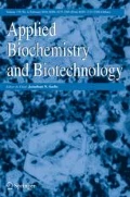
This approach was constructed to appraise the therapeutic effectiveness of a single i.v. dose of osteoblasts generated from co-culturing BM-MSCs with nano-HA, Pt-NPs, or Pt-HA-nanocomposite in osteoporotic rats. MSCs were grown, propagated in culture, and characterized. The effect of the suggested nanoplatforms on the survival, osteogenic differentiation, and mineralization of BM-MSCs was assessed by MTT assay, real-time PCR analysis, and Alizarin red S staining, respectively. Thereafter, the generated osteoblasts were employed for the treatment of ovariectomized rats. Our results revealed that the selected nanoplatforms upregulate the expression of osteogenic differentiation related genes (Runx-2 and BMP-2) significantly and enhance calcium deposition in BM-MSCs after 7 and 21 days, respectively, whereas the in vivo study validated that the infusion of the generated osteoblasts considerably downturn serum BALP, BSP, and SOST levels; upswing OSX level; and regain femur bone mineralization and histoarchitecture. Conclusively, the outcomes of this work provide scientific evidence that transplanting osteoblasts derived from differentiation of BM-MSCs in the presence of nanoplatforms in ovariectomized rats restores bone remodeling balance which constitutes a new hope for the treatment of osteoporosis.


Abbreviations
- BALP:
-
Bone alkaline phosphatase
- BM-MSCs:
-
Bone marrow derived mesenchymal stem cells
- BMP-2:
-
Bone morphogenetic protein 2
- BSP:
-
Bone sialoprotein
- Nano-HA:
-
Nanohydroxyapatite
- OSX:
-
Osterix
- Pt-NPs:
-
Platinum nanoparticles
- Pt-HA-nanocomposite:
-
Platinum-hydroxyapatite-nanocomposite
- Runx-2:
-
Runt-related transcription factor 2
- SOST:
-
Sclerostin
References
Aghebati-Maleki, L., Dolati, S., Zandi, R., Fotouhi, A., Ahmadi, M., Aghebati, A., Nouri, M., Kazem Shakouri, S., & Yousefi, M. (2018). Prospect of mesenchymal stem cells in therapy of osteoporosis: A review. Journal of Cellular Physiology, 234(6), 8570–8578. https://doi.org/10.1002/jcp.27833.
Facts and Statistics | International Osteoporosis Foundation. (n.d.). Retrieved March 19, 2019, from https://www.iofbonehealth.org/facts-statistics
Korpi-Steiner, N., Milhorn, D., & Hammett-Stabler, C. (2014). Osteoporosis in men. Clinical Biochemistry, 47(10–11), 950–959. https://doi.org/10.1016/j.clinbiochem.2014.03.026.
El-Said Hossien, Y. (2014). Osteoporosis knowledge among female adolescents in Egypt. American Journal of Nursing Science, 3(2), 13. https://doi.org/10.11648/j.ajns.20140302.11.
Lin, X., Xiong, D., Peng, Y. Q., Sheng, Z. F., Wu, X. Y., Wu, X. P., et al. (2015). Epidemiology and management of osteoporosis in the people’s republic of China: Current perspectives. Clinical Interventions in Aging, 10, 1017–1033. https://doi.org/10.2147/CIA.S54613.
Knopp-Sihota, J. A., Cummings, G. G., Homik, J., & Voaklander, D. (2013). The association between serious upper gastrointestinal bleeding and incident bisphosphonate use: A population-based nested cohort study. BMC Geriatrics, 13(1), 110–120. https://doi.org/10.1186/1471-2318-13-36.
Zhang, Y., Luo, H., Zhang, Z., Lu, Y., Huang, X., Yang, L., Xu, J., Yang, W., Fan, X., du, B., Gao, P., Hu, G., & Jin, Y. (2010). A nerve graft constructed with xenogeneic acellular nerve matrix and autologous adipose-derived mesenchymal stem cells. Biomaterials, 31(20), 5312–5324. https://doi.org/10.1016/j.biomaterials.2010.03.029.
Hu, L., Yin, C., Zhao, F., Ali, A., Ma, J., & Qian, A. (2018). Mesenchymal stem cells: Cell fate decision to osteoblast or adipocyte and application in osteoporosis treatment. International Journal of Molecular Sciences, 19(2), 360. https://doi.org/10.3390/ijms19020360.
Ichioka, N., Inaba, M., Kushida, T., Esumi, T., Takahara, K., Inaba, K., Ogawa, R., Iida, H., & Ikehara, S. (2002). Prevention of senile osteoporosis in SAMP6 mice by intrabone marrow injection of allogeneic bone marrow cells. Stem Cells, 20(6), 542–551. https://doi.org/10.1634/stemcells.20-6-542.
Wang, Z., Goh, J., De, S. D., Ge, Z., Ouyang, H., Sue, J., et al. (2006). Efficacy of bone marrow–derived stem cells in strengthening osteoporotic bone in a rabbit model. Tissue Engineering, 12(7), 1753–1761. https://doi.org/10.1089/ten.2006.12.1753.
Gutwald, R., Haberstroh, J., Kuschnierz, J., Kister, C., Lysek, D. A., Maglione, M., Xavier, S. P., Oshima, T., Schmelzeisen, R., & Sauerbier, S. (2010). Mesenchymal stem cells and inorganic bovine bone mineral in sinus augmentation: Comparison with augmentation by autologous bone in adult sheep. British Journal of Oral and Maxillofacial Surgery, 48(4), 285–290. https://doi.org/10.1016/j.bjoms.2009.06.226.
Granero-Moltó, F., Weis, J. A., Miga, M. I., Landis, B., Myers, T. J., O’Rear, L., et al. (2009). Regenerative effects of transplanted mesenchymal stem cells in fracture healing. Stem Cells, 27(8), 1887–1898. https://doi.org/10.1002/stem.103.
Tautzenberger, A., Kovtun, A., & Ignatius, A. (2012). Nanoparticles and their potential for application in bone. International Journal of Nanomedicine, 7, 4545–4557. https://doi.org/10.2147/IJN.S34127.
Roohani-Esfahani, S. I., Nouri-Khorasani, S., Lu, Z., Appleyard, R., & Zreiqat, H. (2010). The influence hydroxyapatite nanoparticle shape and size on the properties of biphasic calcium phosphate scaffolds coated with hydroxyapatite-PCL composites. Biomaterials, 31(21), 5498–5509. https://doi.org/10.1016/j.biomaterials.2010.03.058.
Yin, Z., Chen, X., Chen, J. L., Shen, W. L., Hieu Nguyen, T. M., Gao, L., & Ouyang, H. W. (2010). The regulation of tendon stem cell differentiation by the alignment of nanofibers. Biomaterials, 31(8), 2163–2175. https://doi.org/10.1016/j.biomaterials.2009.11.083.
Oh, S., Brammer, K. S., Li, Y. S. J., Teng, D., Engler, A. J., Chien, S., et al. (2009). Stem cell fate dictated solely by altered nanotube dimension. P Natl Acad Sci USA, 160(24), 60.
Amna, T. (2018). Valorization of bone waste of Saudi Arabia by synthesizing hydroxyapatite. Applied Biochemistry and Biotechnology, 186(3), 779–778. https://doi.org/10.1007/s12010-018-2768-5.
Heo, S.-J., Kim, S.-E., Wei, J., Kim, D. H., Hyun, Y.-T., Yun, H.-S., Kim, H. K., Yoon, T. R., Kim, S. H., Park, S. A., Shin, J. W., & Shin, J.-W. (2009). In vitro and animal study of novel nano-hydroxyapatite/poly(epsilon-caprolactone) composite scaffolds fabricated by layer manufacturing process. Tissue engineering. Part A, 15(5), 977–989. https://doi.org/10.1089/ten.tea.2008.0190.
Porcel, E., Liehn, S., Remita, H., Usami, N., Kobayashi, K., Furusawa, Y., Sech, C. L., & Lacombe, S. (2010). Platinum nanoparticles: A promising material for future cancer therapy? Nanotechnology, 21, 085103. https://doi.org/10.1088/0957-4484/21/8/085103.
Park, E. J., Kim, H., Kim, Y., Yi, J., Choi, K., & Park, K. (2010). Inflammatory responses may be induced by a single intratracheal instillation of iron nanoparticles in mice. Toxicology, 275(1–3), 65–71. https://doi.org/10.1016/j.tox.2010.06.002.
Yoshihisa, Y., Honda, A., Zhao, Q. L., Makino, T., Abe, R., Matsui, K., Shimizu, H., Miyamoto, Y., Kondo, T., & Shimizu, T. (2010). Protective effects of platinum nanoparticles against UV-light-induced epidermal inflammation. Experimental Dermatology, 19(11), 100–106. https://doi.org/10.1111/j.1600-0625.2010.01128.x.
Yubao, L., de Groot, K., de Wijn, J., Klein, C. P. A. T., & Meer, S. V. D. (1994). Morphology and composition of nanograde calcium phosphate needle-like crystals formed by simple hydrothermal treatment. J Mater Sci Mater Med, 5, 326–331. https://doi.org/10.1007/BF00058956.
Alhadlaq, A., & Mao, J. J. (2004). Mesenchymal stem cells: Isolation and therapeutics. Stem Cells and Development, 13(4), 436–448. https://doi.org/10.1089/1547328041797552.
Woodbury, D., Schwarz, E. J., Prockop, D. J., & Black, I. B. (2000). Adult rat and human bone marrow stromal cells differentiate into neurons. Journal of Neuroscience Research, 61(4), 364–370. https://doi.org/10.1002/1097-4547(20000815)61:4<364::AID-JNR2>3.0.CO;2-C.
Van Meerloo, J., Kaspers, G. J. L., & Cloos, J. (2011). Cell sensitivity assays: The MTT assay. Methods in Molecular Biology, 237–245, 237–245. https://doi.org/10.1007/978-1-61779-80-5_20.
Yi, C., Liu, D., Fong, C. C., Zhang, J., & Yang, M. (2010). Gold nanoparticles promote osteogenic differentiation of mesenchymal stem cells through p38 MAPK pathway. ACS Nano, 4(11), 6439–6448. https://doi.org/10.1021/nn101373r.
Gori, F., Divieti, P., & Demay, M. B. (2001). Cloning and characterization of a novel WD-40 repeat protein that dramatically accelerates osteoblastic differentiation. Journal of Biological Chemistry, 26(49), 46515–46522. https://doi.org/10.1074/jbc.M105757200.
Huang, G. T.-J., Gronthos, S., & Shi, S. (2009). Mesenchymal stem cells derived from dental tissues vs. those from other sources: Their biology and role in regenerative medicine. Journal of Dental Research, 88(9), 792–806. https://doi.org/10.1177/0022034509340867.
Power, R. A., Iwaniec, U. T., Magee, K. A., Mitova-Caneva, N. G., & Wronski, T. J. (2004). Basic fibroblast growth factor has rapid bone anabolic effects in ovariectomized rats. Osteoporosis International, 15(9), 716–723. https://doi.org/10.1007/s00198-004-1595-4.
Ahmed, H. H., El-Sayed Mahdy, E. S. M., Shousha, W. G., Rashed, L. A., & Abdo, S. M. (2013). Potential role of bone marrow derived mesenchymal stem cells with or without injectable calcium phosphate composite in management of osteoporosis in rat model. International Journal of Pharmacy and Pharmaceutical Sciences, 5(SUPPL 3), 494–504.
Griffin, M. G., Kimble, R., Hopfer, W., & Pacifici, R. (2009). Dual-energy x-ray absorptiometry of the rat: Accuracy, precision, and measurement of bone loss. Journal of Bone and Mineral Research, 8(7), 795–800. https://doi.org/10.1002/jbmr.5650080704.
Carleton, H. M. (Harry M., Drury, R. A. B. (Roger A. B., & Wallington, E. A. (1980). Carleton’s Histological technique. Oxford University Press Oxford.
Chandrasekar, A., Sagadevan, S., & Dakshnamoorthy, A. (2013). Synthesis and characterization of nano-hydroxyapatite (n-HAP) using the wet chemical technique. International Journal of Physical Sciences, 8(32), 1639–1645. https://doi.org/10.5897/IJPS2013.3990.
Zhang, C. Y., Chen, J., Zhuang, Z., Zhang, T., Wang, X. P., & Fang, Q. F. (2012). In situ hybridization and characterization of fibrous hydroxyapatite/chitosan nanocomposite. Journal of Applied Polymer Science, 124, 397–402. https://doi.org/10.1002/app.35103.
Rajathi, F. A. A., & Nambaru, V. R. M. S. (2014). Phytofabrication of nano-crystalline platinum particles by leaves of Cerbera manghas and its antibacterial efficacy. International Journal of Pharma and BioSciences, 5(1), 619–628.
Rajasekharreddy, P., & Rani, P. U. (2014). Biosynthesis and characterization of Pd and Pt nanoparticles using Piper betle L. Plant in a Photoreduction Method. Journal of Cluster Science, 25, 1377–1388. https://doi.org/10.1007/s10876-014-0715-3.
Oreffo, R. O. C., Cooper, C., Mason, C., & Clements, M. (2005). Mesenchymal stem cells lineage, plasticity, and skeletal therapeutic potential. Stem Cell Reviews, 1(2), 169–178. https://doi.org/10.1385/SCR:1:2:169.
Kaur, G., Valarmathi, M. T., Potts, J. D., Jabbari, E., Sabo-Attwood, T., & Wang, Q. (2010). Regulation of osteogenic differentiation of rat bone marrow stromal cells on 2D nanorod substrates. Biomaterials, 31(7), 1732–1741. https://doi.org/10.1016/j.biomaterials.2009.11.041.
White, M. A., & Anderson, R. G. W. (2005). Signaling networks in living cells. Annual Review of Pharmacology and Toxicology, 45(1), 587–603. https://doi.org/10.1146/annurev.pharmtox.45.120403.095807.
Liu, D., Yi, C., Zhang, D., Zhang, J., & Yang, M. (2010). Inhibition of proliferation and differentiation of mesenchymal stem cells by carboxylated carbon nanotubes. ACS Nano, 4(4), 2185–2195. https://doi.org/10.1021/nn901479w.
Porter, A. E., Gass, M., Bendall, J. S., Muller, K., Goode, A., Skepper, J. N., Midgley, P. A., & Welland, M. (2009). Uptake of noncytotoxic acid-treated single-walled carbon nanotubes into the cytoplasm of human macrophage cells. ACS Nano, 3(6), 1485–1492. https://doi.org/10.1021/nn900416z.
Remya, N. S. S., Syama, S., Gayathri, V., Varma, H. K. K., & Mohanan, P. V. V. (2014). An in vitro study on the interaction of hydroxyapatite nanoparticles and bone marrow mesenchymal stem cells for assessing the toxicological behaviour. Colloids and Surfaces B: Biointerfaces, 117, 389–397. https://doi.org/10.1016/j.colsurfb.2014.02.004.
Dong, P., Zhu, D., Deng, X., Zhang, Y., Ma, J., Sun, X., & Liu, Y. (2019). Effect of hydroxyapatite nanoparticles and wedelolactone on osteoblastogenesis from bone marrow mesenchymal stem cells. Journal of biomedical materials research. Part A, 107(1), 145–153. https://doi.org/10.1002/jbm.a.36541.
Rostek, A., Breisch, M., Pappert, K., Loza, K., Heggen, M., Köller, M., Sengstock, C., & Epple, M. (2018). Comparative biological effects of spherical noble metal nanoparticles (Rh, Pd, Ag, Pt, Au) with 4-8 nm diameter. Beilstein Journal of Nanotechnology, 9, 2763–2774. https://doi.org/10.3762/bjnano.9.258.
Komori, T., Kot, M., & Daniel, W. A. (2003). Requisite roles of Runx2 and Cbfb in skeletal development. Journal of Bone and Mineral Metabolism, 21(3), 193–197. https://doi.org/10.1007/s00774-002-0408-0.
Benoit, D. S. W., Collins, S. D., & Anseth, K. S. (2007). Multifunctional hydrogels that promote osteogenic hMSC differentiation through stimulation and sequestering of BMP2. Advanced Functional Materials, 17(13), 2085–2093. https://doi.org/10.1002/adfm.200700012.
Santos, C., Gomes, P. S., Duarte, J. A., Franke, R. P., Almeida, M. M., Costa, M. E. V., & Fernandes, M. H. (2012). Relevance of the sterilization-induced effects on the properties of different hydroxyapatite nanoparticles and assessment of the osteoblastic cell response. Journal of the Royal Society Interface, 9(77), 3397–3410. https://doi.org/10.1098/rsif.2012.0487.
Kim, K., Dean, D., Lu, A., Mikos, A. G., & Fisher, J. P. (2011). Early osteogenic signal expression of rat bone marrow stromal cells is influenced by both hydroxyapatite nanoparticle content and initial cell seeding density in biodegradable nanocomposite scaffolds. Acta Biomaterialia, 7(3), 1249–1264. https://doi.org/10.1016/j.actbio.2010.11.007.
Peng, H., Yin, Z., Liu, H., Chen, X., Feng, B., Yuan, H., Su, B., Ouyang, H., & Zhang, Y. (2012). Electrospun biomimetic scaffold of hydroxyapatite/chitosan supports enhanced osteogenic differentiation of mMSCs. Nanotechnology, 23(48), 485102. https://doi.org/10.1088/0957-4484/23/48/485102.
Zhang, Q., Nguyen, A. L., Shi, S., Hill, C., Wilder-Smith, P., Krasieva, T. B., & Le, A. D. (2012). Three-dimensional spheroid culture of human gingiva-derived mesenchymal stem cells enhances mitigation of chemotherapy-induced oral mucositis. Stem Cells and Development, 21(6), 937–947. https://doi.org/10.1089/scd.2011.0252.
Lu, Z., Roohani-Esfahani, S.-I., Kwok, P. C. L., & Zreiqat, H. (2011). Osteoblasts on rod shaped hydroxyapatite nanoparticles incorporated PCL film provide an optimal osteogenic niche for stem cell differentiation. Tissue Engineering Part A, 17(11–12), 1651–1661. https://doi.org/10.1089/ten.tea.2010.0567.
Pockwinse, S. M., Wilming, L. G., Conlon, D. M., Stein, G. S., & Lian, J. B. (1992). Expression of cell growth and bone specific genes at single cell resolution during development of bone tissue-like organization in primary osteoblast cultures. Journal of Cellular Biochemistry, 49(3), 310–323. https://doi.org/10.1002/jcb.240490315.
STEIN, G. S., & LIAN, J. B. (1993). Molecular mechanisms mediating proliferation/differentiation interrelationships during progressive development of the osteoblast phenotype. Endocrine Reviews, 14(4), 424–442. https://doi.org/10.1210/edrv-14-4-424.
Thompson, D. D., Simmons, H. A., Pirie, C. M., & Ke, H. Z. (1995). FDA guidelines and animal models for osteoporosis. Bone., 17(4), S125–S133. https://doi.org/10.1016/8756-3282(95)00285-L.
Nikolaou, V. S., Efstathopoulos, N., Kontakis, G., Kanakaris, N. K., & Giannoudis, P. V. (2009). The influence of osteoporosis in femoral fracture healing time. Injury, 40(6), 663–668. https://doi.org/10.1016/j.injury.2008.10.035.
Abuohashish, H. M., Ahmed, M. M., Al-Rejaie, S. S., & Eltahir, K. E. (2015). The antidepressant bupropion exerts alleviating properties in an ovariectomized osteoporotic rat model. Acta Pharmacologica Sinica, 36(2), 209–220. https://doi.org/10.1038/aps.2014.111.
Katagiri, T., & Takahashi, N. (2002). Regulatory mechanisms of osteoblast and osteoclast differentiation. Oral Diseases, 8(3), 147–159. https://doi.org/10.1034/j.1601-0825.2002.01829.x.
Sims, N. A., & Martin, T. J. (2014). Coupling the activities of bone formation and resorption: A multitude of signals within the basic multicellular unit. BoneKEy Reports, 3, 1–10. https://doi.org/10.1038/bonekey.2013.215.
Saraç, F., & Saygılı, F. (2007). Causes of high bone alkaline phosphatase. Biotechnology and Biotechnological Equipment, 18(5), 444–448. https://doi.org/10.1080/13102818.2007.10817444.
Jagtap, V. R., Ganu, J. V., & Nagane, N. S. (2011). BMD and serum intact osteocalcin in postmenopausal osteoporosis women. Indian Journal of Clinical Biochemistry, 26(1), 70–73. https://doi.org/10.1007/s12291-010-0074-2.
Chan, B. Y., Fuller, E. S., Russell, A. K., Smith, S. M., Smith, M. M., Jackson, M. T., Cake, M. A., Read, R. A., Bateman, J. F., Sambrook, P. N., & Little, C. B. (2011). Increased chondrocyte sclerostin may protect against cartilage degradation in osteoarthritis. Osteoarthritis and Cartilage, 19(7), 874–885. https://doi.org/10.1016/j.joca.2011.04.014.
Fisher, L., & Fedarko, N. (2007). Six genes expressed in bones and teeth encode the current members of the SIBLING family of proteins. Connective Tissue Research, 44(1), 33–40. https://doi.org/10.1080/713713644.
Ogbureke, K. U. E., & Fisher, L. W. (2005). Renal expression of SIBLING proteins and their partner matrix metalloproteinases (MMPs). Kidney International, 292(3), 905–911. https://doi.org/10.1111/j.1523-1755.2005.00389.x.
Ganss, B., Kim, R. H., & Sodek, J. (1999). Bone sialoprotein. Critical Reviews in Oral Biology and Medicine, 10(1), 79–98.
Lei, Z., Xiaoying, Z., & Xingguo, L. (2009). Ovariectomy-associated changes in bone mineral density and bone marrow haematopoiesis in rats. International Journal of Experimental Pathology, 90(5), 512–519. https://doi.org/10.1111/j.1365-2613.2009.00661.x.
Cenci, S., Weitzmann, M. N., Gentile, M. A., Aisa, M. C., & Pacifici, R. (2000). M-CSF neutralization and Egr-1 deficiency prevent ovariectomy-induced bone loss. Journal of Clinical Investigation, 105(9), 1279–1287. https://doi.org/10.1172/JCI8672.
Nian, H., Ma, M. H., Nian, S. S., & Xu, L. L. (2009). Antiosteoporotic activity of icariin in ovariectomized rats. Phytomedicine, 16(4), 320–326. https://doi.org/10.1016/j.phymed.2008.12.006.
Ferretti, M., Bertoni, L., Cavani, F., Zavatti, M., Resca, E., Carnevale, G., Benelli, A., Zanoli, P., & Palumbo, C. (2010). Influence of ferutinin on bone metabolism in ovariectomized rats. II: Role in recovering osteoporosis. Journal of Anatomy, 2017(1), 48–56. https://doi.org/10.1111/j.1469-7580.2010.01242.x.
Cardinali, D. P., Ladizesky, M. G., Boggio, V., Cutrera, R. A., & Mautalen, C. (2003). Melatonin effects on bone: Experimental facts and clinical perspectives. Journal of Pineal Research, 34(2), 81–87. https://doi.org/10.1034/j.1600-079X.2003.00028.x.
Kotlarczyk, M. P., Lassila, H. C., O’Neil, C. K., D’Amico, F., Enderby, L. T., Witt-Enderby, P. A., & Balk, J. L. (2012). Melatonin osteoporosis prevention study (MOPS): A randomized, double-blind, placebo-controlled study examining the effects of melatonin on bone health and quality of life in perimenopausal women. Journal of Pineal Research, 52(4), 414–426. https://doi.org/10.1111/j.1600-079X.2011.00956.x.
Kim, S. J., Jang, J. D., & Lee, S. K. (2007). Treatment of long tubular bone defect of rabbit using autologous cultured osteoblasts mixed with fibrin. Cytotechnology, 54(2), 115–120. https://doi.org/10.1007/s10616-007-9084-1.
Okabe, Y. T., Kondo, T., Mishima, K., Hayase, Y., Kato, K., Mizuno, M., Ishiguro, N., & Kitoh, H. (2014). Biodistribution of locally or systemically transplanted osteoblast-like cells. Bone & Joint Research, 3(3), 76–81. https://doi.org/10.1302/2046-3758.33.2000257.
Liao, Y. J., Tang, P. C., Chen, Y. H., Chu, F. H., Kang, T. C., Chen, L. R., & Yang, J. R. (2018). Porcine induced pluripotent stem cell-derived osteoblast-like cells prevent glucocorticoid-induced bone loss in Lanyu pigs. PLoS One, 13(8), e0202155. https://doi.org/10.1371/journal.pone.0202155.
Acknowledgments
The authors gratefully acknowledge the financial support of the National Research Centre, Egypt (Grant no. P100528) and the technical support of the Science and Technology Development Fund (STDF), Egypt, through Capacity Building program (Grant no. 4880). Also, the authors express sincere appreciation to Prof. Adel Bakeer kholoussy, Professor of Pathology, Faculty of Veterinary Medicine, Cairo University for his kind cooperation in conducting histological examination in this study.
Funding
This work was financially supported by the National Research Centre, Egypt (grant no. P100528).
Author information
Authors and Affiliations
Corresponding author
Ethics declarations
This study was conducted in strict accordance with the recommendations of the guidelines for the care and use of laboratory animals of the National Research Centre, Egypt. The experimental procedures were endorsed by the Ethical Committee of Medical Research of National Research Centre, Giza, Egypt (Admittance code 15 - 154).
Conflict of Interest
The authors declare that they have no conflict of interest.
Additional information
Publisher’s Note
Springer Nature remains neutral with regard to jurisdictional claims in published maps and institutional affiliations.
Rights and permissions
About this article

Cite this article
Aglan, H.A., Ahmed, H.H., Mahmoud, N.S. et al. Nanotechnological Applications Hold a Pivotal Position in Boosting Stem Cells Osteogenic Activity: In Vitro and In Vivo Studies. Appl Biochem Biotechnol 190, 551–573 (2020). https://doi.org/10.1007/s12010-019-03105-y
Received:
Accepted:
Published:
Issue Date:
DOI: https://doi.org/10.1007/s12010-019-03105-y