Abstract
Purpose
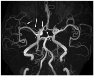
To describe a case of duplicated middle cerebral artery (MCA) combined with ipsilateral accessory MCA, forming a triplicated MCA, associated with the accessory anterior cerebral artery (ACA), forming a triplicated A2 segment of the ACA detected incidentally on magnetic resonance (MR) angiography.
Methods
A 70-year-old woman with internal carotid artery (ICA) stenosis at the origin, which was detected by ultrasound, underwent cranial MR imaging and MR angiography of the intracranial region for an evaluation of brain and cerebral arterial lesions. The MR machine was a 3-Tesla scanner. MR angiography was performed using a standard 3-dimensional time-of-flight technique.
Results
Multiple ischemic white matter lesions are observed. No significant stenotic lesions were observed in intracranial arteries. The right duplicated MCA was originated from right distal ICA. And main MCA was originated from right ICA bifurcation. Right accessory MCA was arisen from the A2 segment of the right ACA. Thus, the right MCA was triplicated. There was also an accessory ACA forming a triplicated ACA at its A2 segment. These findings were clearly identified on partial volume-rendering (VR) images.
Conclusion
We herein report a case of triplicated MCA associated with triplicated ACA. MCA variations are relatively rare, and this is the third case of triplicated MCA reported in relevant English-language literature. To identify multiple cerebral arterial variations, creating partial VR images using MR angiographic source images is useful.


Data availability
No datasets were generated or analysed during the current study.
References
Alnafie MA (2023) Unreported branching pattern of the triplicated anterior cerebral artery associated with multiple bilateral variants of the superior cerebellar artery detected by computed tomography angiography and 3D modeling. Surg Radiol Anat 45:1263–1267. https://doi.org/10.1007/s00276-023-03213-9
Kashtiara A, Beldé S, Schollaert J, Menovsky T (2024) Anatomical variations and anomalies of the middle cerebral artery. World Neurosurg 183:e187–e200. https://doi.org/10.1016/j.wneu.2023.12.052
Kobari M, Ishihara N, Yunoki K, Togashi O, Sato S (1988) Triplication of the middle cerebral artery associated with fenestration of the anterior cerebral artery. Keio J Med 37:429–433. https://doi.org/10.2302/kjm.37.429
Komiyama M, Nakajima H, Nishikawa M, Yasui T (1998) Middle cerebral artery variations: duplicated and accessory arteries. AJNR Am J Neuroradiol 19:45–49
Ota T, Komiyama M (2021) Twig-like middle cerebral artery: embryological persistence or secondary consequences? Interv Neuroradiol 27:584–587. https://doi.org/10.1177/15910199211024077
Otani N, Nawashiro H, Tsuzuki N, Osada H, Suzuki T, Shima K, Nakai K (2010) A ruptured internal carotid artery aneurysm located at the origin of the duplicated middle cerebral artery associated with accessory middle cerebral artery and middle cerebral artery aplasia. Surg Neurol Int 1:51. https://doi.org/10.4103/2152-7806.69378
Teal JS, Rumbaugh CL, Bergeron RT, Segall HD (1973) Anomalies of the middle cerebral artery: accessory artery, duplication, and early bifurcation. Am J Roentgenol Radium Ther Nucl Med 118:567–575. https://doi.org/10.2214/ajr.118.3.567
Uchino A, Nakadate M (2022) Multiple cerebral arterial variations incidentally detected by magnetic resonance angiography: a case report. Surg Radiol Anat 44:411–414. https://doi.org/10.1007/s00276-022-02891-1
Uchino A, Kato A, Takase Y, Kudo S (2000) Middle cerebral artery variations detected by magnetic resonance angiography. Eur Radiol 10:560–563. https://doi.org/10.1007/s003300050960
Uchino M, Kitajima S, Sakata Y, Honda M, Shibata I (2004) Ruptured aneurysm at a duplicated middle cerebral artery with accessory middle cerebral artery. Acta Neurochir (Wien) 146:1373–1374 discussion 1375. https://doi.org/10.1007/s00701-004-0353-x
Uchino A, Nomiyama K, Takase Y, Kudo S (2006) Anterior cerebral artery variations detected by MR Angiography. Neuroradiology 48:647–652. https://doi.org/10.1007/s00234-006-0110-3
Uchino A, Saito N, Okada Y, Nakajima R (2012) Duplicate origin and fenestration of the middle cerebral artery on MR Angiography. Surg Radiol Anat 34:401–404. https://doi.org/10.1007/s00276-012-0936-9
Funding
The authors did not receive support from any organization for the submitted work.
Author information
Authors and Affiliations
Contributions
AU designed the study and drafted the manuscript. AU and KT critically reviewed the manuscript and read and approved the final manuscript.
Corresponding author
Ethics declarations
Ethical approval and consent to participate
All procedures performed in studies involving human participants were in accordance with the ethical standards of the institutional and/or national research committee and with the 1964 Helsinki Declaration and its later amendments or comparable ethical standards.
Consent for publication
The patient gave his written informed consent for the publication of publishing of his data and figures.
Competing interests
The authors declare no competing interests.
Additional information
Publisher’s Note
Springer Nature remains neutral with regard to jurisdictional claims in published maps and institutional affiliations.
Rights and permissions
Springer Nature or its licensor (e.g. a society or other partner) holds exclusive rights to this article under a publishing agreement with the author(s) or other rightsholder(s); author self-archiving of the accepted manuscript version of this article is solely governed by the terms of such publishing agreement and applicable law.
About this article
Cite this article
Uchino, A., Tokushige, K. Triplicated middle cerebral arteries (duplicated and ipsilateral accessory) associated with triplicated anterior cerebral arteries (accessory) diagnosed by magnetic resonance angiography. Surg Radiol Anat (2024). https://doi.org/10.1007/s00276-024-03380-3
Received:
Accepted:
Published:
DOI: https://doi.org/10.1007/s00276-024-03380-3


