Summary
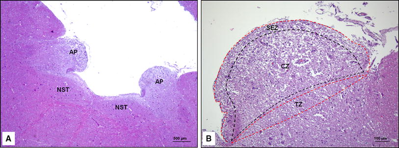
The area postrema of the rabbit, which was perfused with glutaraldehyde and postfixed in osmium tetroxide, was observed under the electron microscope. This area showed neuronal and neuroglial structures similar to those of ordinary cerebral tissue, except for rich blood capillaries, which were surrounded by conspicuous perivascular spaces. Parenchymal cells included a moderate number of small neurons and large numbers of specific astrocyte-like cells. The neuropil consisted of a small number of thin myelinated and many non-myelinated nerve fibers of varying calibers, axo-dendritic synapses, and neuroglial cell processes, leaving no spaces between them. The axons and synaptic terminals contained moderate amounts of granular vesicles, which were similar in size to those found in the hypothalamus and were supposed to contain catecholamine. Glycogen paticles were demonstrated mainly in the cytoplasm of the astrocyte-like cells.
References
Brizzee, K. R.: A comparison of cell structure in the area postrema, supraoptic crest, and intercolumnar tubercle with notes on the neuro-hypophysis and pineal body in the cat. J. comp. Neurol.100, 699–716 (1954).
Cammermeyer, J.: Is the human area postrema a neuro-vegetative nucleus? Acta anat. (Basel)2, 294–320 (1947). Cited fromG. B. Wislocki andE. H. Leduc.
Dempsey, E. W., andG. B. Wislocki: An electron microscopic study of the blood-brain barrier of the rat, employing silver nitrate as a vital stain. J. biophys. biochem. Cytol.1, 245–256 (1955).
De Iraldi, A. P., H. F. Duggan, andE. de Robertis: Adenergic synaptic vesicles in the anterior hypothalamus of the rat. Anat. Rec.145, 521–531 (1963).
De Robeetis, E., andA. P. de Iraldi: Plurivesicular secretory processes and nerve endings in the pineal gland of the rat. J. biophys. biochem. Cytol.10, 361–372 (1961).
Grillo, M. A., andS. L. Palay: Granule-containing vesicles in the autonomic system. Electron microscopy, vol.2, U-1. New York: Academic Press 1962.
Iijima, K., S. Hirakawa, K. Kono, S. Matsuo, andH. Yamada: Fine structure of area postrema of human and several mammals with special reference to neuroglial elements. Bull. Tokyo med. dent. Univ.10, 361–385 (1963).
Kawashima, S.: About the auto-microchopper (McIlwain type). Protein, Nucleic acid. Enzyme7, 795–798 (1962).
King, L. S.: Cellular morphology in the area postrema. J. comp. Neurol.66, 1–21 (1937).
Koelle, G. B.: A proposed dual neurohumoral role of acetylcholine: Its functions at the pre- and post-synaptic sites. Nature (Lond.)190, 208–211 (1961).
Leduc, E. H., andG. B. Wislocki: The histochemical localization of acid and alkaline phosphatases, non-specific esterases and succinic dehydrogenase in the structures comprising the hematoencephalic barrier of the rat. J. comp. Neurol.97, 241–280 (1952).
Palay, S. L., S. M. McGee-Russell, S. Gordon, andM. A. Grillo: Fixation of neural tissues for electron microscopy by perfusion with solution of osmium tetroxide. J. Cell Biol.12, 385–410 (1962).
Sabatini, D. D., K. Bensch, andR. J. Barrnett: Cytochemistry and Electron microscopy. The preservation of cellular ultrastructure and enzymatic activity by aldehyde fixation. J. Cell Biol.17, 19–58 (1963).
Shimizu, N., andS. Ishii: Electron microscopic observation of catecholamine-containing granules in the hypothalamus and area postrema and their changes following reserpine injection. Arch. hist. jap.24, 489–497 (1964).
Shimizu, N., andT. Kumamoto: Histochemical studies on the glycogen of the mammalian brain. Anat. Rec.114, 479–497 (1952).
—, andM. Okada: Histochemical studies of monoamine oxidase of the brain of rodents. Z. Zellforsch.49, 389–400 (1959).
Smart, I.: The subependymal layer of the mouse brain and its cell production as shown by radioautography after thymidine-H3 injection. J. comp. Neurol.116, 325–347 (1961).
Sutherland, E. W., andT. W. Rall: The relation of adenosine-3′5′-phosphate to the action of catecholamines. A Ciba Foundation Symposium on Adrenergic Mechanisms, p. 295–304. London: Churchill 1960.
Vogt, M.: The concentration of sympathin in different parts of the central nervous system under normal conditions and after the administration of drugs. J. Physiol. (Lond.)123, 451–481 (1954).
Wislocki, G. B., andE. H. Leduc: Vital staining of the hematoencephalic barrier by silver nitrate and trypan blue, and cytological comparisons of the neurohypophysis, pineal body, area postrema, intercolumnar tubercle and supraoptic crest. J. comp. Neurol.96, 371–413 (1952).
—, andT. J. Putnam: Note on the anatomy of the area postrema. Anat. Rec.19, 281–287 (1920).
Wolfe, D. E., L. T. Potter, K. C. Richardson, andJ. Axelrod: Localizing tritiated norepinephrine in sympathetic axons by electron microscopic autoradiography. Science138, 440–442 (1962).
Author information
Authors and Affiliations
Rights and permissions
About this article
Cite this article
Shimizu, N., Ishii, S. Fine structure of the area postrema of the rabbit brain. Z.Zellforsch 64, 462–473 (1964). https://doi.org/10.1007/BF01045119
Received:
Issue Date:
DOI: https://doi.org/10.1007/BF01045119


