Abstract
Background
Although roxadustat has been reported to cause central hypothyroidism, the details of the mechanisms and clinical characteristics of patients who are prone to developing hypothyroidism with roxadustat are uncertain.
Case presentation
A 53-year-old man with a 3-year history of hemodialysis due to diabetic kidney disease who had been treated with roxadustat, a hypoxia-inducible factor prolyl hydroxylase inhibitor, for 2 years was admitted to the hospital because of worsening gait disturbance and impaired consciousness. He had also acquired pure red cell aplasia associated with T-cell large granular lymphocytic leukemia and received multiple blood transfusions. Because his serum concentration of thyroid hormones was low, we diagnosed him with hypothyroidism, and his consciousness level recovered to normal with thyroid hormone replacement therapy. Computed tomography revealed a high-intensity atrophic thyroid gland, and magnetic resonance imaging showed diffusely reduced T2 and T1 signals of the pituitary anterior gland. These findings confirmed the accumulation of iron in the pituitary and thyroid glands. Combined pituitary stimulation tests with thyrotropin-releasing hormone, luteinizing hormone-releasing hormone, and corticotropin-releasing hormone revealed that the patient had pan-hypopituitarism. After discontinuation of roxadustat, the patient was treated with another hypoxia-inducible factor prolyl hydroxylase inhibitor, vadadustat. One month after switching medication, a stimulation test with thyrotropin-releasing hormone showed normal responses to thyroid-stimulating hormone. The patient was treated with levothyroxine 50 μg daily without any significant symptoms and is currently under follow-up observation as an outpatient.
Conclusions
We encountered a dialysis patient with roxadustat-induced hypothyroidism associated with transfusion iron overload. To our knowledge, this is the first case to clearly show that roxadustat can impair thyroid-stimulating hormone secretion in repeated thyrotropin-releasing hormone stimulation tests. Because the present patient had received roxadustat for more than 2 years before hypothyroidism became apparent, regular monitoring of the thyroid function may be needed in patients with renal anemia who have been treated with roxadustat, especially those at high risk of thyroid dysfunction.
Background
Transfusion iron overload is a major concern in the management of patients with severe anemia, as iron can form free radicals, and accumulated iron in the body can cause tissue injuries [1]. Iron first accumulates in reticuloendothelial macrophages and later in the liver, pancreas, heart, and endocrine tissue, where it can lead to liver dysfunction, diabetes mellitus, cardiomyopathy, and endocrine disorders, including hypopituitarism and hypothyroidism [2,3,4].
Roxadustat, a hypoxia-inducible factor prolyl hydroxylase inhibitor (HIF-PHI), has a structure similar to that of triiodothyronine [5] and can cross the blood–brain barrier [6]. Therefore, roxadustat may cause hypothyroidism by suppressing thyroid-stimulating hormone (TSH) in the pituitary gland and/or thyrotropin-releasing hormone (TRH) release in the hypothalamus. In fact, some cases of roxadustat-related hypothyroidism have been reported [7,8,9]. However, the regions affected by roxadustat have not been well examined. The clinical characteristics of patients prone to hypothyroidism with roxadustat are also uncertain.
We herein report a hemodialysis patient with transfusional iron overload who developed severe hypothyroidism during treatment with roxadustat and underwent endocrine tests during administration and after discontinuation of roxadustat.
Case presentation
A 53-year-old man with a 3-year history of hemodialysis for diabetic kidney disease was admitted to our hospital because of gait disturbance and impaired consciousness. He also had a 10-year history of acquired pure red cell aplasia associated with T-cell large granular lymphocytic leukemia and had received multiple blood transfusions. In addition, he was being treated for insomnia and restless leg syndrome. He was receiving dulaglutide 0.75 mg/day, carvedilol 2.5 mg/day, sodium zirconium cyclosilicate hydrate 10 g/day (only on non-hemodialysis days), lanthanum carbonate 2250 mg/day, calcium carbonate 1500 mg/day, bixalomer 5.22 g/day, zinc acetate dihydrate 25 mg/day, lansoprazole 15 mg/day, zolpidem tartrate 5 mg, lemborexant 5 mg, mianserin hydrochloride 20 mg, rotigotine 4.5 mg, and maxacalcitol 0.25 μg (only on hemodialysis days). He was also being administered 1500 mg of atovaquone and 400 mg of voriconazole to prevent opportunistic infections. In addition, because the patient required multiple red cell transfusions, 1080 mg of deferasirox was administered to treat iron overload. He had also been taking 150 mg of roxadustat three times weekly for 2 years. The patient had been in his usual state of health for one week before admission. A total of 5 days before admission, he complained of general weakness in his legs, nausea, and fatigue. His weakness worsened to the point he was unable to stand. He became drowsy on the morning of admission.
On admission, he had a Glasgow Coma Scale (GCS) score of 11 (E3, V3, M5). He was 1.62 m tall and weighed 66 kg. An examination of his vital signs revealed an axillary temperature of 35.2 °C, pulse rate of 61 bpm with regular rhythm, blood pressure of 164/90 mmHg, and oxygen saturation of 100% (ambient air). On a physical examination, the patient did not present with obvious paralysis. Complete blood count results were as follows: hemoglobin, 6 g/dL; hematocrit, 18.4%; red blood cell count, 201 × 104/μL, white blood cell count, 9620/μL; platelet count, 18 × 104/μL. Blood chemistry findings were as follows: total protein, 6.2 g/dL; albumin, 3.2 g/dL; alanine aminotransferase, 675 IU/L; aspartate aminotransferase, 614 IU/L; lactate dehydrogenase, 1547 IU/L; creatine kinase, 777 IU/L; calcium, 7.6 mg/dL; phosphate, 8.9 mg/dL; uric acid, 9.2 mg/dl; total cholesterol, 82 mg/dl; ammonia, 2 μg/mL; glucose, 201 mg/dl; transferrin saturation, 98%; ferritin, 41,894 ng/mL; free triiodothyronine (FT3), < 1.5 pg/mL; free thyroxine (FT4), < 0.42 ng/dl; and TSH, 1.146 μU/mL. Tests for thyroid peroxidase and thyroglobulin antibodies were negative. Brain computed tomography (CT) and brain magnetic resonance imaging (MRI) showed no evidence of cerebrovascular disease or traumatic brain injury. These findings indicated that his symptoms were related to hypothyroidism, and he was started on treatment with an initial dose of oral levothyroxine at 25 μg daily, which was increased to 37.5 μg 3 days later. A total of 3 days after the initiation of thyroid hormone replacement therapy, his consciousness level had recovered to normal (GCS score of 15) and he could walk on his own.
He had no known family history of the hemochromatosis and before onset of the pure red cell aplasia red, his serum ferritin (195 ng/mL) and TSAT (35%) were normal. However, over the past 5 years, as the frequency of blood transfusions increased, his serum ferritin level had persisted was over 3000 ng/mL.
In the past year, he had received 112 units of red blood cell transfusion. In addition, transthoracic echocardiography showed left ventricular chamber enlargement and a decreased left ventricular systolic function (left ventricular end-diastolic diameter, 64 mm; left ventricular end-systolic diameter, 56 mm; ejection fraction, 26%), and abdominal MRI showed a low intensity on T2-weighted imaging of the liver, suggesting iron deposition in the heart and liver. Since his serum ferritin level was elevated at 41,894 ng/mL, he was diagnosed with transfusional iron overload.
To examine the effect of iron overload on the endocrine gland function, before the discontinuation of roxadustat, we performed combined pituitary stimulation tests with TRH (500 μg), luteinizing hormone-releasing hormone (LHRH), and corticotropin-releasing hormone (CRH) on day 7 of hospitalization (Table 1). As a result, all peak values of TSH, LH, follicle-stimulating hormone (FSH), adrenocorticotropic hormone (ACTH), and cortisol were suppressed, indicating that the patient had pan-hypopituitarism. MRI showed diffusely reduced T2 and T1 signals in the pituitary anterior gland. Computed tomography (CT) revealed a high-intensity atrophic thyroid gland. These findings confirmed the accumulation of iron in the pituitary and thyroid glands. He showed decreased spontaneous erection and low libido. Although the baseline cortisol level was not suppressed, we treated him with hydrocortisone in a dosage of 10 mg daily. Furthermore, to examine the effect of roxadustat on hypothyroidism in the patent, roxadustat was switched to another HIF-PHI, vadadustat. One month after switching the medication, a pituitary stimulation test with TRH was performed, and a normal TSH response was obtained (Table 2). These findings indicated that central hypothyroidism was induced by treatment with roxadustat. The patient was treated with levothyroxine 50 μg daily without any significant symptoms and is currently under follow-up observation as an outpatient.
Discussion and conclusions
In the present case, transfusional iron overload induced multi-organ involvement, including the thyroid. On admission, the patient had central hypothyroidism. After discontinuation of roxadustat, his TSH secretion returned to the normal state, although the patient required thyroid hormone supplementation. These findings indicate that hypothyroidism was associated with iron deposition-induced dysfunction of the thyroid gland and impaired TSH release, which was induced by roxadustat.
Because HIF-PHI has been reported to improve iron metabolism compared with erythropoiesis-stimulating agents (ESAs) [10], we treated the present patient with roxadustat, which is the first HIF-PHI drug approved for the treatment of renal anemia in Japan. Recent case reports and case series have shown that roxadustat is associated with central hypothyroidism [7,8,9, 11]. Although the exact mechanisms are uncertain, the structural similarity of roxadustat to triiodothyronine (T3) may involve the pituitary gland and/or hypothalamus [5, 6]. Thus, roxadustat may have an affinity for the thyroid hormone receptor beta and is considered to be able to suppress the hypothalamic–pituitary–thyroid axis. In the present case, TRH-stimulated TSH levels were reduced during treatment with roxadustat, and both basal and peripheral TSH levels were normalized after switching from roxadustat to vadadustat. These results clearly show that roxadustat, but not vadadustat, suppressed TSH secretion by the pituitary gland. To our knowledge, this is the first report on the effects of roxadustat on TRH-stimulated TSH secretion.
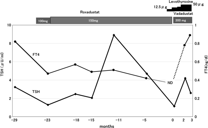
Because no prospective clinical trials have focused on changes in the thyroid function in patients with renal anemia, the clinical characteristics of patients who are prone to develop hypothyroidism with roxadustat are uncertain. In previous cases, clinical manifestations of hypothyroidism were apparent within a few months after the administration of roxadustat (Table 3). In addition, two of the three patents were treated with levothyroxine before treatment with roxadustat. The remaining patient was very elderly, suggesting that it may have the potential to cause thyroid dysfunction. The present patient had received roxadustat for more than 2 years before hypothyroidism became apparent (Fig. 1). The time course of the patient’s presentation could be related to iron deposition in the thyroid, which gradually progressed to hypothyroidism. Therefore, patients with subclinical or asymptotic impairment of the thyroid gland may be at a high risk of developing hypothyroidism with roxadustat, especially in the elderly. It is also well known that hypothyroidism is highly prevalent in chronic kidney disease patients, including those receiving dialysis [12, 13]. Therefore, regular monitoring of the thyroid function may be needed in patients with renal anemia treated with roxadustat.
Tineline of the changes in the thyroid function. The patient took indicated doses of roxadustat three times weekly. After discontinuation of roxadustat, the patient was treated with vadadustat (300 mg daily). Reference ranges of TSH and FT4 were between 0.2 μg and 4.5 µIU/mL and between 0.8 and 1.6 ng/dL, respectively. ND, not detected (< 0.42 ng/dL); TSH, thyroid-stimulating hormone; FT4, free thyroxine
Several drugs used to treat non-thyroid disorders, such as amiodarone, glucocorticoids, and antiepileptic agents, can affect the thyroid function [14]. In addition, drugs used in cancer therapy, e.g., immune checkpoint inhibitors, immunostimulatory cytokines or tyrosine kinase inhibitors were reported to cause thyroiditis [14]. This patient was treated with methotrexate, cyclophosphamide, and bendamustine for the T-cell large granular lymphocytic leukemia. To our knowledge, these drugs have little effect on the thyroid function. In addition, these drugs were discontinued more than 4 years before the admission. Therefore, drugs other than roxadustat had little effect on his thyroid function.
In conclusion, we encountered a dialysis patient with roxadustat-induced hypothyroidism associated with transfusion iron overload. To our knowledge, this is the first study to show that roxadustat can impair TRH-stimulated TSH secretion.
References
Radford-Smith DE, Powell EE, Powell LW. Haemochromatosis: a clinical update for the practising physician. Intern Med J. 2018;48(5):509–16.
Richardson KJ, McNamee AP, Simmonds MJ. Haemochromatosis: pathophysiology and the red blood cell1. Clin Hemorheol Microcirc. 2018;69(1–2):295–304.
McNeil LW, McKee LC, Lorber D, Rabin D. The endocrine manifestations of hemochromatosis. Am J Med Sci. 1983;285:7–13.
McLaren GD, Muir WA, Kellermeyer RW. Iron overload disorders: natural history, pathogenesis, diagnosis, and therapy. Crit Rev Clin Lab Sci. 1983;19(3):205–66.
Yao B, Wei Y, Zhang S, Tian S, Xu S, Wang R, et al. Revealing a mutant-induced receptor allosteric mechanism for the thyroid hormone resistance. Iscience. 2019;20:489–96.
Hoppe G, Yoon S, Gopalan B, Savage AR, Brown R, Case K, et al. Comparative systems pharmacology of HIF stabilization in the prevention of retinopathy of prematurity. Proc Natl Acad Sci USA. 2016;113:E2516–25.
Ichii M, Mori K, Miyaoka D, Sonoda M, Tsujimoto Y, Nakatani S, et al. Suppression of thyrotropin secretion during roxadustat treatment for renal anemia in a patient undergoing hemodialysis. BMC Nephrol. 2021;22(1):104.
Tokuyama A, Kadoya H, Obata A, Obata T, Sasaki T, Kashihara N. Roxadustat and thyroid-stimulating hormone suppression. Clin Kidney J. 2021;14(5):1472–4.
Shi XT, Li M, Cui WX, Chen J, Lu Y, Hu Y. Hypothyrotropin hypothyroidism caused by roxadustat: a case report. Zhonghua Nei Ke Za Zhi. 2022;61(12):1357–9.
Mima A. Hypoxia-inducible factor-prolyl hydroxylase inhibitors for renal anemia in chronic kidney disease: advantages and disadvantages. Eur J Pharmacol. 2021;5(912): 174583.
Haraguchi T, Hamamoto Y, Kuwata H, Yamazaki Y, Nakatani S, Hyo T, Yamada Y, et al. Effect of roxadustat on thyroid function in patients with renal anemia. J Clin Endocrinol Metab. 2023;109(1):1483.
Chonchol M, Lippi G, Salvagno G, Zoppini G, Muggeo M, Targher G. Prevalence of subclinical hypothyroidism in patients with chronic kidney disease. Clin J Am Soc Nephrol. 2008;3(5):1296–300.
Yuasa R, Ohashi Y, Saito A, Tsuboi K, Shishido S, Sakai K. Prevalence of hypothyroidism in Japanese chronic kidney disease patients. Ren Fail. 2020;42(1):572–9.
Burch HB. Drug effects on the thyroid. N Engl J Med. 2019;381(8):749–61.
Acknowledgements
Not applicable.
Funding
None.
Author information
Authors and Affiliations
Contributions
C.Y. and T.U. collected and analyzed the clinical data. Y.H., T.N., K.M., A.M., and M.K. were involved in the clinical care of the patient and helped to edit the manuscript. All authors contributed to the preparation of the manuscript. All authors read and approved the final manuscript.
Corresponding author
Ethics declarations
Ethics approval and consent to participate
All procedures performed in studies involving human participants were in accordance with the ethical standards of the Institutional and/or National Research Committee at which the studies were conducted and with the 1964 Declaration of Helsinki and its later amendments or comparable ethical standards.
Consent for publication
Written informed consent was obtained from the patient for the publication of this case report.
Competing interests
Takashi Uzu: Honoraria, Eli Lilly Japan K.K., AstraZeneca K.K. and Kyowa Kirin Co., Ltd. Novartis Pharma K.K.
Additional information
Publisher’s Note
Springer Nature remains neutral with regard to jurisdictional claims in published maps and institutional affiliations.
Rights and permissions
Open Access This article is licensed under a Creative Commons Attribution 4.0 International License, which permits use, sharing, adaptation, distribution and reproduction in any medium or format, as long as you give appropriate credit to the original author(s) and the source, provide a link to the Creative Commons licence, and indicate if changes were made. The images or other third party material in this article are included in the article's Creative Commons licence, unless indicated otherwise in a credit line to the material. If material is not included in the article's Creative Commons licence and your intended use is not permitted by statutory regulation or exceeds the permitted use, you will need to obtain permission directly from the copyright holder. To view a copy of this licence, visit http://creativecommons.org/licenses/by/4.0/. The Creative Commons Public Domain Dedication waiver (http://creativecommons.org/publicdomain/zero/1.0/) applies to the data made available in this article, unless otherwise stated in a credit line to the data.
About this article
Cite this article
Yamashita, C., Hirai, Y., Nishigaito, T. et al. Roxadustat and transfusional iron overload induced hypothyroidism in a hemodialysis patient: a case report with a literature review. Ren Replace Ther 10, 20 (2024). https://doi.org/10.1186/s41100-024-00537-z
Received:
Accepted:
Published:
DOI: https://doi.org/10.1186/s41100-024-00537-z


