
Abstract
Background
Interface fluid syndrome (IFS) is an unusual complication after laser-assisted in-situ keratomileusis (LASIK). We report the first case of IFS after uncomplicated phacotrabeculectomy in a patient who had undergone LASIK 10 years previously. This case emphasizes the importance of intraocular pressure (IOP) interpretation in eyes that have undergone LASIK.
Case presentation
A 30-year-old woman with a history of LASIK surgery presented to glaucoma clinic due to uncontrolled IOP despite of maximally tolerable medical treatment. After receiving phacotrabeculectomy, IOP decreased to 3 mmHg on the first postoperative day, but again increased up to 21 mmHg and a diffuse corneal edema with cloudy flap interface was demonstrated by slit-lamp microscopy. Corneal edema was sustained even after the IOP was lowered to 14 mmHg. Spectral-domain optical coherence tomography scanning of the cornea revealed a diffuse, thin fluid pocket in the corneal interface. After laser lysis of the scleral flap sutures, IOP was further decreased to 9 mmHg and interface fluid was resolved.
Conclusion
IFS should be considered as a possible cause of postoperative corneal edema despite of low IOP in the eyes that underwent LASIK surgery. Additional IOP lowering may be helpful for resolving the corneal edema.
Background
Interface fluid syndrome (IFS) is an unusual complication after laser-assisted in-situ keratomileusis (LASIK) that is characterized by diffuse fluid accumulation within the flap interface. Although elevation of intraocular pressure (IOP) is the main sign associated with the IFS [1], falsely low IOP readings after LASIK could mimic the condition and delay an accurate diagnosis [2]. We report the first case of IFS after uncomplicated phacotrabeculectomy in a patient who had undergone LASIK 10 years previously. The IFS did not resolve when the IOP was reduced to within the statistically normal range, but it did resolve with further IOP reduction. This case emphasizes the importance of IOP interpretation in eyes that have undergone LASIK.
Case report
A 30-year-old woman was presented to a glaucoma clinic due to uncontrolled IOP. She had undergone bilateral LASIK 10 years previously, and had been treated with oral and topical steroids as well as albendazole for 10 months for uveitis associated with ocular toxocariasis in the left eye. Sub-Tenon injection of triamcinolone acetonide (40 mg) had also been performed 5 months previously.
At the first visit, her visual acuity was 20/200 and the IOP was 30 mmHg in the left eye measured by Goldmann applanation tonometry (GAT). Slit-lamp examination revealed Grade 1 posterior subcapsular opacity, and fundus examination showed glaucomatous change in the optic nerve head. Inflammatory cells were not detected in either the anterior or posterior chamber. Despite maximally tolerable medical treatment, the IOP subsequently increased up to 32 mmHg, and her visual acuity worsened to 20/500 with ongoing glaucomatous optic nerve damage and progression of posterior subcapsular opacity. Phacotrabeculectomy with topically applied mitomycin-C (0.04%) was then performed.
The IOP was 3 mmHg by GAT on the first postoperative day but increased up to 21 mmHg on the following day. Her visual acuity was hand movement and could not be corrected. A diffuse corneal edema with a cloudy flap interface was noted in a slit-lamp examination. At 1 week postoperatively, the IOP had decreased to 14 mmHg after the application of brimonidine (0.2%)/timolol (0.5%) twice daily, but the corneal edema did not resolve. Spectral-domain optical coherence tomography (SD-OCT) scanning revealed a diffuse and thin fluid pocket in the corneal interface region (Fig. 1A). After using an argon laser to perform suture-lysis of the scleral flap on the following day, the IOP decreased to 9 mmHg and the visual acuity improved to 20/150. Resolution of the interface fluid was noted by SD-OCT (Fig. 1B). Central corneal thicknesses were 553.5μm preoperatively and 576.6μm at 14 days postoperatively. Eight months postoperatively, the IOP was maintained at 8 mmHg without using topical IOP-lowering agents, the cornea was clear without any interface haze by detailed slit-lamp examination, and the corrected visual acuity was 20/100.
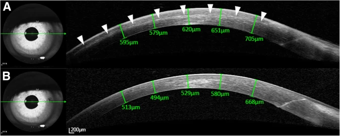
Spectral-domain optical coherence tomography scanning of the cornea before (a) and after (b) performing suture lysis using an argon laser. Note that the diffuse and thin fluid pocket in the corneal interface region (arrowheads) resolved when the intraocular pressure was lowered from 14 to 9 mmHg
Discussions and conclusions
Lowering of IOP commonly results in the resolution of interface fluid that can appear after LASIK [3]. However, the interface fluid in our case did not improve even when the IOP was reduced to within the statistically normal range; instead, a substantial IOP lowering down to a subnormal level was necessary for resolution of this fluid. We speculate that the decreased corneal thickness after LASIK and the ability of the accumulated fluid to absorb shock might have resulted in the underestimation of IOP [4]. This suggests that the target IOP should be lower in eyes that have undergone LASIK to allow for the possibility of falsely low IOP reading. In this case, IOP was measured at central cornea, but IOP measurement peripheral to the LASIK flap is also required for accuracy.
In eyes having a history of LASIK, the possibility of IFS should be considered as a possible cause of postoperative corneal edema even when the IOP is within normal range, particularly when the edema is long-standing and refractory to conventional treatment. An additional IOP lowering beyond the normal range may be helpful for resolving the corneal edema.
Abbreviations
- GAT:
-
Goldmann applanation tonometry
- IFS:
-
Interface fluid syndrome
- IOP:
-
Intraocular pressure
- LASIK:
-
laser-assisted in-situ keratomileusis
- SD-OCT:
-
Spectral-domain optical coherence tomography
References
Bamashmus MA, Saleh MF. Post-LASIK interface fluid syndrome caused by steroid drops. Saudi journal of ophthalmology : official journal of the Saudi Ophthalmological Society. 2013;27(2):125–8.
Duch S, Serra A, Castanera J, Abos R, Quintana M. Tonometry after laser in situ keratomileusis treatment. J Glaucoma. 2001;10(4):261–5.
Ortega-Usobiaga J, Martin-Reyes C, Llovet-Osuna F, Damas-Mateache B, Baviera-Sabater J. Interface fluid syndrome in routine cataract surgery 10 years after laser in situ keratomileusis. Cornea. 2012;31(6):706–7.
Han SB, Woo SJ, Hyon JY. Delayed-onset interface fluid syndrome after laser in situ keratomileusis secondary to combined cataract and vitreoretinal surgery. J Cataract Refract Surg. 2012;38(3):548–50.
Acknowledgments
None.
Funding
This study received no specific grant from any funding agency.
Availability of data and materials
All data and materials are available in this article.
Author information
Authors and Affiliations
Contributions
CYK, YHJ, EJL, JYH, KHP and TWK contributed to conception and design, data acquisition and interpretation of data. CYK and YHJ drafted the article, and all authors approved the final version.
Corresponding author
Ethics declarations
Ethics approval and consent to participate
Not applicable.
Consent for publication
Written informed consent for this case report was obtained from the patient.
Competing interests
The authors declare that they have no competing interests.
Publisher’s Note
Springer Nature remains neutral with regard to jurisdictional claims in published maps and institutional affiliations.
Rights and permissions
Open Access This article is distributed under the terms of the Creative Commons Attribution 4.0 International License (http://creativecommons.org/licenses/by/4.0/), which permits unrestricted use, distribution, and reproduction in any medium, provided you give appropriate credit to the original author(s) and the source, provide a link to the Creative Commons license, and indicate if changes were made. The Creative Commons Public Domain Dedication waiver (http://creativecommons.org/publicdomain/zero/1.0/) applies to the data made available in this article, unless otherwise stated.
About this article

Cite this article
Kim, C.Y., Jung, Y.H., Lee, E.J. et al. Delayed-onset interface fluid syndrome after LASIK following phacotrabeculectomy. BMC Ophthalmol 19, 74 (2019). https://doi.org/10.1186/s12886-019-1077-2
Received:
Accepted:
Published:
DOI: https://doi.org/10.1186/s12886-019-1077-2