Abstract
Background
Circulating total insulin-like growth factor-I (IGF-I) is an established risk factor for prostate cancer. However, only a small proportion of circulating IGF-I is free or readily dissociable from IGF-binding proteins (its bioavailable form), and few studies have investigated the association of circulating free IGF-I with prostate cancer risk.
Methods
We analyzed data from 767 prostate cancer cases and 767 matched controls nested within the European Prospective Investigation into Cancer and Nutrition cohort, with an average of 14-years (interquartile range = 2.9) follow-up. Matching variables were study center, length of follow-up, age, and time of day and fasting duration at blood collection. Circulating free IGF-I concentration was measured in serum samples collected at recruitment visit (mean age 55 years old; standard deviation = 7.1) using an enzyme-linked immunosorbent assay (ELISA). Conditional logistic regressions were performed to examine the associations of free IGF-I with risk of prostate cancer overall and subdivided by time to diagnosis (≤ 14 and > 14 years), and tumor characteristics.
Results
Circulating free IGF-I concentrations (in fourths and as a continuous variable) were not associated with prostate cancer risk overall (odds ratio [OR] = 1.00 per 0.1 nmol/L increment, 95% CI: 0.99, 1.02) or by time to diagnosis, or with prostate cancer subtypes, including tumor stage and histological grade.
Conclusions
Estimated circulating free IGF-I was not associated with prostate cancer risk. Further research may consider other assay methods that estimate bioavailable IGF-I to provide more insight into the well-substantiated association between circulating total IGF-I and subsequent prostate cancer risk.
Background
Prostate cancer is the second most commonly diagnosed cancer and the fifth leading cause of cancer deaths among men worldwide [1]. The global incidence and mortality of prostate cancer are predicted to increase by 80% and to almost double by 2040, respectively [2, 3]. Higher circulating insulin-like growth factor-I (IGF-I) concentration is one of a very small number of established etiological risk factors for prostate cancer. Evidence in favor of a relationship between IGF-I and prostate cancer risk stems principally from a consistent risk association identified in several large prospective observational studies [4, 5]. More recently, additional evidence from both Mendelian randomization (MR) and cis colocalization analyses have identified a shared genetic cause between circulating IGF-I concentrations and prostate cancer risk at the IGFI locus [6, 7].
IGF-I is a growth-promoting peptide hormone, which, following the binding to its cognate receptor, stimulates cell proliferation and survival and decreases apoptosis, thereby increasing the risk of carcinogenesis [8, 9]. IGF-I is produced mainly by the liver (~ 75%) and also locally in many tissues [9]. In the blood circulation, IGF-I is predominantly bound to IGF binding proteins (IGFBPs), particularly IGFBP-3 (accounting for 75–80% of bound IGF-I) [10]. Only a small portion of circulating IGF-I (~ 1%) is free (or unbound) or readily dissociable from IGFBPs [11, 12]. This fraction is hypothesized to be the most bioactive and more readily available to bind to IGF-I receptors on cell surfaces to activate intracellular signaling cascades [13, 14].
The role of IGF-I in prostate cancer risk has been well-characterized from studies of circulating total IGF-I, which includes both bound and free IGF-I [4,5,6,7, 15]. To our knowledge, however, only three prospective studies of prostate cancer risk have measured circulating free IGF-I concentration using immunoradiometric assay (IRMA) [16] or enzyme-linked immunosorbent assay (ELISA) [17, 18], with sample sizes up to 1076 men; they did not identify associations but may not have had the power to detect small to moderate associations.
The present study aimed to examine the association between circulating free IGF-I and subsequent risk of prostate cancer in a case-control study nested within the European Prospective Investigation into Cancer and Nutrition (EPIC) cohort. Using a recently developed ELISA for free IGF-I [19], we measured free IGF-I levels in baseline serum samples from men who subsequently developed prostate cancer and their matched controls [median time to diagnosis: 14 years, interquartile range (IQR) = 2.9] in a large sample (767 pairs or n = 1534). We assessed the association of free IGF-1 with prostate cancer risk overall and by time to diagnosis, and also with risk by tumor subtypes according to histological grade, tumor stage, and aggressiveness.
Methods
Study population
EPIC is a large prospective multicenter cohort study that recruited more than 521,000 participants (153,426 men), who were predominantly white and aged 35–70 years old, from 23 centers in 10 European countries (Denmark, France, Germany, Greece, Italy, the Netherlands, Norway, Spain, Sweden and the United Kingdom) [20]. At the recruitment visit between 1992 and 1999, information on diet, lifestyle, medical history, and anthropometric measurements were recorded. Fasting or non-fasting blood samples were also collected from 387,889 individuals (137,000 men), and are stored at local centers and at the International Agency for Research on Cancer – World Health Organization (IARC-WHO) in Lyon, France. The current study included male participants with baseline blood samples from Germany, Italy, the Netherlands, Spain and the United Kingdom, which were collected according to a standardized protocol. Plasma, serum, erythrocytes, and buffy coat were separated by centrifugation and aliquoted into 28 straws for storage, until required for laboratory analysis. All of the serum samples for this study were thawed for removal from storage straws and refrozen, and then thawed again for aliquotting in preparation for assaying.
Ascertainment of prostate cancer cases and controls
Information on cancer incidence, tumor characteristics, vital status, and cause of death was ascertained through population-based cancer registries in Italy, the Netherlands, Spain and the United Kingdom, and by active follow-up with different sources in Germany, including health insurance records, municipality registries, and hospital- or physician-based cancer and pathology registries. Prostate cancer cases were identified as men diagnosed with first incident prostate cancer based on the International Classification of Diseases 10th revision code (ICD-10: C61) [21], after blood collection and before the end of follow-up in 2013 (i.e. fourth round of EPIC endpoint follow-up, known as phase 4). These cases were matched one-to-one with controls who were randomly selected among male cohort participants who were free of cancer (excluding non-melanoma skin cancer) and alive at the time of diagnosis of the index case, using an incidence density sampling protocol. Matching variables were study center, length of follow-up (± 6 months), age at blood collection (± 6 months), time at blood collection (± 1 h) and fasting duration at blood collection (< 3 h, 3–6 h or > 6 h). The present analyses included 767 cases with 767 matched controls.
Information on histological grade and tumor stage at diagnosis was available for 641 (83.6%) cases and 406 (52.9%), respectively. For histological grade, there were 545 low-intermediate grade (Gleason score < 8, or grade coded in the recruitment center as well, moderately or poorly differentiated) and 96 high grade (Gleason score ≥ 8, or grade coded in the recruitment center as undifferentiated). For tumor stage, 273 cases were clinically localized (tumor-node-metastasis (TNM) staging score of T0-T2 and N0/Nx and M0, or stage coded in the recruitment center as localized) and 134 cases were clinically advanced (T3-T4 and/or N1-N3 and/or M1, or stage coded in the recruitment center as metastasis). Death from prostate cancer (n = 38) was defined as prostate cancer recorded as the underlying cause on the death certificate. We also further classified aggressive prostate cancer (n = 229) as those which were clinically advanced and/or high grade and/or prostate-specific antigen (PSA) > 20 ng/ml at diagnosis based on the definition from the European Association of Urology [22], and/or those who died from prostate cancer.
Measurement of circulating free IGF-I and other analytes
Serum free IGF-I concentrations were assayed, with blinding to case-control status, in the laboratory of Dr. Michael Pollak at McGill University in Montreal, Canada, using ELISA (Ansh Labs, Webster, TX, USA) in 2021–2022. This assay is referred to as a highly sensitive two-site or “sandwich” method that directly detects free IGF-I that is bound between the first capture antibody immobilized on the microtiter plate and the second detection antibody specific for free IGF-I [11, 19]. Bound IGF-I is not detected since the epitope of IGF-I in IGF-I/IGFBPs complexes is concealed. Free IGF-I was measured in duplicate for each sample and mean values were used for analyses. All measures below the lower limit of detection (LOD, 0.33 ng/mL) (n = 525, 34%) were set to be 0.165 ng/mL, which is the midpoint between 0 and the LOD. The inter- and intra-batch coefficients of variation for this assay were 4.73% and 1.49%, respectively.
Measurements of serum total IGF-I, IGFBP-1, IGFBP-2, IGFBP-3, IGF-II, testosterone and sex hormone binding globulin (SHBG) concentrations were performed using ELISA or electrochemiluminescence immunoassay, as described in detail elsewhere [23, 24]. Total IGF-I was assayed with the elimination of IGFBPs using an acid ethanol precipitation step. Free testosterone concentrations were estimated using a formula based on the law mass action from measured total testosterone and SHBG concentrations [25, 26], assuming a constant albumin concentration of 43 g/L [23].
Statistical analyses
Differences in selected participant characteristics between prostate cancer cases and controls were compared using chi-squared tests for categorical variables and t-tests for continuous variables. Given the left truncated distribution for free IGF-I owing to concentrations below the LOD, correlations between free IGF-I and other members of the IGF axis analytes (IGFBP-1, IGFBP-2, IGFBP-3, IGF-II) and sex hormones (testosterone, free testosterone, SHBG) were estimated using Spearman’s rank tests. Differences in free IGF-I concentrations by categories of selected participant characteristics among controls and cases were assessed using analysis of covariance, adjusted for laboratory batch, and age at blood collection, body mass index (BMI) and/or recruitment center. The concentrations of free IGF-I and other IGF axis analytes were presented as geometric means with 95% confidence interval (CI).
The association between circulating free IGF-I concentrations and overall prostate cancer risk was estimated using logistic regression models conditioned on the matching factors and adjusted for laboratory batch. Circulating free IGF-I concentrations were modelled in fourths (based on quartile cut-points defined among controls) (Table 1), and as a continuous variable. Linear trends for the associations of free IGF-I with risk were calculated across the medians within each fourth of free IGF-I. These analyses were repeated by time to diagnosis (≤ 14 and > 14 years), fasting status and BMI (< 30 kg/m2 and ≥ 30 kg/m2) as well as by tumor subtype of histological grade, tumor stage and aggressiveness. Similarly, the associations of circulating total IGF-I concentrations with risks for overall and aggressive prostate cancer were tested.
All statistical analyses were conducted using Stata 17.0 (Stata Corp LP, College Station, TX). Findings were plotted using “ggplot2” package in R 4.1.1.
Results
Participants’ characteristics
The present analyses included 767 incident prostate cancer cases and 767 matched controls, with mean age at blood collection of 55 (standard deviation = 7.1) years old. For cases, the mean age at diagnosis was 69 years and the median time from blood collection to diagnosis was 14 (IQR = 2.9) years. No material differences in selected characteristics were found between men who developed prostate cancer and men who did not (Table 2).
Total IGF-I concentration was higher in cases than controls (geometric means = 19.2 nmol/L, 95% CI: 18.89–19.57 vs. 18.64, 18.32–18.98), while no differences in free IGF, IGFBPs or IGF-II concentrations were observed (Supplemental Table 1). Overall, free IGF-I concentration was modestly positively correlated with total IGF-I concentration (Spearman’s rank correlation coefficient, ρ = 0.230) and the ratio of total IGF-I to IGFBP-3 (ρ = 0.250), but not with other IGFBPs, IGF-II or sex hormone concentrations (Supplementary Tables 2 and Supplementary Fig. 1).
Table 3 shows the differences in free IGF-I concentrations across selected characteristics among controls and cases. Men who had higher BMI, blood collected at an earlier time of day, higher alcohol consumption, who fasted before blood collection, or who were current smokers tended to have lower free IGF-I concentration, in both controls and cases.
IGF-I and prostate cancer
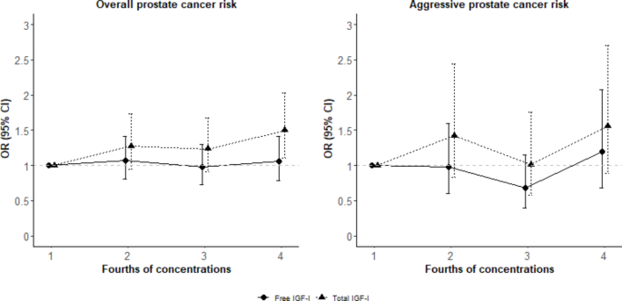
Figure 1; Table 1 show the adjusted associations of circulating free and total IGF-I concentrations with prostate cancer risk. Higher free IGF-I concentration (in fourths or as a continuous variable) was not associated with total prostate cancer risk [Odds ratio (OR) = 1.00 per 0.1 nmol/L increase, 95% CI: 0.99, 1.02]. Similarly, there were no significant associations between free IGF-I concentration and total prostate cancer risk when stratified by time to diagnosis, fasting status and BMI (< 30 kg/m2 and ≥ 30 kg/m2) (Supplementary Table 3). Also, higher free IGF-1 concentration was not associated with prostate cancer risk when analyses were repeated by histological grade, tumor stage or aggressiveness. Higher circulating total IGF-I concentration was associated with higher overall (OR = 1.18 per 5 nmol/L increase, 95% CI: 1.05, 1.33) and possibly aggressive prostate cancer risks (OR = 1.25, 95% CI: 1.00, 1.56).
Adjusted associations of circulating free and total IGF-I concentrations with overall and aggressive prostate cancer risks in EPIC Models conditioned on the matching variables: center, follow-up time, fasting status, age at blood collection and time at blood collection, and adjusted for laboratory batch (only for free IGF-I)
Discussion
In this matched nested case-control study with a long duration of follow-up from blood collection among European men, we found no association of circulating free IGF-I concentration measured using a recently developed ELISA with prostate cancer risk, and these findings did not vary by time to diagnosis or tumor subtype. In contrast, we observed a positive association of total IGF-I concentration with total prostate cancer risk, as we and others have previously reported in EPIC and other studies [4,5,6,7, 15].
Our null findings for free IGF-I are consistent with the two previous nested case-control studies of free IGF-I and prostate cancer [16, 17]. In the Physician’s Health Study (PHS), Mucci et al. measured fasting or non-fasting plasma free IGF-I concentration in 545 matched case-control pairs using a different ELISA and found no association with prostate cancer risk [17]. In the European Randomized Study of Screening for Prostate cancer, Janssen et al. used IRMA to assay serum free IGF-I (201 pairs) and found no difference in free IGF-I concentrations between prostate cancer cases and matched controls [16]. In addition to these studies of overall prostate cancer, in a case-only study in the PHS and Health Professionals Follow-Up Study, Ma et al. found no evidence for an association of free IGF-I with risk of lethal compared to nonlethal prostate cancer (524 nonlethal, 434 lethal cases) [18].
Higher circulating total IGF-I concentration is one of a limited number of established causal and potentially modifiable risk factors for prostate cancer risk, with strong evidence from both large prospective observational and genetic study designs [4,5,6,7, 15]. Higher circulating free IGF-I, which may reflect the bioactive form of total IGF-I, has been suggested as one possible mechanism driving the observed association of total IGF-I with prostate cancer risk. Previous studies have identified cancer-promoting properties for free-IGF-I, including mitotic and antiapoptotic effects [27], motivating the present study. Nevertheless, we did not observe an association between circulating free IGF-I concentration and prostate cancer, despite the positive association for circulating total IGF-I concentration in the same sample.
Our null findings for circulating free IGF-I may relate to the complexity of IGF signaling; IGFBPs can both enhance and inhibit IGF-I signaling [9]. The prostate also produces IGFs, IGFBPs and IGFBP proteases locally [10, 28]. Therefore, circulating free IGF-I may not be a good predictor of intra-prostatic IGF-I bioactivity. While circulating free IGF-I concentration might not be relevant for prostate cancer risk, this does not exclude the possible biological effect of free IGF-I in prostate tissue on prostate cancer development. Future studies using assays that quantify IGF1 receptor activation or expression in prostate tissue may help to further understand relationships between IGF-I signaling and prostate cancer.
Our null results for free but not total IGF-I might reflect the inherent difficulty in measuring free IGF-I due to its short half-life (1–2 min) in the circulation, in contrast to circulating total IGF-I (up to 24 h) [14]. It is also possible that IGF-I released from the IGFBP-bound IGF-I complex immediately binds to its receptor once IGFBPs are cleaved or bind to the target cell surface [10], and thus this bioavailable IGF-I cannot be well estimated based on free IGF-I in blood samples. While about one-third of participants had free IGF-I below the LOD, coefficients of variation were low and duplicate measurements were very highly correlated, implying that measurement error in between-individual variations of current assay is likely to be modest. Additionally, we observed a modest but highly significant positive correlation for free IGF-I with total IGF-I, consistent with a previous study [17].
Several limitations in the present analyses need to be acknowledged. Although we included a large sample in the overall analyses, the statistical power and thus our ability to detect associations in the stratified analyses was more limited. The number of prostate cancer deaths was also modest. Levels of circulating free IGF-I concentrations might have been affected by storage time since the serum samples used for IGF-I measurements in 2021–2022 had been stored since recruitment in the 1990s, nonetheless we observed associations for total IGF-I concentrations using the same samples. Also, the estimate of free IGF-I in this study includes both free and readily dissociable IGF-I [11, 19]; however, readily dissociable IGF-I, unlike stably bound IGF-I, may also have biological relevance. Additionally, our study considered free IGF-I concentration for each individual measured only at a single timepoint. Given the low proportion of free IGF-I in circulation (~ 1%), even modest measurement error may induce considerable attenuation of risk estimates where single measurements may not adequately capture average concentrations over the medium to long term. Furthermore, we measured free IGF-I concentration in circulating blood samples. Previous studies have suggested there may be effects of locally accumulated free IGF-I in tissues on risk for prostate cancer [27, 29], which we did not estimate in the present study. Although participants in the present study had lower concentrations in IGF axis analytes than other studies, the magnitude of the association between circulating total IGF-I concentration and prostate cancer was similar across studies [4,5,6,7]. Finally, our study analyzed white men, and thus our findings may not be generalizable to other populations.
Conclusions
In conclusion, this study did not find evidence of an association of higher circulating free IGF-I, measured using a recently developed sandwich ELISA, with subsequent risk of prostate cancer overall or by follow-up duration and prostate tumor characteristics including histological grade, tumor stage and aggressiveness. Further research may consider other assays that estimate the bioavailability of circulating IGF-I, as well as methods for measuring free IGF-I in prostate tissue, to deepen the understanding of potential pathways and mechanisms for the substantiated association between circulating total IGF-I and subsequent prostate cancer development and progression.
Data Availability
For information on how to submit an application for gaining access to EPIC data, please follow the instructions at http://epic.iarc.fr/access/index.php.
References
Sung H, Ferlay J, Siegel RL, Laversanne M, Soerjomataram I, Jemal A, et al. Global Cancer Statistics 2020: GLOBOCAN estimates of incidence and mortality worldwide for 36 cancers in 185 countries. CA Cancer J Clin. 2021;71(3):209–49.
Rawla P. Epidemiology of prostate Cancer. World J Oncol. 2019;10(2):63–89.
Withrow D, Pilleron S, Nikita N, Ferlay J, Sharma S, Nicholson B, et al. Current and projected number of years of life lost due to prostate cancer: a global study. Prostate. 2022;82(11):1088–97.
Knuppel A, Fensom GK, Watts EL, Gunter MJ, Murphy N, Papier K, et al. Circulating insulin-like growth factor-I concentrations and risk of 30 cancers: prospective analyses in UK Biobank. Cancer Res. 2020;80(18):4014–21.
Travis RC, Appleby PN, Martin RM, Holly JMP, Albanes D, Black A, et al. A meta-analysis of individual participant data reveals an association between circulating levels of IGF-I and prostate cancer risk. Cancer Res. 2016;76(8):2288–300.
Watts EL, Fensom GK, Smith Byrne K, Perez-Cornago A, Allen NE, Knuppel A, et al. Circulating insulin-like growth factor-I, total and free testosterone concentrations and prostate cancer risk in 200 000 men in UK Biobank. Int J Cancer. 2021;148(9):2274–88.
Watts EL, Perez-Cornago A, Fensom GK, Smith-Byrne K, Noor U, Andrews CD et al. ,. Circulating insulin-like growth factors and risks of overall, aggressive and early-onset prostate cancer: a collaborative analysis of 20 prospective studies and Mendelian randomization analysis. Int J Epidemiol. 2022;dyac124
Weroha SJ, Haluska P. The insulin-like growth factor system in cancer. Endocrinol Metab Clin North Am. 2012;41(2):335–50.
LeRoith D, Holly JMP, Forbes B. The insulin-like growth factors: ligands, binding proteins and receptors. Mol Metab. 2021:101245.
Allard JB, Duan C. IGF-binding proteins: why do they exist and why are there so many? Front Endocrinol (Lausanne). 2018;9:117.
Frystyk J. Exercise and the growth hormone-insulin-like growth factor axis. Med Sci Sports Exerc. 2010;42(1):58–66.
Yu H, Mistry J, Nicar MJ, Khosravi MJ, Diamandis A, van Doorn J, et al. Insulin-like growth factors (IGF-I, free IGF-I and IGF-II) and insulin-like growth factor binding proteins (IGFBP-2, IGFBP-3, IGFBP-6, and ALS) in blood circulation. J Clin Lab Anal. 1999;13(4):166–72.
Talia C, Connolly L, Fowler PA. The insulin-like growth factor system: a target for endocrine disruptors? Environ Int. 2021;147:106311.
Baxter RC. IGF binding proteins in cancer: mechanistic and clinical insights. Nat Rev Cancer. 2014;14(5):329–41.
Larsson SC, Carter P, Vithayathil M, Kar S, Mason AM, Burgess S. Insulin-like growth factor-1 and site-specific cancers: a mendelian randomization study. Cancer Med. 2020;9(18):6836–42.
Janssen JA, Wildhagen MF, Ito K, Blijenberg BG, Van Schaik RH, Roobol MJ, et al. Circulating free insulin-like growth factor (IGF)-I, total IGF-I, and IGF binding protein-3 levels do not predict the future risk to develop prostate cancer: results of a case-control study involving 201 patients within a population-based screening with a 4-year interval. J Clin Endocrinol Metab. 2004;89(9):4391–6.
Mucci LA, Stark JR, Pollak MN, Li H, Kurth T, Stampfer MJ, et al. Plasma levels of acid-labile subunit, free insulin-like growth factor-I, and prostate cancer risk: a prospective study. Cancer Epidemiol Biomarkers Prev. 2010;19(2):484–91.
Ma C, Wang Y, Wilson KM, Mucci LA, Stampfer MJ, Pollak M, et al. Circulating insulin-like growth factor 1-related biomarkers and risk of lethal prostate cancer. JNCI Cancer Spectr. 2022;6(1):pkab091.
AnshLabs, Free. IGF-I ELISA. [cited 2022 Nov 15]. Available from https://www.anshlabs.com/wp-content/uploads/inserts/AL122.pdf.
Riboli E, Hunt KJ, Slimani N, Ferrari P, Norat T, Fahey M, et al. European prospective investigation into Cancer and Nutrition (EPIC): study populations and data collection. Public Health Nutr. 2002;5(6b):1113–24.
World Health Organization. International statistical classification of diseases and related health problems: 10th revision. [cited 2013 Dec 31]. Available from http://apps.who.int/classifications/icd10/browse/2010/en.
Chang AJ, Autio KA, Roach M, Scher HI. High-risk prostate cancer-classification and therapy. Nat Rev Clin Oncol. 2014;11(6):308–23.
Watts EL, Perez-Cornago A, Fensom GK, Smith-Byrne K, Noor U, Andrews CD, et al. Circulating free testosterone and risk of aggressive prostate cancer: prospective and mendelian randomisation analyses in international consortia. Int J Cancer. 2022;151(7):1033–46.
Watts EL, Perez-Cornago A, Appleby PN, Albanes D, Ardanaz E, Black A, et al. The associations of anthropometric, behavioural and sociodemographic factors with circulating concentrations of IGF-I, IGF-II, IGFBP-1, IGFBP-2 and IGFBP-3 in a pooled analysis of 16,024 men from 22 studies. Int J Cancer. 2019;145(12):3244–56.
Vermeulen A, Verdonck L, Kaufman JM. A critical evaluation of simple methods for the estimation of free testosterone in serum. J Clin Endocrinol Metab. 1999;84(10):3666–72.
Bartsch W. Interrelationships between sex hormone-binding globulin and testosterone, 5 alpha-dihydrotestosterone and oestradiol-17 beta in blood of normal men. Maturitas. 1980;2(2):109–18.
Grimberg A. Mechanisms by which IGF-I may promote cancer. Cancer Biol Ther. 2003;2(6):630–5.
Oxvig C. The role of PAPP-A in the IGF system: location, location, location. J Cell Commun Signal. 2015;9(2):177–87.
Ahearn TU, Peisch S, Pettersson A, Ebot EM, Zhou CK, Graff RE, et al. Expression of IGF/insulin receptor in prostate cancer tissue and progression to lethal disease. Carcinogenesis. 2018;39(12):1431–7.
Acknowledgements
We thank the National Institute for Public Health and the Environment (RIVM), Bilthoven, the Netherlands, for their contribution and ongoing support to the EPIC Study.
The coordination of EPIC-Europe is financially supported by International Agency for Research on Cancer (IARC) and also by the Department of Epidemiology and Biostatistics, School of Public Health, Imperial College London which has additional infrastructure support provided by the NIHR Imperial Biomedical Research Centre (BRC).
The national cohorts are supported by: Danish Cancer Society (Denmark); Ligue Contre le Cancer, Institut Gustave Roussy, Mutuelle Générale de l’Education Nationale, Institut National de la Santé et de la Recherche Médicale (INSERM) (France); German Cancer Aid, German Cancer Research Center (DKFZ), German Institute of Human Nutrition Potsdam-Rehbruecke (DIfE), Federal Ministry of Education and Research (BMBF) (Germany); Associazione Italiana per la Ricerca sul Cancro-AIRC-Italy, Compagnia di SanPaolo and National Research Council (Italy); Dutch Ministry of Public Health, Welfare and Sports (VWS), Netherlands Cancer Registry (NKR), LK Research Funds, Dutch Prevention Funds, Dutch ZON (Zorg Onderzoek Nederland), World Cancer Research Fund (WCRF), Statistics Netherlands (The Netherlands); Health Research Fund (FIS) - Instituto de Salud Carlos III (ISCIII), Regional Governments of Andalucía, Asturias, Basque Country, Murcia and Navarra, and the Catalan Institute of Oncology - ICO (Spain); Swedish Cancer Society, Swedish Research Council and County Councils of Skåne and Västerbotten (Sweden); Cancer Research UK (14136 to EPIC-Norfolk; C8221/A29017 to EPIC-Oxford), Medical Research Council (1000143 to EPIC-Norfolk; MR/M012190/1 to EPIC-Oxford) (United Kingdom).
Funding
This study was supported by grants from the Cancer Research United Kingdom (grant no. C8221/A29017). ELW was supported by the Intramural Research Program of the NIH at the National Cancer Institute.
Author information
Authors and Affiliations
Contributions
Conceptualization, resources, funding acquisition (TJK, RCT); Data curation (TSC, UN, MP, YW); Formal analysis (TSC); Validation (TSC, YW); Methodology (TSC, TJK, KSB, RCT); Supervision (TJK, KSB, RCT); Writing original draft (TSC). All authors read and reviewed the manuscript, and approved the final manuscript.
Corresponding author
Ethics declarations
Competing interests
The authors declare no competing interests.
Abbreviations
BMI body mass index.
CI confidence interval.
ELISA enzyme-linked immunosorbent assay.
EPIC European Prospective Investigation into Cancer and Nutrition.
IGFBP IGF binding protein.
IGF-I insulin-like growth factor-I.
IRMA immunoradiometric assay.
IQR interquartile range.
LOD limit of detection.
MR Mendelian randomization.
PSA prostate-specific antigen.
SHBG sex hormone binding globulin.
TNM tumor-node-metastasis.
Ethics approval and consent to participate
All participants provided written informed consent to participate in the EPIC study. The EPIC study was approved by the Ethics Committee of the International Agency for Research on Cancer (IARC) and all EPIC centers. All methods were carried out in accordance with relevant guidelines and regulations.
Consent for publication
NA.
Additional information
Publisher’s Note
Springer Nature remains neutral with regard to jurisdictional claims in published maps and institutional affiliations.
Electronic supplementary material
Below is the link to the electronic supplementary material.
Rights and permissions
Open Access This article is licensed under a Creative Commons Attribution 4.0 International License, which permits use, sharing, adaptation, distribution and reproduction in any medium or format, as long as you give appropriate credit to the original author(s) and the source, provide a link to the Creative Commons licence, and indicate if changes were made. The images or other third party material in this article are included in the article’s Creative Commons licence, unless indicated otherwise in a credit line to the material. If material is not included in the article’s Creative Commons licence and your intended use is not permitted by statutory regulation or exceeds the permitted use, you will need to obtain permission directly from the copyright holder. To view a copy of this licence, visit http://creativecommons.org/licenses/by/4.0/. The Creative Commons Public Domain Dedication waiver (http://creativecommons.org/publicdomain/zero/1.0/) applies to the data made available in this article, unless otherwise stated in a credit line to the data.
About this article
Cite this article
Cheng, T.S., Noor, U., Watts, E. et al. Circulating free insulin-like growth factor-I and prostate cancer: a case-control study nested in the European prospective investigation into cancer and nutrition. BMC Cancer 24, 676 (2024). https://doi.org/10.1186/s12885-023-11425-w
Received:
Accepted:
Published:
DOI: https://doi.org/10.1186/s12885-023-11425-w


