
Abstract
This chapter is focused on automatic microscopy techniques. It covers a practical approach to different possible automation in microscopes. All the techniques presented in this chapter aim at capturing images with the best quality as well as do it automatically. The main section (Sect. 7.5) of the chapter focuses on slide scanning and it covers all the techniques necessary to do it such as stage motorization, automatic slide scanning approaches, and autofocus of the samples. The rest of the chapter is devoted to illumination control, and its implementation (Sect. 7.3); image calibration using basic tools (Sect. 7.4) and finally preprocessing techniques to improve image quality reducing the noise introduced by the camera or the illumination defects (Sect. 7.6). In addition to the theoretical explanation, the sections include implementation examples for clarification purposes.
Access this chapter
Tax calculation will be finalised at checkout
Purchases are for personal use only
References
Bay, H., Ess, A., Tuytelaars, T., Van Gool, L.: Speeded-up robust features (SURF). Comput. Vis. Image Underst. 110(3), 346–359 (2008)
Boddeke, F., Van Vliet, L., Young, I.: Calibration of the automated z-axis of a microscope using focus functions. J. Microsc. 186(3), 270–274 (1997)
Brenner, J.F., Dew, B.S., Horton, J.B., King, T., Neurath, P.W., Selles, W.D.: An automated microscope for cytologic research a preliminary evaluation. J. Histochem. Cytochem. 24(1), 100–111 (1976)
European Committee for Standardization: Water quality—Guidance standard for the identification, enumeration and interpretation of benthic diatom samples from running waters. Technical report (2004)
Forster, B., Van De Ville, D., Berent, J., Sage, D., Unser, M.: Complex wavelets for extended depth-of-field: a new method for the fusion of multichannel microscopy images. Microsc. Res. Tech. 65(1–2), 33–42 (2004)
Groen, F.C., Young, I.T., Ligthart, G.: A comparison of different focus functions for use in autofocus algorithms. Cytometry: J. Int. Soc. Anal. Cytol. 6(2), 81–91 (1985)
ImageJ plugins website. https://imagej.net/Extended_Depth_of_Field (2010). Accessed 25 Apr 2019
Jain, A.K.: Fundamentals of Digital Image Processing. Prentice Hall, Englewood Cliffs, NJ (1989)
Kloster, M., Esper, O., Kauer, G., Beszteri, B.: Large-scale permanent slide imaging and image analysis for diatom morphometrics. Appl. Sci. 7(4), 330 (2017)
Li, J.: Autofocus searching algorithm considering human visual system limitations. Opt. Eng. 44(11), 113201 (2005)
Lowe, D.G.: Distinctive image features from scale-invariant keypoints. Int. J. Comput. Vis. 60(2), 91–110 (2004)
Magee, D., Treanor, D., Crellin, D., Shires, M., Smith, K., Mohee, K., Quirke, P.: Colour normalisation in digital histopathology images. In: Proceedings of the Optical Tissue Image analysis in Microscopy, Histopathology and Endoscopy (MICCAI Workshop), vol. 100. Daniel Elson, London (2009)
Marty, G.D.: Blank-field correction for achieving a uniform white background in brightfield digital photomicrographs. BioTechniques 42(6), 716–720 (2007)
Meiniel, W., Olivo-Marin, J.C., Angelini, E.D.: Denoising of microscopy images: a review of the state-of-the-art, and a new sparsity-based method. IEEE Trans. Image Process. 27(8), 3842–3856 (2018)
Narra, P., Zinger, D.S.: An effective led dimming approach. In: Conference Record of the 2004 IEEE Industry Applications Conference, 2004. 39th IAS Annual Meeting, vol. 3, pp. 1671–1676. IEEE, New York (2004)
Papini, A.: A new algorithm to reduce noise in microscopy images implemented with a simple program in Python. Microsc. Res. Tech. 75(3), 334–342 (2012)
Pech-Pacheco, J.L., Cristóbal, G., Chamorro-Martinez, J., Fernández-Valdivia, J.: Diatom autofocusing in brightfield microscopy: a comparative study. In: Proceedings 15th International Conference on Pattern Recognition. ICPR-2000, vol. 3, pp. 314–317. IEEE, New York (2000)
Perez, L., Wang, J.: The effectiveness of data augmentation in image classification using deep learning (2017). Preprint. arXiv:1712.04621
Pertuz, S., Puig, D., Garcia, M.A.: Analysis of focus measure operators for shape-from-focus. Pattern Recognit. 46(5), 1415–1432 (2013)
Pizer, S.M., Amburn, E.P., Austin, J.D., Cromartie, R., Geselowitz, A., Greer, T., ter Haar Romeny, B., Zimmerman, J.B., Zuiderveld, K.: Adaptive histogram equalization and its variations. Comput. Vis. Graph. Image Process. 39(3), 355–368 (1987)
Preibisch, S., Saalfeld, S., Tomancak, P.: Globally optimal stitching of tiled 3D microscopic image acquisitions. Bioinformatics 25(11), 1463–1465 (2009)
Reddy, G.D., Cotton, R.J., Tolias, A.S., Saggau, P.: Random-access multiphoton microscopy for fast three-dimensional imaging. In: Membrane Potential Imaging in the Nervous System and Heart, pp. 455–472. Springer, New York (2015)
Redmon, J.: Darknet: Open source neural networks in C (2013–2016). http://pjreddie.com/darknet/
Redmon, J., Farhadi, A.: YOLO9000: better, faster, stronger (2016). Preprint. arXiv:1612.08242
Reinhard, E., Adhikhmin, M., Gooch, B., Shirley, P.: Color transfer between images. IEEE Comput. Graph. Appl. 21(5), 34–41 (2001)
Sanchez, C., Cristóbal, G., Bueno, G., Blanco, S., Borrego-Ramos, M., Olenici, A., Pedraza, A., Ruiz-Santaquiteria, J.: Oblique illumination in microscopy: a quantitative evaluation. Micron 105, 47–54 (2018)
Santos, A., Ortiz de Solórzano, C., Vaquero, J.J., Pena, J., Malpica, N., Del Pozo, F.: Evaluation of autofocus functions in molecular cytogenetic analysis. J. Microsc. 188(3), 264–272 (1997)
Smith, S.W., et al.: The Scientist and Engineer’s Guide to Digital Signal Processing. California Technical Publishing, San Diego, CA (1997)
Sternberg, S.: Biomedical image processing. Computer 16(1), 22–34 (1983). https://doi.org/10.1109/MC.1983.1654163
Wu, Q., Merchant, F., Castleman, K.: Microscope Image Processing, 548 pp. Academic (2008). https://www.ebook.de/de/product/7100910/microscope_image_processing.html
Wu, Q., Merchant, F., Castleman, K.: Microscope Image Processing. Elsevier, Amsterdam (2010)
Yang, S.J., Berndl, M., Ando, D.M., Barch, M., Narayanaswamy, A., Christiansen, E., Hoyer, S., Roat, C., Hung, J., Rueden, C.T., et al.: Assessing microscope image focus quality with deep learning. BMC Bioinf. 19(1), 77 (2018)
Yazdanfar, S., Kenny, K.B., Tasimi, K., Corwin, A.D., Dixon, E.L., Filkins, R.J.: Simple and robust image-based autofocusing for digital microscopy. Opt. Express 16(12), 8670–8677 (2008)
Yeo, T., Ong, S., Sinniah, R., et al.: Autofocusing for tissue microscopy. Image Vis. Comput. 11(10), 629–639 (1993)
Zuiderveld, K.: Graphics gems IV. In: Contrast Limited Adaptive Histogram Equalization, pp. 474–485. Academic Press Professional, Inc., San Diego, CA (1994)
Author information
Authors and Affiliations
Corresponding author
Editor information
Editors and Affiliations
Appendix
Appendix
1.1 Microscope Control Application
An application with a graphical user interface (GUI) has been developed to control the prototype described in this chapter. Most of the application has been developed using Python programming language, although some algorithms have been developed in C to improve the performance. OpenCV library has been used to manage image processing operations and PyQt for the GUI design. In this section, the most important implemented functionalities are reviewed.
First, in Fig. 7.18 the main window of the designed GUI is presented. Basically, it is divided into two different sections, the camera viewer and the control panel. In the first one, the user is capable to visualize in real time the images captured by the camera, according to the established camera settings. In the control panel section, through GUI elements such as buttons, text fields, or check boxes, the user can manage and configure some options related to the camera or the motorized stage, as well as to run the implemented algorithms and procedures. Also, under the camera viewer, some information about the stage position and the camera focusing value is presented.

Main window of the GUI
In the next sections, the different GUI elements are briefly described, along with its functionality.
1.2 General Configuration
In Fig. 7.19, a detail view of the general configuration section is shown. In this panel, the user can manage the next functions:
Enabling and disabling the camera view: Using the Show and Not show buttons, the user can enable and disable the camera view. When the Not show button is enabled, the camera panel change into a black window.
Fig. 7.19 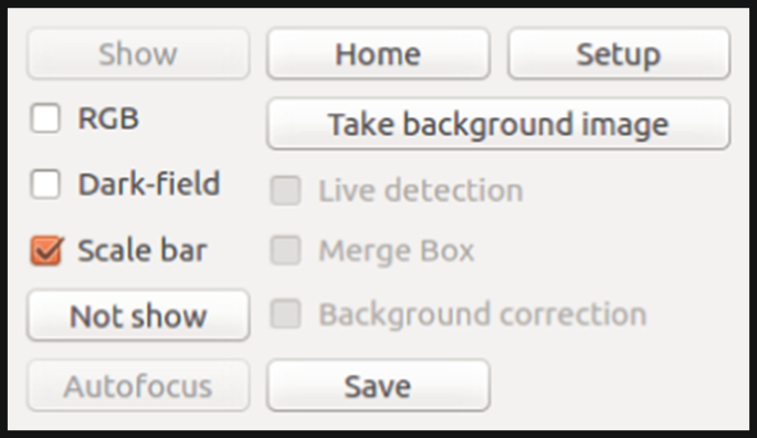
General configuration panel
Color mode: The RGB check box allows swapping between the gray scale and RGB color mode, for cameras that support this feature.
Illumination mode: As explained in Sect. 7.3.2, using an LCD several illumination modes can be achieved. The Dark-field check box allows the user to switch between brightfield and darkfield illumination modes.
Background correction: In Sect. 7.6.2, different methods to correct illumination defects are described. In this application, this correction is done dividing the original image by an unfocused image. The background image can be obtained automatically through Take background image button. After doing that, the live background correction can be activated enabling the Background correction check box. Then, the camera view panel shows the corrected image instead of the original captured image.
Save images: The application allows the user to capture an individual image at any time, through the Save button. The file name, image format, and location can be selected in a folder browser window.
Autofocus: An implementation of the autofocus algorithm based on Lorentzian function described in Sect. 7.5.3.2 is included, which required no more than three images. The Autofocus button allows the user to use this approach at any time.
Live diatoms detection: A trained diatom detector has been included. It is based on YOLO framework [23, 24], whose speed and accuracy allows making inference in real time, even in modest devices. The user can enable this feature through the Live detection check box. Then, in the camera panel, for each detected diatom a bounding box will be overlaid. Additionally, another algorithm has been included to merge possible overlapping bounding boxes. This feature can be activated enabling the Merge Box check box.
Setup and Home: Using the Setup button, the user can define the “Home” position. Then, the Home button automatically moves the stage to this position.
Scale bar: A useful tool commonly required by diatomists and other experts is the possibility to add a scale bar to the images. Enabling the Scale bar check box, a calibrated scale bar will be added to an image corner.

1.3 Stage Control
The motorized stage control panel is presented in Fig. 7.20. In this section, the user can configure the motion parameters and carry out the manual displacements through the sample.

Motorized stage control
First, the distance measurement unit can be switched between mm and μm. Then, the user can move the stage using the arrow buttons for X and Y axes and Z− or Z+ buttons for Z axis. For this case, the movement step can be configured between 5, 20, or 100 (mm or μm depending on the unit selected). Furthermore, if the user needs more accuracy, the desired step size can be set into the text fields for each axis. The Move button performs then a relative displacement taking into account the introduced values.
1.4 Scanning and Processing Functions
A set of automated functionalities can be found in the last panel (Fig. 7.21), which are briefly described:
Objective selection: In addition to the stage, the objective nosepiece is motorized too, so the automatic change between objectives has been included. To perform this action, the user only has to select the desired objective from the list and the procedure will automatically start.
Fig. 7.21 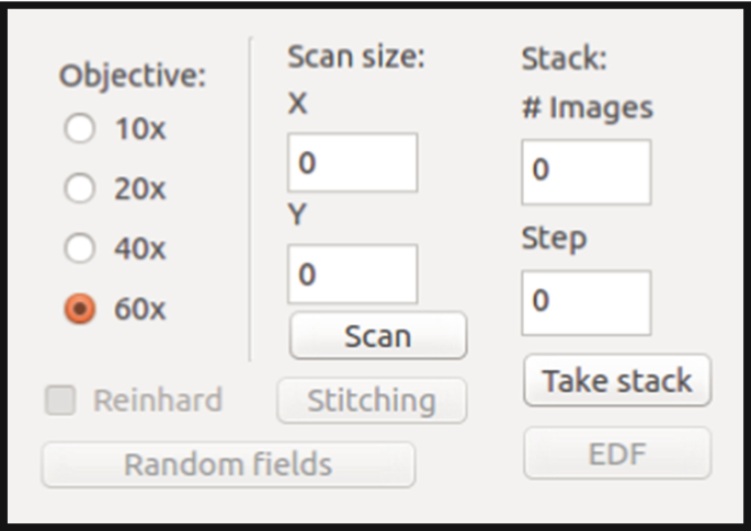
Scanning and processing settings
Sequential scanning: A sequential scanning algorithm has been implemented, following the pattern described in Sect. 7.5.2.1. The user only has to set the number of FOVs for X and Y axes and then click the Scan button. During the scanning, the autofocus algorithm is executed for each FOV, as well as a background correction. Also, for color images, a color normalization through Reinhard algorithm [12, 25] can be applied enabling Reinhard check box.
Random scanning: For certain applications, it is desirable not to acquire the FOVs according to a predefined pattern, as discussed in Sect. 7.5.2.2. For this reason, a random scanning procedure has been included to complement the sequential one. Clicking on the Random fields button, the algorithm will take as many FOVs as defined in the configuration file. In this case, due to the possible large differences in the focusing point between two consecutive FOVs, the two-phase autofocus algorithm described in Sect. 7.5.3.2 is applied for each step.
Stitching: The images in the sequential scanning procedure are captured with a certain overlap between them. This fact allows to perform a stitching algorithm to combine all individual FOVs into a high resolution image. Once the sequential scanning is finished, the user can carry out the stitching through the Stitching button.
Stack acquisition and multifocus fusion: In addition to capture images along X and Y axes, it is possible to acquire them at Z axis, that is, a stack of images at different focusing points. The user has to select the number of images to capture (# Images) and the distance between them (Step) and then click on the Take stack button to start the procedure at the current Z position. Also, once the stack is already captured, a multifocus fusion algorithm can be applied, clicking on EDF button that is an implementation of the Extended Depth of Field technique [5, 7].

Rights and permissions
Copyright information
© 2020 Springer Nature Switzerland AG
About this chapter

Cite this chapter
Sánchez, C., Ruiz-Santaquiteria Alegre, J., Espinosa Aranda, J.L., Salido, J. (2020). Automatization Techniques. Slide Scanning. In: Cristóbal, G., Blanco, S., Bueno, G. (eds) Modern Trends in Diatom Identification. Developments in Applied Phycology, vol 10. Springer, Cham. https://doi.org/10.1007/978-3-030-39212-3_7
Download citation
DOI: https://doi.org/10.1007/978-3-030-39212-3_7
Published:
Publisher Name: Springer, Cham
Print ISBN: 978-3-030-39211-6
Online ISBN: 978-3-030-39212-3
eBook Packages: Biomedical and Life SciencesBiomedical and Life Sciences (R0)