
Abstract
Porcine epidemic diarrhea virus (PEDV), a member of the genus Alphacoronavirus, has resulted in significant economic losses in the European, Asian, and North American swine industries in previous years. PEDV infection causes acute diarrhea/vomiting, dehydration, and high morbidity and mortality in seronegative neonatal piglets. In this chapter, materials and methods for performing immunohistochemistry (IHC) for the detection of PEDV antigens in frozen or formalin-fixed, paraffin-embedded (FFPE) tissues are provided. In IHC of frozen tissues where viral antigens are well preserved, the use of specific antibodies labeled with fluorescence dyes provides excellent advantages and convenience, resulting in high sensitivity and specificity of IHC and reduction of operation time. In IHC of FFPE tissues where tissue or cell morphology is well preserved, the use of specific antibodies labeled with enzymes, such as alkaline phosphatase, also gives rise to significant advantages in defining the correlation of viral antigens with histopathologic lesions. PEDV antigens in frozen tissues are visualized as green staining in the cytoplasm of infected cells by fluorescent dyes conjugated with antibodies when activated by exciting light of a specific wavelength under a fluorescence microscope. In FFPE tissues, PEDV antigens are visualized as red staining in the cytoplasm of infected cells by the deposition of the substrate chromogen, Fast Red.
You have full access to this open access chapter, Download protocol PDF
Key words
1 Introduction
The family Coronaviridae can be divided into four genera: Alphacoronavirus, Betacoronavirus, Gammacoronavirus, and Deltacoronavirus. Porcine epidemic diarrhea virus (PEDV), a member of the genus Alphacoronavirus, causes acute diarrhea, vomiting, dehydration, and high mortality in neonatal piglets, resulting in significant economic losses, initially only seen in the European and Asian swine industries over the last three decades. Recently, however, PEDV was first reported in the USA in May 2013 in Iowa [1]. Since then, the virus has rapidly spread nationwide [2] and to other countries in North America, including Canada and Mexico. The PED epidemic in the USA, from April 2013 to present, has led to a substantial loss of piglets (more than 10 % of US swine population). Because of similar clinical and pathogenic features between PEDV and another Alphacoronavirus, transmissible gastroenteritis virus (TGEV), or a Deltacoronavirus, porcine deltacoronavirus (PDCoV), differential laboratory tests are required for their diagnosis [3–5]. Reverse transcription -polymerase chain reaction (RT-PCR ) or quantitative RT-PCR (qRT-PCR) is useful for the rapid differential diagnosis; however, detection of viral antigens in tissues is essential for confirming each viral infection .
Viral antigens in frozen, fixed cells, or tissues can be detected by immunohistochemistry (IHC) (or immunohistochemical staining) using specific antibodies labeled with fluorescent dyes, such as Alexa Fluor®488 and fluorescein isothiocyanate (FITC), or enzymes, such as alkaline phosphatase (AP) and peroxidase. Immunofluorescence (IF) staining is the first immunohistochemical staining method but is still widely used in veterinary and medical diagnosis . With fundamentality of antigen -antibody and antibody-antibody binding reactions, antigens are visualized by fluorescent dyes conjugated with antibodies when activated by an exciting light of a specific wavelength (499–519 nm for Alexa Fluor®488 and 494–521 nm for FITC), under a fluorescence microscope. Due to the high sensitivity, specificity, and convenience in using Alexa Fluor®488 in frozen tissues, this chapter details an IF staining method for the rapid and precise detection of PEDV antigens in PEDV-infected, frozen tissues, contributing to verification of the tissue sites of PEDV replication in infected pigs. A combination of PEDV-specific anti-sera as antigen detection antibody and secondary antibody conjugated with Alexa Fluor®488 is applied in the IF staining method. However, because of suboptimal conditions of tissue or cell morphology in frozen tissues, this staining method limits investigation of the correlation of PEDV antigens with histopathologic lesions.
To compensate for the limitation of IF staining in frozen tissues, additional IHC staining method using enzyme-labeled antibodies, i.e., immunoenzymological staining, in formalin-fixed, paraffin-embedded (FFPE) tissues is also provided in this chapter. After adding a substrate of enzyme, such as Fast Red, it generates insoluble particles that can be localized in cells or tissues under light microscope. Compared to the IF staining in frozen tissues, the IHC in FFPE tissues has more accurate localization of antigens with a better contrast ratio, contributing to defining the correlation of PEDV antigen -positive cells with severity of histopathologic lesions caused by PEDV, such as intestinal villous atrophy.
Only one serotype of PEDV has been reported from different countries [4]. There has been no evidence of cross-reactivity of PEDV with TGEV [6, 7]. The use of hyperimmune anti-sera or polyclonal antibodies against PEDV in IHC staining is likely able to detect geographically different strains of PEDV and differentiate them from TGEV in tissues [8, 9], but with a potential disadvantage of inducing background or false signals. To improve the sensitivity and specificity of IHC for the detection of PEDV antigens in tissues, the use of monoclonal antibodies to structural proteins of PEDV, such as spike (S) or membrane (M) protein, has been preferred [10, 11]. Prior to their application on the tissues, potential cross-reactivity of monoclonal antibodies to TGEV and PDCoV can be tested by more sensitive assays, such as enzyme-linked immunosorbent assay, immunoblotting, and immunoprecipitation, compared to IHC.
Tissue tropism of PEDV is related to the expression of aminopeptidase N (APN), a 150 kDa glycosylated transmembrane protein identified as the cellular receptor , on porcine small intestinal villous enterocytes [12]. PEDV-infected enterocytes rapidly undergo acute necrosis, leading to marked villous atrophy in the small but not large intestine [10]. In PEDV-infected nursing pigs, major histologic lesions include acute diffuse, severe atrophic enteritis , and mild vacuolation of superficial epithelial cells and subepithelial edema in cecum and colon [8–10, 13]. PEDV antigens are observed mainly in villous enterocytes of the small (duodenum to ileum) and large intestines (except rectum) [1, 8, 10]. Occasionally, a few PEDV-positive cells were detected in the intestinal crypt cells or the Peyer’s patches [1, 8–10]. No lesions were seen in the spleen, liver, lung, kidney, and mesenteric lymph node of orally and/or intranasally infected piglets [8]. Lung tissue of oronasally infected pigs was negative for PEDV antigen [1, 8–10]. PEDV antigens were not detected in other major organs, such as the pylorus, tonsils, liver, and kidneys. However, a recent study reported the replication of PEDV in swine pulmonary macrophages in vitro and in vivo [14].
Epidemic PEDV strains are highly enteropathogenic and acutely infect villous epithelial cells of the entire small and large intestines, but the jejunum and ileum are the primary sites of infection . To detect PEDV antigen in tissues and evaluate the pathogenicity of PEDV strains in pigs, the jejunum and ileum are the most critical tissue samples for performing IHC.
2 Materials
2.1 Solutions and Reagents (See Note 1 )
-
1.
Primary antibody: Monoclonal antibody 6C8-1 against the spike protein of PEDV strain DR13 kindly provided by Dr. Daesub Song, Korea Research Institute of Bioscience and Biotechnology, Daejeon, Korea.
-
2.
Secondary antibody: Goat anti-mouse IgG antibody conjugated with Alexa Fluor®488 (Invitrogen, Carlsbad, CA, USA) for IF staining in frozen tissues; goat anti-mouse IgG antibody labeled with alkaline phosphatase (Dako, Glostrup, Denmark) for IHC in FFPE tissues.
IF staining in frozen tissues
-
1.
10% (w/v) Sucrose solution (pH 7.2).
-
2.
Tissue -Tek® O.C.T™ Compound (Sakura Finetek USA, Inc., Torrance, CA, USA).
-
3.
Liquid nitrogen.
-
4.
25% (v/v) Acetone in ethanol.
-
5.
Prolong® Gold Antifade Mountant with 4′,6-diamidino-2-phenylindole, dihydrochloride (DAPI) (Invitrogen).
-
6.
1× Universal Blocking Reagent (Biogenex, Fremont, CA).
-
7.
1× Phosphate-buffered saline (PBS) (pH 7.4).
-
8.
1× PBS (pH 7.4) containing Tween 20, 0.1 % (Sigma Aldrich, St. Louis, MO, USA) (PBTS).
IHC in FFPE tissues
-
1.
Xylene (Sigma Aldrich).
-
2.
100, 95, 70, and 50 % ethanol.
-
3.
3 % (v/v) glacial acetic acid in deionized water.
-
4.
Proteinase K (Invitrogen, Carlsbad, CA, USA).
-
5.
1× Universal Blocking Reagent (Biogenex).
-
6.
1× PBS (pH 7.4).
-
7.
1× PBS (pH 7.4) containing Tween 20, 0.1 % (Sigma Aldrich, St. Louis, MO, USA) (PBTS).
-
8.
Fast Red (Roche Applied Science, Mannheim, Germany).
-
9.
0.1 M Tris buffer (pH 8.2).
-
10.
Gill’s or Mayer’s hematoxylin (Sigma Aldrich).
-
11.
Ultramount Permanent Aqueous Mounting Medium (Dako).
-
1.
2.2 Equipment/Tools
-
1.
Glass slide-staining dishes or jars.
-
2.
Slide racks and trays.
-
3.
Humidified chamber tray with lid.
-
4.
Adjustable pipettors with tips.
-
5.
Barrier Dako Pen (Dako).
-
6.
4 °C Refrigerator.
-
7.
37 °C Incubator.
-
8.
Microscope slides: Superfrost™ Plus Gold Slides (Fisher Scientific, PA, USA).
-
9.
Glass cover slip (Fisher Scientific).
-
10.
Rocker platform shaker.
-
11.
Kimberly-Clark® Kimwipes™ (Kimberly-Clark Corporation, Irving, Texas, USA).
-
12.
Vortex mixer.
IF staining in frozen tissues only
-
1.
−80 °C freezer.
-
2.
Cryostat.
-
3.
Glass dropping pipettes.
-
4.
Fluorescence microscope.
IHC in FFPE tissues only
-
1.
Microtome.
-
2.
60 °C Oven.
-
3.
10 ml Syringes.
-
4.
0.9 μm Syringe filters.
-
5.
Permount permanent mounting media (Fisher Scientific).
-
6.
Light microscope.
-
1.
3 Methods
3.1 Tissue or Slide Preparation
3.1.1 IF Staining in Frozen Tissues
One to three pieces (2–3 cm per piece) of duodenum, proximal jejunum, mid-jejunum, distal jejunum, ileum, cecum, and colon are collected and perfused with 100–400 ml of 10 % sucrose solution, depending on the size of the immersed tissue . The tissues are trimmed, embedded in OCT compound, and frozen at −80 °C or by liquid nitrogen. Using a cryostat (−20 to −25 °C), tissue sections are cut at 5 to 10 μm thickness and mounted on Superfrost™ Plus Gold Slides. The tissue slides can be stored at −70 to −80 °C for several months. At the beginning of IF staining, the slides are dried for 30 min to 1 h at room temperature (RT). The slides are then fixed and dehydrated with 25 % acetone for 30 min to 1 h at RT.
3.1.2 IHC in FFPE Tissues
FFPE tissue sections are cut at 3 to 4 μm thickness using a microtome and mounted on Superfrost™ Plus Gold Slides in a 55 °C water bath. Tissue slides are dried at 60 °C for 1 h or at 37 °C for 3–4 h or overnight.
3.2 Deparaffinization and Rehydration of Tissue Section Slides (IHC in FFPE Tissues Only)
-
1.
Place slides of FFPE tissues (Sect. 3.1.2) in a slide rack for 15 min at 60 °C.
-
2.
Place the slides in the rack into xylene for 20 min at RT to remove the paraffin.
-
3.
Place the slides in 100 % ethanol for 5 min to rehydrate tissues through a graded ethanol series (100–50 %; the following steps 3–6). Repeat through two changes of fresh 100 % ethanol, 5 min for each.
-
4.
Place the slides in 95 % ethanol for 10 min.
-
5.
Place the slides in 70 % ethanol for 7 min.
-
6.
Place the slides in 50 % ethanol for 7 min.
-
7.
Move the slides into deionized water and wash at RT for 1 min.
-
8.
Move the slides into PBS and immerse at RT for 7 min (see Note 2 ).
3.3 Antigen Retrieval (IHC in FFPE Tissues Only)
-
1.
Lay the deparaffinized, rehydrated tissue slides (Sect. 3.2) across a horizontal slide tray or humidified chamber tray.
-
2.
Gently and carefully blot the slides with Kimwipes around the tissue , as well as the non-charged slide surface.
-
3.
Surround the tissue with a hydrophobic barrier using the Dako Pen.
-
4.
Apply 300–500 μl of 3 % glacial acetic acid to quench endogenous alkaline phosphatase and incubate at RT for 20 min.
-
5.
Apply 300–500 μl of proteinase K (100 μg/ml) to the tissue sections with the hydrophobic barrier and incubate at 37 °C for 30 min.
-
6.
Rinse the slides in PBS for 5 min on a rocker platform shaker. Repeat through three changes of fresh PBS, 5 min for each.
3.4 Immunohisto-chemical Staining Protocol
3.4.1 IF Staining in Frozen Tissues
-
1.
Place the fixed, dehydrated tissues (Sect. 3.1.1) in PBS at RT for 5 min to rehydrate (see Note 2 ).
-
2.
Lay the tissue slides across a horizontal slide tray or humidified chamber tray.
-
3.
Gently and carefully blot the slides with Kimwipes around the tissue , as well as the non-charged slide surface.
-
4.
Surround the tissue with a hydrophobic barrier using the Dako Pen.
-
5.
Apply 300–500 μl of 1× Universal Blocking Reagent to the tissue within the hydrophobic barrier and incubate at 37 °C for 30 min.
-
6.
Drain the slides and place them on a horizontal surface.
-
7.
Apply the primary antibody (diluted 1:200 in PBTS) enough to cover the tissue section (200–300 μl) and incubate in a humidified chamber at 4 °C overnight (see Note 3 ).
-
8.
Rinse the slides gently with PBS on a rocker platform shaker at RT for 5 min. Repeat through three changes of fresh PBS, 5 min for each.
-
9.
Apply the Alexa Fluor®488-labeled secondary antibody (diluted 1:200 in PBTS) enough to cover the tissue section (200–300 μl) and incubate in a humidified chamber at 37 °C for 1 h (see Note 3 ).
-
10.
Rinse the slides gently with PBS on a rocker platform shaker at RT for 5 min. Repeat through three changes of fresh PBS, 5 min for each.
-
11.
Gently blot the slides with Kimwipes around the tissue and place horizontally.
-
12.
Apply 3–5 drops of Prolong® Gold Antifade Mountant (with DAPI) to the tissue section using a glass dropping pipette and immediately put a cover slip on (see Note 4 ).
-
13.
The slides are ready to be evaluated under fluorescence microscope. They need to be stored in a dark area until evaluation. PEDV antigens will appear to be green or as fluorescent staining in the cytoplasm of infected cells (Fig. 1). Cell nuclei are stained blue with DAPI.
Fig. 1 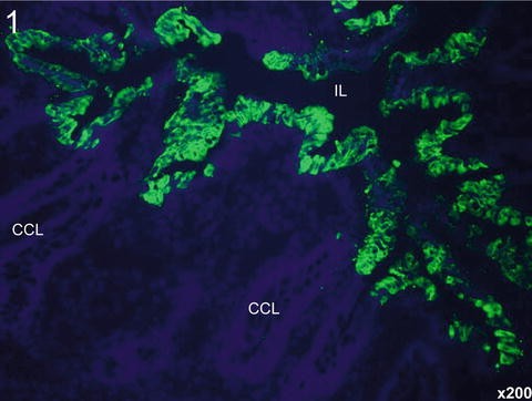
Detection of PEDV antigens (green staining) in the cytoplasm of enterocytes lining atrophied villi by immunofluorescence staining in frozen jejunal tissues using a monoclonal antibody specific for the spike protein of PEDV and secondary antibody conjugated with Alexa Fluor®488. Original magnification ×200. IL intestinal lumen, CCL crypt cell layer. Nuclei were stained with blue-fluorescent 4′,6-diamidino-2-phenylindole, dihydrochloride

3.4.2 IHC in FFPE Tissues
-
1.
When antigen retrieval procedure is completed (Sect. 3.3), drop 300–500 μl of 1× Universal Blocking Reagent on the tissue within the hydrophobic barrier and incubate at 37 °C for 30 min.
-
2.
Drain the slides and place them on a horizontal surface.
-
3.
Apply the primary antibody (diluted 1:200 in PBTS) enough to cover the tissue section (200–300 μl) and incubate in a humidified chamber at 4 °C overnight (see Note 3 ).
-
4.
Rinse the slides gently with PBS on a rocker platform shaker at RT for 5 min. Repeat through three changes of fresh PBS, 5 min for each.
-
5.
Apply the AP-labeled secondary antibody (diluted 1:200 in PBTS) enough to cover the tissue section (200–300 μl) and incubate in a humidified chamber at 37 °C for 1 h (see Note 3 ).
-
6.
Rinse the slides gently with PBS on a rocker platform shaker at RT for 5 min. Repeat through three changes of fresh PBS, 5 min for each.
-
7.
For step 6, add 1 tablet of Fast Red in 2 ml of 0.1 M Tris buffer (pH 8.2), depending on the number of the tissue sections, and dissolve by a vortex mixer (see Note 5 ).
-
8.
Drain the slides and place them on a horizontal surface.
-
9.
Apply the Fast Red solution enough to cover the tissue section (300–500 μl) and incubate in a humidified chamber at RT for 30–60 min (see Note 6 ).
-
10.
Place the slides in a rack and rinse well in distilled water. Three changes, 2 min each.
-
11.
Tissue sections are counterstained in a glass dish with Gill’s hematoxylin at RT for 10 min.
-
12.
Rinse the slides thoroughly in tap water for 5 min, and move into deionized water.
-
13.
Drain the slides and place them on a horizontal surface.
-
14.
Apply 2–4 drops of Permanent Aqueous Mounting Medium to the tissue section (see Note 7 ), and immediately put a cover slip on (see Note 8 ).
-
15.
The slides are ready to be evaluated under light microscope. PEDV antigens will appear as a red precipitate in the cytoplasm of infected cells (Fig. 2). Cell nuclei are stained blue with hematoxylin.
Fig. 2 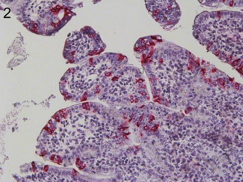
Detection of PEDV antigens (red staining) in the cytoplasm of enterocytes lining atrophied villi by immunohistochemical staining in formalin-fixed, paraffin-embedded jejunal tissues using a monoclonal antibody specific for the spike protein of PEDV and secondary antibody conjugated with alkaline phosphatase. Original magnification ×200. Immunohistochemistry. Fast Red. Gill’s hematoxylin counterstaining

4 Notes
-
1.
Use of fresh reagents is recommended. A large amount of washing buffer, 1× PBS, is needed, because complete washing is critical to reduce background and increase true signals.
-
2.
Throughout immunostaining procedures, the tissues should stay rehydrated. Adequate antigen -antibody or antibody-antibody binding reaction is not expected in dried tissues, resulting in poor or weak staining results or a high level of background staining. It is also critical for a comparative immunostaining study in multiple different tissues.
-
3.
The optimal dilutions of primary and secondary antibodies should be tested and selected in both frozen and FFPE tissue conditions.
-
4.
Instead of plastic pipette tips, the use of glass dropping pipette will reduce the number of bubbles in the mounting medium as applied to the tissue sections.
-
5.
When the Fast Red tablet is completely dissolved, the solution can be filtered via 0.9 μm syringe filter and used to reduce an irregular deposition of Fast Red on the tissues or background.
-
6.
The color development, including intensity of true or false signals, in all tissue slides tested should be frequently monitored under the microscope. Wipe the non-charged slide surface with Kimwipes before putting the tissue slides on the microscope.
-
7.
Gently drop the mounting medium so as not to create bubbles. The mounted slides need to be evaluated as soon as possible, because bubbles can be created spontaneously in the mounted medium.
-
8.
To make the stained slides permanent, a large amount of mounting medium can be applied to the tissues so that the entire section is covered. Place slides horizontally in a 60 °C oven for 30 min to allow the medium to harden. Remove the slides from the oven, and allow them to cool at RT. Dip the slides in xylene and cover slip with permount permanent mounting medium (Fisher Scientific).
References
Stevenson GW, Hoang H, Schwartz KJ, Burrough EB, Sun D, Madson D, Cooper VL, Pillatzki A, Gauger P, Schmitt BJ, Koster LG, Killian ML, Yoon KJ (2013) Emergence of Porcine epidemic diarrhea virus in the United States: clinical signs, lesions, and viral genomic sequences. J Vet Diagn Invest 25(5):649–654
Cima G (2013) Fighting a deadly pig disease. J Am Vet Med A 243(4):467–470
Jung K, Saif LJ (2015) Porcine epidemic diarrhea virus infection: Etiology, epidemiology, pathogenesis and immunoprophylaxis. Vet J 204(2):134–143
Saif LJ, Pensaert MP, Sestak K, Yeo SG, Jung K (2012) Coronaviruses. In: Zimmerman JJ, Karriker LA, Ramirez A, Schwartz KJ, Stevenson GW (eds) Diseases of swine, 10th edn. Iowa State University, Wiley-Blackwell, pp 501–524
Jung K, Hu H, Eyerly B, Lu Z, Chepngeno J, Saif LJ (2015) Pathogenicity of 2 porcine deltacoronavirus strains in gnotobiotic pigs. Emerg Infect Dis 21(4):650–654
Hofmann M, Wyler R (1989) Quantitation, biological and physicochemical properties of cell culture-adapted porcine epidemic diarrhea coronavirus (PEDV). Vet Microbiol 20(2):131–142
Pensaert MB, Debouck P, Reynolds DJ (1981) An immunoelectron microscopic and immunofluorescent study on the antigenic relationship between the coronavirus-like agent, CV 777, and several coronaviruses. Arch Virol 68(1):45–52
Debouck P, Pensaert M, Coussement W (1981) The pathogenesis of an enteric infection in pigs, experimentally induced by the coronavirus-like agent, Cv-777. Vet Microbiol 6(2):157–165
Sueyoshi M, Tsuda T, Yamazaki K, Yoshida K, Nakazawa M, Sato K, Minami T, Iwashita K, Watanabe M, Suzuki Y et al (1995) An immunohistochemical investigation of porcine epidemic diarrhoea. J Comp Pathol 113(1):59–67
Jung K, Wang Q, Scheuer KA, Lu Z, Zhang Y, Saif LJ (2014) Pathology of US porcine epidemic diarrhea virus strain PC21A in gnotobiotic pigs. Emerg Infect Dis 20(4):662–665
Kim O, Chae C, Kweon CH (1999) Monoclonal antibody-based immunohistochemical detection of porcine epidemic diarrhea virus antigen in formalin-fixed, paraffin-embedded intestinal tissues. J Vet Diagn Invest 11(5):458–462
Li BX, Ge JW, Li YJ (2007) Porcine aminopeptidase N is a functional receptor for the PEDV coronavirus. Virology 365(1):166–172
Coussement W, Ducatelle R, Debouck P, Hoorens J (1982) Pathology of experimental CV777 coronavirus enteritis in piglets. I. Histological and histochemical study. Vet Pathol 19(1):46–56
Park JE, Shin HJ (2014) Porcine epidemic diarrhea virus infects and replicates in porcine alveolar macrophages. Virus Res 191:143–152
Acknowledgements
Salaries and research support were provided by state and federal funds appropriated to the Ohio Agricultural Research and Development Center, The Ohio State University. This work was supported by a grant from the OARDC SEEDS, Grant # OAOH1536.
Author information
Authors and Affiliations
Corresponding author
Editor information
Editors and Affiliations
Rights and permissions
Copyright information
© 2016 Springer Science+Business Media New York
About this protocol
Cite this protocol
Jung, K. (2016). Immunohistochemical Staining for Detection of Porcine Epidemic Diarrhea Virus in Tissues. In: Wang, L. (eds) Animal Coronaviruses. Springer Protocols Handbooks. Humana Press, New York, NY. https://doi.org/10.1007/978-1-4939-3414-0_2
Download citation
DOI: https://doi.org/10.1007/978-1-4939-3414-0_2
Published:
Publisher Name: Humana Press, New York, NY
Print ISBN: 978-1-4939-3412-6
Online ISBN: 978-1-4939-3414-0
eBook Packages: Springer Protocols