Abstract
In the present work, we used the ultraviolet–Visible (UV–Vis) spectroscopy technique to find the optical band gap of zinc phthalocyanine nanoparticles (ZnPc-NP) experimentally. Moreover, we used a time-dependent density functional theory (TDDFT) to simulate the UV–Vis absorption spectrum of ZnPc molecule in gas and solution phases. The ZnPc-NP absorption spectrum shows a shift toward higher energies compared to the bulk ZnPc. The simulated UV–Vis and the experimental nanoparticle’s spectrum were found to have a good agreement. The ZnPc energy band gap from the DFT calculations shows how it’s possible to get wider range of energy band gap for the ZnPc. The ZnPc-NP’s size and shape were examined using the transmission electron microscope (TEM).
Introduction
Preparation and characterization of nanostructure systems during the past decades has got scholars’ attention due to their interesting optical, electronic and chemical properties (Weller 1996; Alivisatos 1996). Scholars focused on studying the nanoparticles derived from metals and inorganic semiconductors, due to their significant electrical and optical properties compared to bulk materials (Murray et al. 1993; Braum et al. 1996; Micic et al.1995; Heath et al. 1994).
As the dimension of a material gets smaller, the physical and optical properties were observed to change; such as the absorption spectrum of the material (Chen et al. 1994). We can observe this effect obviously in exotic colors, which is reflected in their absorption and emission spectra; this effect was attributed to the gradual conversion of the material edges into sharp and discrete molecular (excitonic) absorption bands; therefore, band gap becomes more larger, which is also known as the size quantization effect (Smith and Nie 2010).
The control of material’s band gap energy, is an important issue in the semiconductor properties and nanoparticles applications (Dabbousi et al.1995; Colvin et al.1994). The theoretical band gap can be represented by Eq. 1 (Yoffe 1993):
where E bulkg is the bulk band gap. R, m *e , m *h and ε are the radius of the nanoparticle, electron’s effective mass, hole’s effective mass, and the dielectric constant, respectively. The third term in Eq. 1 arises due to the columbic attraction which is usually ignored in a high dielectric constant medium. Equation 1 shows that as the material’s size gets smaller, its band gap energy gets higher. This formula is used to estimate the band gap for an isotropic spherically nanostructures. Therefore, values obtained for the energy band gap from Eq. 1 are usually over estimated (Li et al. 2001).
Zinc phthalocyanine (ZnPc) is an organic semiconductor compound (Eley 1948), which is used as a photosensitizer for Photodynamic therapy (PDT) (Fadel et al. 2010; Ping et al. 2016); and in optoelectronic applications such as organic light-emitting diodes (OLED) (Run-Da et al. 2013; Heo et al. 2013) and organic photovoltaics (OPVs) (Kim and Bard 2004). The size effect on the electrical and optical properties of metal-free phthalocyanine nanoparticles have been studied using the physical method such as mechanical grinding, it’s found that as the material’s size gets smaller their photoconductivity efficiency was improved (Saito et al. 1991). However, fabrication of very tiny homogeneous nanoparticles of MPc’s by using the physical methods (grinding method) is not feasible. Therefore, other methods have been developed to fabricate a tiny size of organic nanoparticles such as liquid phase direct precipitation (LPDP) (Chen et al. 1991), laser ablation (Tamaki et al. 2002) and liquid–liquid interface recrystallization techniques (LLIRCT) [Patil et al. 2007].
The spectroscopic study of metal phthalocyanine compounds (MPc) gives very useful information about the charge transfer between the central metal and the phthalocyanine ligand, as a result of the overlapping between their energy wave functions. One of the easiest ways to determine the optical energy band gap of the material is using their absorption spectrum.
In the present work, we studied the zinc phthalocyanine bulk and nanoparticles thin film’s energy band gap using a simple technique, which is the UV–Vis absorption spectrum technique. The objective of this work is to synthesize the zinc phthalocyanine nanoparticles (ZnPc-NP) to study the size effect on the absorption spectrum and calculate their band gap energy. Additionally, density functional theory (DFT) calculations were used to find the minimum energy and optimized geometry for ZnPc molecule, and to simulate the expected UV–Vis absorption spectrum. Nanoparticles’s size and shape were examined using the transmission electron microscope (TEM) images.
Experimental and theoretical work
Synthesis of zinc phthalocyanine nanoparticles (ZnPc-NP)
Zinc phthalocyanine (ZnPc) (99% purity) and cetyltrimethylammonium bromide (CTAB) were purchased from Sigma-Aldrich, through a local chemical provider. The surfactant was added to (150 ml) distilled water and stirred at 50 °C until a transparent solution was observed. ZnPc (0.05 g) was dissolved in 17 ml concentrated sulfuric acid (98%), then the solution was added dropwisely, until a dark blue colloidal solution was obtained into an aqueous surfactant solution in an ice-methanol-water bath to keep the temperature of the solution at 0 °C, the solution was under vigorous magnetic stirring. The resulted solution was washed to neutral with water using an ultra-filter. A trace amount of sulfuric acid was removed by anion exchange resin (Wang et al. 1999). The precipitate of the MPc nanoparticles was washed with water and acetone to remove any residual surfactants. ZnPc nanoparticles were dispersed into Dimethylformamide (DMF) and then spin coated at low speed (500 rpm for 40 s) over clean glass substrates, to fabricate thin films. The thin films were annealed at 200 °C to remove the solvent.
Characterization
The UV–Vis spectrum was measured using Shimadzu UV–Vis spectrometer. UV–Vis is an easy and straightforward technique to determine the band gap energy of organic π conjugated systems. The optical band gap can be estimated according to Eq. 2 (Costa et al. 2016):
where E g represents the optical energy band gap (in eV) and λ th represents the threshold wavelength (in nm), obtained from the onset of the absorption spectrum (Bhadwal et al. 2014). The transmission electron microscope (TEM) images were prepared on carbon grids and scanned using Hitachi H-7000, to examine the size and the shape of our nanoparticles.
Theoretical calculations
The DFT Calculations were carried out using Gaussian09 package (Frisch et al. 2010). The applied basis sets are 6-31G(d) and the pseudo-potentials LANL2DZ. The 6-31G(d) (H, C, and N atoms) sets has the polarization function, which it is suitable for atoms that fill s and p atomic orbitals. The pseudo-potentials LANL2DZ is used for heavy atoms such as Zn atom. The ZnPc structure has been optimized using the B3LYP function. To investigate the UV spectra we have employed time-dependent density functional theory TDDFT on the optimized structure in gas and DMF solvent. Moreover, we have reported the energy of the molecular orbitals and then calculated the ΔE (gap) between the frontier HOMO–LUMO orbitals and between molecular orbitals of main peaks of the spectra.
Results and discussion
Organic molecules of phthalocyanine and their derivatives show optical characteristics because of their ring structure. They have two types of energy bands; B-band (γ or Soret band) and Q-band (α band in porphyrin) (Stillman and Nyokong 1989). The zinc phthalocyanine bulk absorption spectrum and the zinc phthalocyanine nanoparticles thin films are shown in Fig. 1. For bulk material, the B-band shows the maximum peak at 332.5 nm. The peak at 618 nm in the Q-band energy was assigned to the first π–π* transition on the phthalocyanine macrocycle (Davidson 1982) and the shoulder at 676 nm could be assigned to an exciton state (Mack and Stillman 1997). ZnPc-NP absorption spectrum shows a shift of the absorption peaks toward the short wavelength region (blue shift). In the B-band region, the 332.5 nm peak was shifted to the 330.5 nm (Δλ = 2 nm); while for the 618 nm peak had been shifted to 605.5 nm in the nanoparticles spectrum, which is considered as the largest shift in the wavelengths (Δλ = 12.5 nm), among all of them. The blue shift of λ can be described like “quantum size effect” in semiconductor nanoparticles (Laurs and Heiland 1987). The 676 nm shoulder was developed into a clear, broad peak (677–691 nm). ZnPc-NP absorption spectrum shows the broadening spectrum compared to the bulk ZnPc, which could be attributed to a change in the type of molecules stacking due to the formation of the nanoparticles (Masuhara et al. 2003).
The optical band gap was determined from the analysis of the absorption spectrum as described by Tauc plot using the formula (El-Nhass et al. 2001)
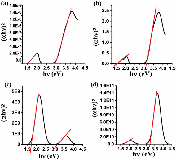
where hv is the energy of incident photons and E g is the value of the optical band gap corresponding to transitions indicated by the value of n. \(\alpha_{\text{o}}\) is a constant which depends on the transition probability. A best linear fit was found for n = 0.5 which indicates an allowed direct transitions in the material (Kim et al. 2012). The extrapolation of the Tauc plot to the abscissa gives the value of the optical band gap. The values of the energy band gaps for the bulk material and the nanoparticles of ZnPc in the two regions are shown in Fig. 2a, b. The DFT data was used to figure out the band gap energy and threshold wavelength. The Tauc plot extracted from DFT calculations, in gas phase and in DMF solvent are shown in Fig. 2c, d. Band gap values that have been extracted from the experimental and the theoretical data at and threshold wavelengths are listed in Table 1.
The ZnPc nanoparticles average size and shape were determined by using transmission electron microscope (TEM). Almost an oval geometry (non-spherical shape) of the nanoparticles was observed. Furthermore, clustering and aggregation effect was observed in our samples as shown in Fig. 3, left. The average calculations of the nanoparticle’s diameter were in the order of 6.5 ± 0.3 nm as shown in Fig. 3, right
Density functional theory (DFT) study on ZnPc was performed, to find the estimated ZnPc band gap for a single molecule in gaseous and solution (DMF) phase, for comparison purposes with the experimental nanoparticle’s band gap. The optimized structure of ZnPc is shown in Fig. 4 which was a planar structure.
In the Q-band the electronic transition occurs from HOMO, which has an electronic density mainly located on the phthalocyanine molecule, to the LUMO, which has a small electronic density on Zn–N bond as shown in Fig. 5. The calculated energy gap is E g = 2.23 eV. The B-band electronic transition occurs between HOMO-4/LUMO orbitals, E g = 3.91 eV as shown in Fig. 6. The electrostatic potential surface and contours of ZnPc is shown in Fig. 7, where the δ+ and δ− charges are residing on Zn and on N-atoms, respectively. The electrostatic potential surfaces can give us an idea about how the ZnPc molecules are stacking in the nanostructure system. The most probable aggregation in the system is the H-aggregation due to high energy shift in the absorption spectrum and the information provided by the electrostatic potential distribution on the ZnPc molecule.
The UV–Vis simulation was performed after optimizing the molecule geometry at minimum energy (no imaginary frequency) or transition states (a single imaginary frequency) as shown in Fig. 8. The simulated UV–Vis shape has a good agreement with the experimental spectra, the B-band and Q-band were located in the same range of wavelength as we observed in the experimental spectra. The position of the maximum peaks is listed in Table 2. Furthermore, the associated calculations of the density of states of the ZnPc are shown in Fig. 9 in gas phase (a) and of the DMF solvent (b) which shows the virtual and occupied orbitals.
Conclusions
The UV–Vis technique was used to determine the band gap energy for bulk ZnPc and ZnPc nanoparticles (ZnPc-NP) thin films. The shift in the absorption position was considered as an evident of the formation of the nanoparticles. Nanoparticle’s band gap energy recorded higher values at both B-band and Q-band (3.13 and 1.55 eV, respectively) compared to the values for similar bands of bulk ZnPc (2.97 and 1.53 eV, respectively). Therefore, ZnPc-NP’s absorption spectrum shows a shift toward the higher energy photons; the 332.5 nm peak was shifted to the 330.5 nm (B-band region); and the 618 nm peak had been shifted to 605.5 nm (Q-band region). DFT calculations using the DFT-B3LYP method was used to find the band gap of ZnPc molecule and compared with the experimental value; DFT calculations show that the maximum tuned band width could reach 2.23 eV. The simulated UV–Vis has a good agreement in shape and position of B-band and Q-band with the experimental measurements. Transmission electron microscope (TEM) was used to examine the ZnPc nanoparticles size, they have an average diameter of 6.5 ± 0.3 nm. Controlling the band gap of the organic semiconductors is an important issue, for applications such as light emitting diode, organic photovoltaic devices and the photodynamic therapy as a photosensitizer.
References
Alivisatos AP (1996) Semiconductor clusters, nanocrystals, and quantum dots. Science 271:933–937. doi:10.1126/science.271.5251.933
Bhadwal AS, Tripathi RM, Gupta RK, Kumar N, Singha RP, Shrivastav A (2014) Biogenic synthesis and photocatalytic activity of CdS nanoparticles. RSC Adv 4:9484–9490. doi:10.1039/c3ra46221h
Braum PV, Osenar P, Stupp SI (1996) Semiconducting superlattices templated by molecular assemblies. Nature 380:325–328. doi:10.1038/380325a0
Chen W, McLendon G, Marchetti A, Rehm JM, Michal IF, Myers C (1994) Size dependence of radiative rates in the indirect band gap material AgBr. J Am Chem Soc 116:1585–1586. doi:10.1021/ja00083a060
Chen HZ, Pan C, Wang M (1991) Characterization and photoconductivity study of TiOPc nanoscale particles prepared by liquid phase direct reprecipitation. Nanostruct Mater 11:523–530. doi:10.1016/s0965-9773(99)00338-4
Colvin VC, Schlamp MC, Alivisatos AP (1994) Light-emitting diodes made from cadmium selenide nanocrystals and a semiconducting polymer. Nature 370:354–357. doi:10.1038/370354a0
Costa JCS, Taveira RJS, Lima CFRAC, Mendes A, Santos LMNBF (2016) Optical band gaps of organic semiconductor materials. Opti Mat 58:51–60. doi:10.1016/j.optmat.2016.03.041
Dabbousi BO, Bawendi MG, Onitsuka O, Rubner MF (1995) Electroluminescence from CdSe quantum-dot/polymer composites. Appl Phys Lett 66:1316. doi:10.1063/1.113227
Davidson AT (1982) The effect of the metal atom on the absorption spectra of phthalocyanine films. J Chem Phys 77:162. doi:10.1063/1.443636
El-Nhass MM, Soliman HS, Metwally HS, Farid AM, Farag AAM, El Shazly AA (2001) Optical properties of evaporated iron phthalocyanine(FePc) thin films. J Opti 30:121–129. doi:10.1007/bf03354732
Eley DD (1948) Phthalocyanines as semiconductors. Nature 162:819. doi:10.1038/162819a0
Fadel M, Kassab K, Fadeel DA (2010) Zinc phthalocyanine-loaded PLGA biodegradable nanoparticles for photodynamic therapy in tumor-bearing mice. Las Med Sci 25:283–292. doi:10.1007/s10103-009-0740-x
Frisch MJ et al. (2010) Gaussian 09, Revision B.01, Gaussian, Inc., Wallingford CT
Heath JR, Shiang JJ, Alivisatos AP (1994) Germanium quantum dots: optical properties and synthesis. J Chem Phys 101:1067. doi:10.1063/1.467781
Heo I, Kim J, Yim S (2013) Effect of incorporated PVP/Ag nanoparticles on ZnPc/C60 organic solar cells. J Nanosci Nanotechnol 13:4316–4319. doi:10.1166/jnn.2013.7451
Kim JY, Bard AJ (2004) Organic donor/acceptor heterojunction photovoltaic devices based on zinc phthalocyanine and a liquid crystalline perylene diimide. Chem Phys Lett 383:11–15. doi:10.1016/j.cplett.2003.10.132
Kim HJ, Kim JW, Lee HH, Lee B, Kim JJ (2012) Initial growth mode, nanostructure, and molecular stacking of a ZnPc:C60 bulk heterojunction. Adv Funct Mater 22:4244–4248. doi:10.1002/adfm.201200778
Laurs H, Heiland G (1987) Electrical and optical properties of phthalocyanine films. Thin Solid Films 149:129–142. doi:10.1016/0040-6090(87)90288-4
Li L, Hu J, Yang W, Alivisatos AP (2001) Band gap variation of size- and shape-controlled colloidal CdSe quantum rods. Nano Lett 1:349–351. doi:10.1021/nl015559r
Mack J, Stillman MJ (1997) Assignment of the optical spectra of metal phthalocyanine anions. Inorg Chem 36:413–425. doi:10.1021/ic960737i
Masuhara H, Nakanishi H, Sasaki K (2003) Single organic nanoparticles. Springer-Verlag, Berlin. ISBN 978-3-642-62429-2
Micic OI, Sprague JR, Curtis CT, Jones KM, Machol JL, Nozik AJ, Giessen H, Fluegel B, Mohs G, Peyghambarian N (1995) Synthesis and characterization of InP, GaP, and GaInP2 quantum dots. J Phys Chem 99:7754–7759. doi:10.1021/j100019a063
Murray CB, Norris DJ, Bawendi MG (1993) Synthesis and characterization of nearly monodisperse CdE (E = sulfur, selenium, tellurium) semiconductor nanocrystallites. J Am Chem Soc 115:8706–8715. doi:10.1021/ja00072a025
Patil KR, Sathaye SD, Hawaldar R, Sathe BR, Mandale AB, Mitra A (2007) Copper phthalocyanine films deposited by liquid-liquid interface recrystallization technique (LLIRCT). J Colloid Interface Sci 315:747–752. doi:10.1016/j.jcis.2007.07.031
Ping J, Peng H, Duan W, You F, Song M, Wang Y (2016) Synthesis and optimization of ZnPc-loaded biocompatible nanoparticles for efficient photodynamic therapy. J Mater Chem B 4:4482–4489. doi:10.1039/c6tb00307a
Run-Da G, Shou-Zhen Y, Peng W, Yi Z, Shi-Yong L, Yu C (2013) Increased performance of an organic light-emitting diode by employing a zinc phthalocyanine based composite hole transport layer. Chin Phys B 22:127304. doi:10.1088/1674-1056/22/12/127304
Saito T, Kawanishi T, Kakuta A (1991) Photocarrier generation process of phthalocyanine particles dispersed in a polymer: effects of pigment particle size, polymer matrix and addition of fine γ-alumina particles. Jpn J Appl Phys A 30:1182. doi:10.1143/jjap.30.l1182
Smith AM, Nie N (2010) Semiconductor nanocrystals: structure, properties, and band gap engineering. Acc Chem Res 43:190–200. doi:10.1021/ar9001069
Stillman MJ, Nyokong T (1989) Phthalocyanines, properties and applications, ed. by C.C. Leznoff and ABP. Lever; Chapt. 3, VCD, New York, ISBN 1-56081-916-2
Tamaki Y, Asahi T, Masuhara H (2002) Nanoparticle formation of vanadyl phthalocyanine by laser ablation of its crystalline powder in a poor solvent. J Phys Chem A 106:2135–2139. doi:10.1021/jp012518a
Wang Y, Deng K, Gui L, Tang Y, Zhou J, Cai L, Qiu J, Ren D, Wang Y (1999) Preparation and characterization of nanoscopic organic semiconductor of oxovanadium phthalocyanine. J Coll Interface Sci 213:270–272. doi:10.1006/jcis.1999.6132
Weller H (1996) Self-organized superlattices of nanoparticles. Angew Chem Int Ed Engl 35:1079–1081. doi:10.1002/anie.199610791
Yoffe AD (1993) Low-dimensional systems: quantum size effects and electronic properties of semiconductor microcrystallites (zero-dimensional systems) and some quasi-two-dimensional systems. Adv Phys 42(173):266. doi:10.1080/00018739300101484
Acknowledgements
We would like to submit our gratitude to Dr. Anas Lataifeh, Dr. Salim Khalil and Dr. Ghassab Al-Mazaideh from chemistry department; for their assistant and the their valued comments.
Author information
Authors and Affiliations
Corresponding author
Rights and permissions
Open Access This article is distributed under the terms of the Creative Commons Attribution 4.0 International License (http://creativecommons.org/licenses/by/4.0/), which permits unrestricted use, distribution, and reproduction in any medium, provided you give appropriate credit to the original author(s) and the source, provide a link to the Creative Commons license, and indicate if changes were made.
About this article
Cite this article
Hamam, K.J., Alomari, M.I. A study of the optical band gap of zinc phthalocyanine nanoparticles using UV–Vis spectroscopy and DFT function. Appl Nanosci 7, 261–268 (2017). https://doi.org/10.1007/s13204-017-0568-9
Received:
Accepted:
Published:
Issue Date:
DOI: https://doi.org/10.1007/s13204-017-0568-9


