Abstract
The adaptation of sponges to freshwater environments was a major event in the evolutionary history of this clade. The transition from a marine environment to freshwater ecosystems entailed a great number of adaptations to more unstable habitats, such as the ability to form resistance gemmules as a defense mechanism against environmental adversity. However, data on the parameters that modulate hatching and growth of these animals are scarce. In the present study, the growth response capacity of Ephydatia fluviatilis (Porifera: Spongillidae) has been evaluated in relation to both water alkalinity and light intensity. The results obtained revealed a positive association between the growth capacity of this freshwater sponge and high alkalinity values. On the other hand, exposure to light, regardless of its intensity, affected the development and distribution of the symbionts, which in turn, corresponds to a higher growth rate of the sponge. The obtained data suggest an explanation for the greater distribution of this species in alkaline environments. The results of this work also shed light on the importance of the symbiosis phenomenon in E. fluviatilis.
Introduction
Having its origins dated back to more than 630 million years ago (Ehrlich et al. 2018; Schuster et al. 2018), the phylum Porifera constitutes one of the earliest metazoan groups on the planet still extant. The structural simplicity and its phenotypic plasticity are possibly the key factors for the success of this phylum, which counts for 9531 valid species (de Voogd et al. 2023), although it is considered that this number could be as high as 15,000 species (Degnan et al. 2015). Likewise, the great adaptive radiation of sponges has allowed them to colonize virtually any aquatic environment.
Despite this, the only clade of sponges that has adapted to inhabit freshwater areas is the family Spongillidae Gray 1867 (Kenny et al. 2020). The members from this family have been able to occupy almost every kind of freshwater environment with a global distribution (Manconi and Pronzato 2008). This has led to significant changes in their physiology, as they have necessarily adapted to a more volatile environment. Therefore, they have adopted strategies of tolerance to adverse ecological conditions, like abrupt temperature changes (Schill et al. 2006) and hypoxic conditions (Reiswig and Miller 1998). To accommodate such circumstances, these sponges are able to form gemmules, resistance structures made up of a protective spiculated cover that stores in its interior a large number of totipotent cells, known as thesocytes (Calheira et al. 2019).
Most freshwater sponges present seasonal cycles of gemulation, germination and growth, all in accordance with the physical and chemical patterns of the biotope (Melão and Rocha 1999). Among these, some parameters such as temperature and illumination (Benfey and Reiswig 1982), food availability (Frost 1991) and water level stand out. However, data on the effect of the water conditions themselves in relation to the growth capacity of these animals are scarce. One of these conditions that have not been really studied is the alkalinity of the water. As most sponges from this family are associated to both lotic and lentic ecosystems, this parameter may influence the growth ability of these animals. Consequently, it may alter the natural functioning of freshwater habitats, as these animals not only play an ecological role as filter-feeding consumers, but also on the recycling of nutrients (Bart et al. 2019) and on certain biogeochemical cycles (Tréguer et al. 2021).
One of the most interesting features of the ecology of the sponges is their potential as hosts for a huge diversity of symbionts (Webster and Thomas 2016). Mutualistic interactions between metazoans and numerous groups of autotrophic organisms have been extensively studied, and appear to be especially relevant in more primitive animal clades, like cnidarians, platyhelminths, certain mollusks and urochordates (Hirose 2015; Jäckle et al. 2019; Rosset et al. 2021; Rola et al. 2022). The importance of symbiotic interactions is particularly important in sponges, and, in fact, it is presumed to be one of the bases of their evolutionary success (Taylor et al. 2007a, b), given the presence of cellular receptors of various types on their surface, together with their complex innate immune system (Degnan 2015), elements that facilitate host recognition by their microbial symbionts (Usher 2008). All these factors seem to indicate that the genetic basis necessary for the establishment of these mutualistic relationships has been present in sponges since the dawn of this phylum. The abundance of symbionts is such that they can constitute up to 38% of the total biomass of the freshwater sponges (Laport et al. 2019) but can represent proportions up to the 50% of the biomass of the animal in the case of marine sponges (Anteneh et al. 2022). This is why the role of sponges goes far beyond the individual organism, as the holobiont per se forms a rich ecosystem with enormous functional diversity. Despite the joint importance of the different organisms in the sponge microbiome, possibly one of the most beneficial functional groups for the host is the photoautotrophs. Thanks to these symbioses, sponges can incorporate a remarkable part of the products of their host’s photosynthetic metabolism (Matsunaga 2018), both by the input of photoassimilated carbon (Taylor et al. 2007a, b), as well as nitrogen (Rix et al. 2020). In addition, sponges benefit from oxygen production as a by-product of symbiont photosynthesis. Such a relationship is not unilateral, as photobionts also benefit from their host, not only thanks to the protection against external adversities (Pröschold and Darienko 2020), but also by the generation of CO2 as a result of the sponge’s metabolism (Achlatis et al. 2019).
The endosymbiotic relationship between sponges and photoautotrophic organisms has been studied especially in freshwater sponges. Unlike their marine counterparts, whose main interaction is with cyanobacteria (Carrier et al. 2022), freshwater sponges are mostly associated with eukaryotic photobionts (Chernogor et al. 2013). Although they can host a wide range of prokaryotic symbionts (Gernert et al. 2005; Keller-Costa et al. 2014), no evidence of symbiosis between cyanobacteria and freshwater sponges has been found under normal conditions (Wilkinson 1987; Adams 2000; Annenkova et al. 2011). The importance of photobionts in freshwater sponges has been questioned on numerous occasions (Wilkinson 1980; Jensen and Pedersen 1994; Hall et al. 2021), as most of these species are associated with shallow and stagnant waters, behaving mostly as scyophilic metazoans (De Santo and Fell 1996). However, the incorporation of photobionts has been shown to induce a higher growth rate in these animals (Frost et al. 1997; Skelton and Strand 2013), given the translocation of nutrients from the symbiont to the host.
The aim of the present study was to determine the growth capacities under different simulated environmental conditions in Ephydatia fluviatilis (Linnaeus, 1759), a freshwater sponge. This species, like the other representatives of the gender Ephydatia, is known for its wide cosmopolitan distribution (Erpenbeck et al. 2020), and it is usually found in brackish water bodies (Kohn et al. 2020), but also in alkaline fresh waters (Poirrier 1974; Gaino et al. 2012). Its ability to produce gemmules that can withstand freezing temperatures of around − 80 ºC (Leys et al. 2019), and the ease of isolation of its green algae symbionts make this species optimal for the purpose of this experiment. The growth capacity of this metazoan will be verified under different alkalinity conditions, a characteristic parameter of the lotic ecosystems of the island of Mallorca, where the presence of this species has been verified. On the other hand, the response of E. fluviatilis to the exogenous incorporation of symbionts in different light conditions will be evaluated. All this will allow to test not only the role that these organisms may have on sponges in their early germination stages, but also the response of the host to the presence of potential photobionts, in order to infer the importance of symbiosis in situations of different light intensity.
Material and methods
Sample collection and purification of the gemmules
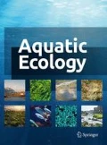
The sponge tissue samples were collected on the month of July of 2021, in the pools of the Comafreda stream, also known as Torrent des Guix (Mallorca Island, Spain, 39º 48′ N, 2º 54′ E). The specimens were found in several ponds along transect, although samples were only collected from six individuals located in one of the pools (39º 48′ 02′′ N, 2º 54′ 16.7′′ E), at an altitude of 260 m above sea level (Fig. 1).

Satellite image of the area where the sponge samples were collected
The selected specimens were all embedded in the limestone rock walls of the road, at a depth of between 0 and 70 cm (Fig. 2). The physical and chemical parameters of turbidity, luminosity, pH, conductivity, temperature, and dissolved oxygen, among others, were measured in each of the areas where sponges were present. These measurements were taken with a Hanna HI 9828 portable multiparameter apparatus. Six tissue samples were taken from the six specimens in the pool by scraping with a scalpel and were stored in 2 mL plastic tubes, together with water from the pool itself. All these samples were stored in cold storage for about 3 h, and after this period, they were kept in the dark at 4 ºC. The entire sample collection process was minimally intrusive to the animals. Scientific authorization for the study was obtained from the Ministry of Environment and Territory from the Government of the Balearic Islands (SEN 0576/2020).

Different specimens of the freshwater sponges present in the pool. a Specimen without gemmules. b Specimen partially gemmulated. Green tissue corresponds to the portion of the individuals occupied by green algae symbionts, while the whitish section is covered by the gemmules of the animal. Scale of the bars: 1 cm. (color figure online)
For the taxonomic identification of the sponge species in the area, some samples from the collected tissue were used. The method followed was based on purely morphological criteria, based on the structure of the animal’s spicules. For their fixation and subsequent microscopic identification, the methodology described by Hajdu et al. (2013) was followed. For the isolation and subsequent culture of the gemmules in both experiments, a modification of the protocol of Leys et al. (2019) for the treatment of E. fluviatilis was followed. For this, mechanical disaggregation of the sponge tissue was performed to separate the gemmules. All gemmules were then resuspended in a solution of H2O2 1% v/v to remove non-viable gemmules. After that all the putatively functional gemmules were stored at 4 ºC in dark conditions, in order to avoid its premature hatching.
Cultivation and maintenance of the sponges
Prior to conduct alkalinity and light experiments, it was necessary to ensure the viability of the gemmules, in order to verify their correct germination. Therefore, those viable gemmules that were preserved in cold were transferred to Petri dishes. Each plate contained 30 mL of medium M (Rasmont 1961).
Following the recommendations described by Leys et al. (2019), once the gemmules were deposited on the plates, they were kept in dark conditions at a temperature of 20 ± 2 °C, awaiting germination. When all the gemmules hatched, they were fed every two days, inoculating 15 μl of an autoclaved suspension of Escherichia coli CECT 101 at a final concentration of 103 CFU ml–1. To prevent the accumulation of toxic metabolites, the medium was changed regularly every two days.
Effect of alkalinity
In order to determine the growth capacity of the sponges based on the alkalinity of the culture medium, the transfer of the sponges fixed to new 85 mm divided Petri dishes was carried out. To study the effect of this parameter, individuals were divided into four groups according to the alkalinity of the medium: low (1 mEq L–1), medium (2.5 mEq L–1), moderate (4 mEq L–1) and high (5 mEq L–1). These values were selected as the range of values for the natural occurrence of E. fluviatilis oscillates between 0.379 and 3.993 mEq L–1 (Poirrier 1974). Two replicates of each treatment were carried out.
For this, a modification of the M medium was carried out to adjust the carbonate/bicarbonate/CO2 balance. Thus, different concentrations of HCO3− and CO32− were added to the culture medium to achieve the desired alkalinity of each level studied (Table 1), which were calculated by using the equation proposed by Millero (1995). All of this was done by using the carb command from the seacarb package in R program. In turn, a certain concentration of NaCl was added to each of the culture plates to balance the Na+ ion concentration at 300 mg mL–1, considered the optimum for E. fluviatilis growth (Francis et al. 1982). The sponges were incubated at 22 °C, with a photoperiod of 16:8 h at an irradiance of 6 μE m–2 s–1. The duration of treatment was 11 days. Between these, the growth capacity of the specimens was monitored three times a week.
Effect of light
Isolation and cultivation of symbionts
For the exogenous inoculation of the symbiont green algae, they were first isolated from one of the sponges’ tissue samples preserved at 4 ºC. Following the methodology proposed by Hall et al. (2021), mechanical homogenization of the sponge fragment was performed using a mortar previously sterilized with ethanol, to which 500 μl of BBM medium (Stein 1980)–used in the culture of Chlorella spp., the most common symbiont of E. fluviatilis—had been previously added.
Once the purification of the sample was performed, based on sequential cycles of centrifugation, the green pellet obtained was resuspended in a new Eppendorf tube with 500 μl of BBM, and transferred to an Erlenmeyer flask containing the same culture medium. In turn, the vessel was incubated in agitation under irradiance conditions of 80 μE m–2 s–1, with a photoperiod of 16:8 h, considered optimal for the growth of Chlorella spp. (Amini Khoeyi et al. 2012), the expected symbiont in these sponges. The addition of ampicillin at concentration of 0.1 mg ml–1 to the medium was carried out to ensure the absence of prokaryotic contamination.
Incubation of the holobiont
To measure the effect of the symbiosis on the growth of sponges under various irradiance conditions, a selection of the fixed sponges was carried out in a manner analogous to that of the alkalinity treatment. In this case, the culture medium for all treatment plates was medium M.
The levels to be evaluated for the independent variable were direct exposure to light (75 μE m–2 s–1), in penumbra conditions (5.75 μE m–2 s–1) and in absolute darkness. Prior to symbiont inoculation, and to ensure the absence of effect of potential photobionts intrinsically present in these structures, all gemmules were incubated in dark conditions at 20 ± 2 °C. Again, two replicates were carried out for each treatment. Once all the gemmules were fixed, inoculation of the symbionts was carried out in each of the plates. Therefore, 1 mL of the culture was transferred into liquid BBM medium in exponential growth phase two weeks after seeding, at which time the total Chlorella-like cell density was 9.63 · 104 cells mL–1 —for density counting, a direct unit count was carried out from 10 μL of the culture—. The final concentration of algae in each plate was around 6 · 103 cells mL–1. Prior to the algae inoculation, the sponges were maintained for 3 days after their hatching, until the development of a functional aquiferous system (Leys et al. 2019).
After the inoculation of the algae cells into the cultures, the light experiment started, and was carried out for 14 days in their respective irradiance conditions. The area of each sponge was measured every 3 days. To ensure the desired levels of light intensity, this parameter was evaluated by means of a portable radiometer Delta OHM HD2302.0. Unlike the measurement of the effect of alkalinity, in this case the sponges were not fed nor the media with the symbionts were changed during the experiment.
Determination of the areas
For the estimation of the area of the porifera during the progression of both experiments, the different specimens were photographed by means of a HAYEAR 5MP USBP 2.0 C-Mount camera coupled to a Leica S8AP0 binocular loupe. Five times area measurements were taken, including the initial area of the individuals, prior to the start of the different treatments. A number between 25 and 30 individuals were measured for each treatment of both experiments. The surface area of the organisms was determined using ImageJ image processing software.
Sample fixation and epifluorescence microscopy
In turn, for the evaluation of the presence of symbionts in the samples, the sponges were mounted and stained with DAPI, considering the autofluorescence capacity of chlorophyll when irradiated with wavelengths of the PAR spectrum corresponding to green and blue.
Therefore, sponges were isolated de novo in 85 mm Petri dishes in a 4% solution in paraformaldehyde mixed with 25% Holtfreter medium. These samples were incubated overnight at 4ºC. After incubation, the isolated sponges were stained by adding 25 μL of DAPI at a concentration of 0.01 mg mL–1, which allowed the cellular genetic material to be observed. After this, the samples were left to stand in the dark for 5 min. Subsequently, mounting was carried out with a drop of glycerol, leaving the samples ready for observation with the Leica DM2500 epifluorescence microscope, using A and I.3 cube filters—DAPI and blue light, respectively. Photographs were taken with a Leica DFC420C camera.
Statistical analyses
All statistical analyses associated with the comparison of the surface area of the sponges were evaluated by means of R-based programming. Welch’s ANOVA test with Games–Howell post hoc analysis was used for the evaluation of the growth rate and the projected area in the alkalinity study, as well as Kruskal–Wallis and Dunn’s test for the determination of these parameters in the experiment of the effect of luminosity. For the analysis of the occupation of the sponge tissue by the algae cells it was used a one-way ANOVA test.
Results
In situ parameters and species identification
The physical and chemical analysis of the pool from which the sponge samples were extracted is exposed in Table 2. The water from the basin was brackish, and its pH was relatively basic, with reducing conditions in the entire pond. Moreover, although exposed to certain illumination, the overall light intensity in the surface was low, more typical from penumbra conditions—as those to which the sponges were subjected in the luminosity experiment.
The six sponges from the pool were distributed on the surface of the limestone walls, adopting encrusting shapes. The morphological analysis of the spicules verified that the species present in the basin was E. fluviatilis. The megasclere oxeas had an average length of 352 ± 71 μm, and a thickness of 13.8 ± 1.6 μm. However, the specific character that allows discerning this species is based on its birotulate gemmuloscleres, which presented 15 rays per rotule, in addition to a spiniform prolongation on the trunk (Evans and Montagnes 2019). Their average length was 58.6 ± 2.1 μm, with a diameter of 25.4 ± 5.1 μm.
Effect of alkalinity
A strong positive correlation was observed between increasing alkalinity values with respect to total sponge growth. This trend could be seen not only in the total growth of the gemmules, but also during the entire progression of the experiment (Fig. 3).

a Progression of the average projected area (vertical bars indicate standard error) among the different alkalinity treatments. b Comparison between the average growth rate of the different treatments. * Indicates significant differences respect to the other alkalinity treatments (Welch’s ANOVA test, p < 0.001). Values are expressed as mean ± S.E.M
The group subjected to the high alkalinity treatment manifested a significantly greater increase in size compared to specimens from the other treatments (p < 0.001, F3, 103 = 8.9959), reaching an average value of 1.03 ± 0.49 mm2. On the other hand, the medium (2.5 mEq L–1) and moderate (4 mEq L–1) alkalinity groups did not present statistically significant differences in final surface area—0.88 ± 0.51 mm2 and 0.91 ± 0.54 mm2, respectively. In fact, it can be observed how their growth progression followed a practically identical trend throughout the experiment. Regarding the low alkalinity treatment (1 mEq L–1), a comparatively reduced growth rate was maintained compared to the other experimental groups. The average area obtained by the specimens of this group was 0.73 ± 0.33 mm2.
Checking the daily growth rate of the different specimens is checked, it has been verified that the high alkalinity treatment was the one that presented the most remarkable growth with respect to the other groups (p < 0.001, F3, 101 = 9.1146), up to 0.08 ± 0.03 mm2 per day. No statistically significant differences were observed between the values of this rate in the other treatments.
Light effect
As evidenced by epifluorescence observations (Fig. 4), only the sponges from the groups exposed to light showed Chlorella-like cells attached to their tissue, while none of the specimens subjected to the dark treatment showed the presence of these photobionts.

Comparison between E. fluviatilis specimens after 14 days of treatment under epifluorescence microscopy. a, d high light (75 μE m–2 s–1). b, e penumbra (5.75 μE m–2 s–1). c, f dark. Upper images show the sponges under optical microscopy, lower images reflect epifluorescence of symbionts when irradiated with blue wave light (red). Scale of the bars: 100 μm. (color figure online)
At the same time, it is worth noting the difference in the distribution of symbionts between the groups of sponges depending on the light intensity to which they were exposed. While those treated under conditions of 75 μE m–2 s–1 presented a more homogeneous organization of Chlorella-like cells throughout their structure, specimens subjected to a dim light intensity (5.75 μE m–2 s–1) showed a greater tendency of aggregation in the periphery of the gemmule, as well as in the outer perimeter of the sponge (Fig. 4). Furthermore, there was a significantly greater symbiont density in the sponges subjected to higher light intensity than the ones exposed to penumbra (p < 0.005) (Fig. 5). Although a minority compared to the sponge-associated forms, a large number of free Chlorella-like cells were also evident in the medium in both light treatments, in contrast to those subjected to dark conditions.

Percentage of occupation of the symbiont algae inside the sponge tissue. *Indicates significant indicates differences between treatments (Kruskal–Wallis test, p < 0.001). Values are expressed as mean ± S.E.M
Regarding growth parameters, it was possible to determine a larger final area in the light treatments, either high intensity or in penumbra conditions (p < 0.001, χ22 = 17.46) compared to sponges incubated in the dark. The differences in size between the two groups of light-exposed individuals, with final surface areas of 1.77 ± 0.89 mm2 and 1.21 ± 0.61 mm2, respectively, are attributable to the difference in surface area of the gemmules at the beginning of the experiment. The treatments of sponges subjected to both high light intensity and penumbra conditions show a parallel growth trend (Fig. 6). On the other hand, the initial progression of the individuals not exposed to light is of decreasing size, recovering a little growth capacity throughout the experiment, although at a lower rate than the other two groups. All these data can be corroborated with the analysis of daily growth, which reveals a superior faculty of development in the presence of light exposure (p < 0.001, χ22 = 49.81).

a Progression of the average projected area between the different light exposure treatments after inoculation of the symbionts. b Comparison between the average growth rate of the different treatments. SEM ± standard deviation. * Indicates significant differences respect to the other light treatments (ANOVA test, p < 0.001). Values are expressed as mean ± S.E.M
Discussion
Throughout the evolution toward to the freshwater environment, sponges from the family Spongillidae have followed adaptive paths peculiar to other representatives of their phylum, such as their ability to gemmulate in response to environmental adversity (Cáceres 1997). Further understanding of the underlying factors behind this peculiar phenomenon would allow determining to determine not only the ecological importance of modulating this quiescent state for these animals individually, but on the holobiont they comprise (Clark et al. 2021).
Ephydatia fluviatilis, the model species used in this study, although it has been recorded in both lentic and lotic environments (Li et al. 2018), is usually more frequently distributed in ecosystems of lotic character (Waterston and Lyster 1979; Didžiulis 2012; Evans and Montagnes 2019), such as the Comafreda torrent, where not only samples have been extracted, but also the occurrence of this taxon has been documented for the first time. This also constitutes the second register of it in the freshwater ecosystem of the Balearic archipelago (Travesset 1991). A higher prevalence and growth capacity of E. fluviatilis has been suggested in reducing environments—that is, of negative redox potential—(Evans and Montagnes 2019), conditions that were present in the sampling area, with an average value of − 61.9 mV pH–1 at pH 8.1.
Alkalinity is a chemical characteristic of the waters of the Balearic archipelago, which, in the case of the inland water bodies of Majorca, ranges between 1.18 and 5.30 mEq–1 (Moyà and Ramón 1981). The results show a positive correlation between this parameter and increased sponge development, as high water alkalinity (5 mEq L–1) favors faster sponge growth.
Our data not only increases the highest tolerable alkalinity range previously proposed for this species, located at 4 mEq L–1 or 4.6 mEq L–1, according to Poirrier (1974) and Old (1932), respectively; but also indicates the importance of higher alkalinity values for E. fluviatilis, as they enhance its growth. This factor could explain the ecological preference of this species for carbonate-rich river bodies (Pisera and Sáez 2003). In addition to alkalinity, the high content of calcium cations in the stream due to its karst nature is probably beneficial for these sponges (Økland and Økland 1996), as this is an essential element in membrane permeability (Belas et al. 1989).
Ion incorporation is essential for freshwater sponge, as it allows the maintenance of internal homeostasis (Senatore et al. 2016). It has been shown that the species from the family Spongillidae exhibit, in addition, a highly selective capacity for ionic regulation of their body, comparable to that observed in vertebrate epithelia (Adams et al. 2010). If such a regulatory capacity is considered, it could be suggested that increased alkalinity is able to act as a modulator of salt uptake, by allowing not only a more efficient buffering of the pH of the medium, but also a higher solubility of calcium cations (Boyd et al. 2016). The latter factor, as indicated, would act as a signal transducer that, among other essential functions, would allow to efficiently modulating the flow of nutrients into the sponge (Elliot and Leys 2010; Leys and Hill 2012). Future studies on the physiological effects of the ionic concentration could contribute to a better understanding of the global importance of alkalinity in these metazoans.
Apart from the effect of alkalinity in sponge growth, the role of symbiotic interaction by exogenous inoculation of chlorophytes present on the surface of the original sponge tissue has been evaluated. The importance of symbiosis with photosynthetic eukaryotes in members of the family Spongillidae has been repeatedly questioned (Wilkinson 1980; Sitte and Eschbach 1992), being restricted to the classes Trebouxiophyceae and Chlorophyceae (Chlorophyta) and Eustigmatophyceae (Ochrophyta). In the case of the species from the genus Ephydatia, only photobionts from the division Chlorophyta—more specifically, Chlorella-like cells—have been documented (Pröschold and Darienko 2020), which could indicate the beginning of a more restricted coevolution, as it has been have suggested (Geraghty et al. 2021).
In spite of not empirically verifying a greater growth in conditions of higher light intensity, not only a different density, but also an unequal qualitative distribution between both types of holobiont has been verified. Under conditions of exposure to peak light of 75 μE m–2 s–1, symbiont algae were more homogeneously distributed throughout the sponge tissue. In contrast, although there was also an aggregation of Chlorella-like cells around the treated gemmules under penumbra conditions, a greater arrangement can be observed at the periphery of the sponge. On the other hand, in specimens treated under dark conditions no such endosymbionts were observed inside the tissue.
The uneven distribution of Chlorella-like cells in the sponge tissue is explained by the establishment of the symbiosis itself. The incorporation of the algae occurs, in the first instance, by filtration—in fact, several experiments have revealed that the uptake of the symbionts by the sponges can become effective in about 4 h (Imsiecke 1993; Hall et al. 2021). After this, algae cells are retained in the collars of the choanocytes, as well as inside the pinacocytes of the outer layer of the animal (Saller 1989). It has been suggested that it is at this point that molecular recognition by both participants in the symbiosis occurs. As far as the sponge is concerned, there is overexpression of certain genes involved in immune recognition (Geraghty et al. 2021), as well as genes associated with oxidative stress (Hall et al. 2021), together with different permeases to mediate the transport of photoassimilates to the host (Grozdanov and Hentschel 2007). This fact has previously been verified in the establishment of symbiosis between these chlorophytes and other potential hosts such as Paramecium spp. (Kodama et al. 2014) or Hydra viridissima (Ishikawa et al. 2016). In turn, the ability of Chlorella sp. to directly secrete the glucose produced into host cells for their benefit has also been evidenced (Fischer et al. 1989).
The transmission of the symbionts will occur by transfer of vacuoles between the different cells. These vesicles, known as perialgal vacuoles, contain a single symbiont cell (Reisser and Wiessner 1984), which will be able to divide autonomously. It is considered that, within a period of 6 h, all the cells of the sponge will be able to present these vacuoles, especially in the mesohyl (Saller 1990). On the other hand, there is also another type of vesicle potentially containing the algae, although these have a digestive function. Their activity will be carried out either in case of dysfunction of the symbionts themselves, or in situations where these organisms do not provide a benefit to the host (Ereskovsky et al. 2022). It is speculated that this is the reason for the absence of symbionts in sponge subjected to dark conditions, since they are unable to carry out photosynthesis; they represent an energetic expense for their host, so they will be phagocyted. This would also explain the scarcity of free Chlorella-like cells in the dark treatments, as they would have been ingested by the sponge as the only available carbon source. Thus, digestion of the symbionts is the alternative for the host in situations of metabolic stress, in conditions where it can no longer take advantage of the symbiosis. The underlying mechanism behind the sponge’s discrimination of photobiont utility in different situations remains unknown.
It has been shown that the distribution of symbionts in E. fluviatilis is not arbitrary, as in general, amoebocytes carrying Chlorella-like cells are distributed in the cortical region of the animal (Gaino et al. 2003). This distribution would allow a greater light uptake, thus favoring the growth of the holobiont. Our results also indicate a strong presence of Chlorella-like cells in the interior of the sponge, surrounding the gemmules, as well as in the periphery of the animal, factor more evident in sponges subjected to penumbra conditions. The reason for the higher density of symbionts in individuals exposed to higher light intensity is probably multifactorial. On the one hand, it is to be understood that, given the autonomous character of Chlorella-like individual replication with respect to its host (Saller 1990), when exposed to its optimum light intensity they see their growth favored compared to penumbra conditions (Metsoviti et al. 2019). For its part, the greater aggregation of symbionts in the case of maximum irradiance could be analogous to the difference in the distribution of plant chloroplasts as a function of light intensity (Maai et al. 2020). This would also explain the greater extensive tendency toward the periphery of photobionts in sponges treated under penumbra conditions, allowing maximum utilization of available light (Wada 2013). It is also suggested that the increased aggregation of Chlorella-like cells around the gemmule could be a mechanism mediated by the sponge to prevent from damage by excessive light exposure, as documented in Paramecium bursaria (Summerer et al. 2009).
The results obtained indicate a higher growth rate of the individuals exposed to light, which were also the only ones that presented associated photobionts in their tissue. Regardless of the light intensity—whether it was optimal for the symbiont, 75 μE m–2 s–1; or in penumbra conditions, 5.75 μE m–2 s–1—, the establishment of the link between E. fluviatilis and Chlorella-like symbionts could be verified. Therefore, based on the data obtained in this study, the establishment of the holobiont only takes place in situations of illumination of sponges, which is consistent with the observations made by Wilkinson (1980).
Thus, in case of light exposure, the symbiosis with Chlorella sp. is particularly important for E. fluviatilis, since the translocation of photoassimilates by this alga supplies all the metabolic demands of its host—since no external carbon source was provided during the whole experiment—, thus stimulating its growth. It is estimated that the contribution of glucose from the photobiont can range from 9 to 17% of the fixed carbon (Pröschold and Darienko 2020). Such nutrient translocation efficiency is comparable to values observed in interactions of Chlorella spp. with other species from the family Spongillidae, such as Spongilla lacustris (Fischer et al. 1989). This fact raises the evolutionary importance of such a symbiotic interaction for this family of sponges, as suggested by numerous studies (Jensen and Pedersen 1994; O’Brien et al. 2019; Hall et al. 2021).
However, compared to other endosymbiosis events between Chlorella-like organisms and other eukaryotes, it appears that the interaction with E. fluviatilis is not as efficient (Wilkinson 1984). For example, the rate of photoassimilate transport by this genus of trebouxiophyceae and Hydra viridissima can range from 25 to 30% (Cook 1983). This last relationship also evidences a greater control of the symbiont by the cnidarian host (Bosch 2012), since the establishment of symbiosis is obligatory for Chlorella individuals in case of coexistence with H. viridissima. It should be emphasized that this hydrozoan is able to express a large diversity of genes exclusively in this interaction (Hamada et al. 2018), revealing a higher degree of coevolution between both species compared to the symbiosis between Chlorella and freshwater sponges (Ereskovsky et al. 2022).
Despite this apparent lower symbiotic efficiency, the ecology of E. fluviatilis needs to be considered for a correct understanding of the establishment of this relationship. This species, like so many other freshwater sponges, manifests a clear sciaphile tendency (De Santo and Fell 1996). This preferential distribution is not arbitrary, since it has been shown that the formation of gemmules tends to take place in dark conditions (Brønsted and Brønsted 1953), which guarantees survival by cryptobiosis in situations of environmental stress. The absence of light, on the other hand, limits the photosynthetic capacity of the symbionts. Although this is not a problem for other species in which such an endosymbiotic relationship has been documented, such as the hydrozoans of the genus Hydra, it should be remembered that these have the capacity for autonomous movement, ability not present in sponges. The data obtained in this experiment suggest that the establishment of aposymbiosis between E. fluviatilis and Chlorella spp. takes place, preferentially, in the case of being able to obtain a direct benefit from the host—glucose supply. However, significant growth has also been observed in the absence of exogenous inoculation of symbionts during the analysis of the effect of alkalinity, which experimentally corroborates the observations carried out in the field (Wilkinson 1980; Evans and Montagnes 2019). All these data support that the establishment of the symbiosis is not essential for the growth of E. fluviatilis under nutrient availability. However, in situations of light exposure it becomes an extremely important factor for the sponges, as it can mediate the transition from a heterotrophic filtering lifestyle to one dependent on photosynthesis mediated by its symbionts.
Final considerations
The results of this study have revealed the important role of alkalinity on the growth of sponges. Fulfilling the initial hypothesis, a positive association between this physicochemical parameter and the development of E. fluviatilis was observed, which explains the ecological tendency of this metazoan to grow in carbonate-rich waters.
On the other hand, the study of the effect of light intensity on the holobiont has revealed a differential effect on growth. Although it is true that no evidence was found of a higher rate of development in conditions of higher light intensity, it was possible to determine an unequal distribution of the symbionts in cases of exposure to different intensities. This fact, together with the absence of growth in the specimens incubated in the dark, indicates an important regulatory role of light not only in the arrangement of the symbionts, but also in the capacity of the sponges to take advantage of them. In conclusion, the data obtained support the relative importance of the aposymbiosis phenomenon in E. fluviatilis, as well as the role of the discriminatory capacity of the metazoan in the establishment of an endosymbiotic association as peculiar as that between sponges and chlorophytes.
References
Achlatis M, Schönberg CHL, van der Zande RM, LaJeunesse TC, Hoegh-Guldberg O, Dove S (2019) Photosynthesis by symbiotic sponges enhances their ability to erode calcium carbonate. J Exp Mar Biol Ecol 516:140–149
Adams EDM (2000) Symbiotic interactions. In: Whitton BA, Potts M (eds) The ecology of cyanobacteria. Their diversity through space and time. Springer Science, Dordrecht, pp 523–552
Adams EDM, Goss GG, Leys SP (2010) Freshwater sponges have functional, sealing epithelia with high transepithelial resistance and negative transepithelial potential. PLoS ONE 5(11):e1540
Amini Khoeyi Z, Seyfabadi J, Ramezanpour Z (2012) Effect of light intensity and photoperiod on biomass and fatty acid composition of the microalgae. Chlorella Vulgaris Aquac Int 20(1):41–49
Annenkova NV, Lavrov DV, Belikov SI (2011) Dinoflagellates associated with freshwater sponges from the ancient Lake Baikal. Protist 162(2):222–236
Anteneh Y, Yang Q, Brown MH, Franco CMM (2022) Factors affecting the isolation and diversity of marine sponge-associated bacteria. Appl Microbiol Biotechnol 106(3):3
Bart MC, de Vet SJ, de Bakker DM, Alexander BE, van Oevelen D, van Loon EE, van Loon JJWA, de Goeji JM (2019) Spiculous skeleton formation in the freshwater sponge Ephydatia fluviatilis under hypergravity conditions. PeerJ 6:e6055
Belas FJ, Francis JC, Poirrier MA (1989) Effects of calcium, magnesium, and sodium on growth of Ephydatia fluviatilis (Porifera: Spongillidae). Trans Am Micros Soc 108(2):139
Benfey TJ, Reiswig HM (1982) Temperature, pH, and photoperiod effects upon gemmule hatching in the freshwater sponge, Ephydatia muelleri (Porifera, Spongillidae). J Exp Zool 221(1):13–21
Bosch TCG (2012) What Hydra has to say about the role and origin of symbiotic interactions. Biol Bull 223:78–84
Boyd CE, Tucker CS, Somridhivej B (2016) Alkalinity and hardness: critical but elusive concepts in aquaculture. J World Aquac Soc 47(1):6–41
Brøndsted A, Brøndsted HV (1953) The effect of symbiontic zoochlorellae on the germination rate of gemmules of Spongilla lacustris (L.). Dansk Naturhistorisk Forening Videnskabelige Meddelelser 115:133–144
Cáceres CE (1997) Dormancy in invertebrates. Invertebr Biol 116(4):371–383
Calheira L, Lanna E, Pinheiro U (2019) Tropical freshwater sponges develop from gemmules faster than their temperate-region counterparts. Zoomorphology 138:425–436
Carrier TJ, Maldonado M, Schmittmann L, Pita L, Bosch TCG, Hentschel U (2022) Symbiont transmission in marine sponges: reproduction, development, and metamorphosis. BMC Biol. https://doi.org/10.1186/s12915-022-01291-6
Chernogor L, Denikina N, Kondratov I, Solovarov I, Khanaev I, Belikov S, Erlich H (2013) Isolation and identification of the microalgal symbiont from primmorphs of the endemic freshwater sponge Lubomirskia baicalensis (Lubomirskiidae, Porifera). Eur J Phycol 48(4):497–508
Clark CM, Hernandez A, Mullowney MW, Fitz-Henley J, Li E, Romanowsky SB, Pronzato R, Manconi R (2021) Relationship between bacterial phylotype and specialized metabolite production in the culturable microbiome of two freshwater sponges. ISME Commun 2:22
Cook CB (1983) Metabolic interchange in algae-invertebrate symbiosis. Int Rev Cytol 14:177–210
De Santo EM, Fell PE (1996) Distribution and ecology of freshwater sponges in connecticut. Hydrobiologia 341:81–89
De Voogd NJ, Alvarez B, Boury-Esnault N et al. (2023) World Porifera Database. https://www.marinespecies.org/porifera. Accessed 2022 December 21
Degnan BM (2015) The surprisingly complex immune gene repertoire of a simple sponge, exemplified by the NLR genes: a capacity for specificity? Dev Comp Inmunol 48(2):269–274
Degnan BM, Adamska M, Richards GS, Larroux C, Leininger S, Bergum B, Calcino A, Taylor K, Nakanishi N, Degnan SM (2015) Porifera. In: Wanninger A (ed) Evolutionary developmental biology of invertebrates 1. Springer, Vienna, pp 65–106
Didžiulis V (2012) Freshwater sponges in the river Thames. The Reading Naturalist 64:34–40
Ehrlich H, Wysokowski M, Żóltowska-Aksamitowska S, Petrenko I, Jesionowski T (2018) Collagens of poriferan origin. Mar Drugs 16(3):79
Elliott GRD, Leys SP (2010) Evidence for glutamate, GABA and NO in coordinating behaviour in the sponge, Ephydatia muelleri (Demospongiae, Spongillidae). J Exp Biol 213:2310–2321
Ereskovsky A, Rinkevich B, Somorjai IML (2022) Adult stem cells host intracellular symbionts: the Poriferan archetype. In: Ballarin L, Rinkevich B, Hobmayer B (eds) Advances in aquatic invertebrate stem cell research. From basic research to innovative applications. MDPI, Switzerland, pp 65–94
Erpenbeck D, Galitz A, Wörheide G, Albrecht C, Pronzato R, Manconi R (2020) Having the balls to colonize – the Ephydatia fluviatilis group and the origin of (ancient) lake “endemic” sponge lineages. J Great Lakes Res 46(5):1140–1145
Evans KL, Montagnes DJS (2019) Freshwater sponge (Porifera: Spongillidae) distribution across a landscape: environmental tolerances, habitats, and morphological variation. Invertebr Biol 00:e12258
Fischer A, Meindl D, Loos E (1989) Glucose excretion by the symbiotic Chlorella of Spongilla lacustris. Planta 179(2):251–256
Francis JC, Poirrier MA, LaBiche RA (1982) Effects of calcium and salinity on the growth rate of Ephydatia fluviatilis (Porifera: Spongillidae). Hydrobiologia 89:225–229
Frost TM (1991) Porifera. In: Thorp JH, Covich AP (eds) Ecology and classification of North America freshwater invertebrates. Academic Press, New York, pp 95–124
Frost TM, Graham LE, Elias JE, Haase MJ, Kretchmer DW, Kranzfelder JA (1997) A yellow-green algal symbiont in the freshwater sponge, Corvomeyenia everetti: convergent evolution of symbiotic associations. Freshw Biol 38:395–399
Gaino E, Rebora M, Corallini C, Lancioni T (2003) The life-cycle of the sponge Ephydatia fluviatilis (L.) living on the reed Phragmites australis in an artificially regulated lake. Hydrobiologia 495:127–142
Gaino E, Scoccia F, Piersanti S, Rebora M, Bellucci LG, Ludovisi A (2012) Spicule records of Ephydatia fluviatilis as a proxy for hydrological and environmental changes in the shallow Lake Trasimeno (Umbria, Italy). Hydrobiologia 679(1):139–153
Geraghty S, Koutsouveli V, Hall C, Chang L, Sacristan-Soriano O, Hill M, Riesgo A, Hill A (2021) Establishment of host–algal endosymbioses: genetic response to symbiont versus prey in a sponge host. Genome Biol Evol. https://doi.org/10.1093/gbe/evab252
Gernert C, Glöckner FO, Krohne G, Hentschel U (2005) Microbial diversity of the freshwater sponge Spongilla lacustris. Microb Ecol 50(2):206–212
Grozdanov L, Hentschel U (2007) An environmental genomics perspective on the diversity and function of marine sponge-associated microbiota. Curr Opin Microbiol 10(3):215–220
Hajdu E, Peixinho S, Fernandez JCC (2013) Bahia Marine Sponges: Laboratory and field guide. Editorial Museu Nacional do Rio de Janeiro, Brasil
Hall C, Camilli S, Dwaah H, Kornegay B, Lacy C, Hill MS, Hill A (2021) Freshwater sponge hosts and their green algae symbionts: a tractable model to understand intracellular symbiosis. PeerJ 9:e1065
Hamada M, Schröder K, Bathia J, Kürn U, Fraune S, Khalturina M, Khalturin K, Shinzato C, Satoh N, Bosch TCG (2018) Metabolic co-dependence drives the evolutionarily ancient Hydra-Chlorella symbiosis. Elife 7:e35122
Hirose E (2015) Ascidian photosymbiosis: diversity of cyanobacterial transmission during embryogenesis. Genesis 53:121–131
Imsiecke G (1993) Ingestion, digestion, and egestion in Spongilla lacustris (Porifera, Spongillidae) after pulse feeding with Chlamydomonas reinhardtii (Volvocales). Zoomorphology 113(4):233–244
Ishikawa M, Yuyama I, Shimizu H, Nozawa M, Ikeo K, Gojobori T (2016) Different endosymbiotic interactions in two Hydra species reflect the evolutionary history of endosymbiosis. Genome Biol Evol 8(7):2155–2163
Jäckle O, Seah BKB, Tietjen M, Gruber-Vodicka HR (2019) Chemosynthetic symbiont with a drastically reduced genome serves as primary energy storage in the marine flatworm Paracatenula. PNAS 116(17):8505–8514
Jensen KS, Pedersen MF (1994) Photosynthesis by symbiotic algae in the freshwater sponge. Spongilla Lacustris Limnol Oceanogr 39(3):551–561
Keller-Costa T, Jousset A, van Overbeek L, van Elsas JD, Costa R (2014) The freshwater sponge Ephydatia fluviatilis harbours diverse Pseudomonas species (Gammaproteobacteria, Pseudomonadales) with broad-spectrum antimicrobial activity. PLoS ONE 9(2):e88429
Kenny NJ, Francis WR, Rivera-Vicéns RE, Juravel K, de Mendoza A, Díez-Vives C, Lister R, Bezares-Calderón LA, Grombacher L, Roller M, Barlow LD, Camilli S, Ryan JF, Wörheide G, Hill A, Riesgo A, Leys SP (2020) Tracing animal genomic evolution with the chromosomal-level assembly of the freshwater sponge Ephydatia muelleri. Nat Commun. https://doi.org/10.1038/s41467-020-17397-w
Kodama Y, Suzuki H, Dohra H, Sugii M, Kitazume T, Yamaguchi K, Shigenobu S, Fujishima M (2014) Comparison of gene expression of Paramecium bursaria with and without Chlorella variabilis symbionts. BMC Genom 15(1):183
Kohn T, Wiegand S, Boedeker C, Rast P, Heuer A, Jetten MSM, Schüler M, Becker S, Rohde C, Müller R-W, Brümer F, Rohde M, Engelhardt H, Jogler M, Jogler C (2020) Planctopirus ephydatiae, a novel Planctomycete isolated from a freshwater sponge. Syst Appl Microbiol 43(1):126022
Laport MS, Pinheiro U, Rachid CTCdC (2019) Freshwater sponge Tubella variabilis presents richer microbiota than marine sponge species. Front Microbiol 10:2799
Leys SP, Grombacher L, Hill A (2019) Hatching and freezing gemmules from the freshwater sponge Ephydatia muelleri. Protocols. io.
Leys SP, Hill A (2012) The physiology and molecular biology of sponge tissues. Adv Mar Biol 62:1–56
Li R, Nitsche F, Arndt H (2018) Mesoscale investigations based on microsatellite analysis of the freshwater sponge Ephydatia fluviatilis in the River-Sieg system (Germany) reveal a genetic divergence. Conserv Genet 19(4):959–968
Maai E, Nishimura K, Takisawa R, Nakazaki T (2020) Diurnal changes in chloroplast positioning and photosynthetic traits of C4 grass finger millet. Plant Prod Sci 23(4):477–389
Manconi R, Pronzato R (2008) Global diversity of sponges (Porifera: Spongillina) in freshwater. In: Balian EV, Lévêque C, Segers H, Martens K (eds) Freshwater animal diversity assessment. Developments in hydrobiology. Springer, Dordrecht, pp 27–33
Matsunaga S (2018) Planimal cells: artificial photosynthetic animal cells inspired by endosymbiosis and photosynthetic animals. Cytologia 83(1):3–6
Melão MGG, Rocha O (1999) Biomass and productivity of the freshwater sponge Metania spinata (Carter, 1881) (Demospongiae: Metaniidae) in a Brazilian reservoir. Hydrobiologia 390:1–10
Metsoviti MN, Papapolymerou G, Karapanagiotidis IT, Katsoulas N (2019) Effect of light intensity and quality on growth rate and composition of Chlorella vulgaris. Plants 9(1):31
Millero FJ (1995) Thermodynamics of the carbon dioxide system in the oceans. Geochim Cosmochim Acta 59(4):661–667
Moyà G, Ramón G (1981) Contribución al conocimiento de la mineralización de las aguas de los embalses de Cúber y gorg blau y de sus principales aportes. Boll Soc Hist Nat Balears 25:21–30
O’Brien PA, Webster NS, Miller DJ, Bourne DG (2019) Host-microbe coevolution: applying evidence from model systems to complex marine invertebrate holobionts. Mbio. https://doi.org/10.1128/mBio.02241-18
Økland KA, Økland J (1996) Freshwater sponges (Porifera: Spongillidae) of Norway: distribution and ecology. Hydrobiologia 330(1):1–30
Old MC (1932) Taxonomy and distribution of the fresh-water sponges (Spongillidae) of Michigan. Pap Mich Acad Sci 15:439–477
Pisera A, Sáez A (2003) Paleoenvironmental significance of a new species of freshwater sponge from the Late Miocene Quillagua Formation (N Chile). J South Am Earth Sci 15(8):847–852
Poirrier MA (1974) Ecomorphic variation in gemmoscleres of Ephydatia fluviatilis Linnaeus (Porifera: Spongillidae) with comments upon its systematics and ecology. Hydrobiologia 44:337–347
Pröschold T, Darienko T (2020) Choricystis and Lewiniosphaera gen. nov (Trebouxiophyceae, Chlorophyta), two different green algal endosymbionts in freshwater sponges. Symbiosis 82:175–188
Rasmont R (1961) Une technique de culture des sponges d’eau douce en milieu control. Ann Soc R Zool Belg 91:147–155
Reisser W, Wiessner W (1984) Autotrophic eukaryotic freshwater symbionts. In: Linskens H, Heslop-Harrison J (eds) Cellular Interactions. Encyclopedia of Plant Physiology. Springer, Berlin, pp 59–74
Reiswig HM, Miller TL (1998) Freshwater sponge gemmules survive months of anoxia. Invertebr Biol 117(1):1–8
Rix L, Ribes L, Coma R, Jahn MT, de Goeji JM, van Oevelen D, Escrig S, Meibom A, Hentschel U (2020) Heterotrophy in the earliest gut: a single-cell view of heterotrophic carbon and nitrogen assimilation in sponge-microbe symbioses. ISME J 14(10):2554–2567
Rola M, Frankenbach S, Bleidissel S, Sickinger C, Donath A, Frommlet JC, Greve C, Serôdio J, Preisfeld A, Melo Clavijo J, Christa G (2022) Cladobranchia (Gastropoda, Nudibranchia) as a promising model to understand the molecular evolution of photosymbiosis in animals. Front Mar Sci. https://doi.org/10.3389/fmars.2021.745644
Rosset SL, Oakley CA, Ferrier-Pagès C, Suggett DJ, Weis VM, Davy SK (2021) The molecular language of the cnidarians-dinoflagellate symbiosis. Trends Microbiol 29(4):320–333
Saller U (1989) Microscopical aspects on symbiosis of Spongilla lacustris (Porifera, Spongillidae) and green algae. Zoomorphology 108(5):291–296
Saller U (1990) Formation and construction of asexual buds of the freshwater sponge Radiospongilla cerebellata (Porifera, Spongillidae). Zoomorphology 109:295–301
Schill RO, Pfannkuchen M, Fritz G, Köhler H, Brümmer F (2006) Quiescent gemmules of the freshwater sponge, Spongilla lacustris (Linnaeus, 1759), contain remarkably high levels of Hsp70 stress protein and hsp70 stress gene mRNA. J Exp Zool 305(5):449–457
Schuster A, Vargas S, Knapp IS, Pomponi SA, Toonen RJ, Erpenbeck D, Wörheide G (2018) Divergence times in demosponges (Porifera): first insights from new mitogenomes and the inclusion of fossils in a birth-death clock model. BMC Evol Biol 18:114
Senatore A, Raiss H, Le P (2016) Physiology and evolution of voltage-gated calcium channels in early diverging animal phyla: cnidaria, placozoa porifera and ctenophora. Front Physiol. https://doi.org/10.3389/fphys.2016.00481
Sitte P, Eschbach S (1992) Cytosymbiosis and its significance in cell evolution. In: Dietmar B, Esser K, Kubitzki K, Runge M, Ziegler H (eds) Progress in Botany, 53. Springer, Berlin, pp 29–43
Skelton J, Strand M (2013) Trophic ecology of a freshwater sponge (Spongilla lacustris) revealed by stable isotope analysis. Hydrobiologia 709(1):227–235
Stein JR (1980) Handbook of phycological methods. culture methods and growth measurements. Cambridge University Press, Cambridge
Summerer M, Sonntag B, Hörtnagl P, Sommaruga R (2009) Symbiotic ciliates receive protection against UV damage from their algae: a test with Paramecium bursaria and Chlorella. Protist 160(2):233–243
Taylor MW, Radax R, Steger D, Wagner M (2007a) Sponge-associated microorganisms: evolution, ecology, and biotechnological potential. Microbiol Mol Biol Rev 71(2):295–347
Taylor MW, Thacker RW, Hentschel U (2007b) Evolutionary insights from sponges. Science 316(5833):1854–1855
Travesset A (1991) Presència d’Ephydatia fluviatilis (Porifera: Spongillidae) en un torrent de Mallorca. Boll Soc Hist Nat Balears 34:97–98
Tréguer PJ, Sutton JN, Brzezinski M, Charette MA, Devries T, Dutzkiewicz S, Ehlert C, Hawkings J, Leynaert A, Mei Liu S, Monferrer NL, López-Acosta M, Maldonado M, Rahman S, Ran L, Rouxel O (2021) The biogeochemical cycle of silicon in the modern ocean. Biogeosciences 18:1269–1289
Usher KM (2008) The ecology and phylogeny of cyanobacterial symbionts in sponges. Mar Ecol 29:178–192
Wada M (2013) Chloroplast movement. Plant Sci 210:177–182
Waterston AR, Lyster IHJ (1979) The macrofauna of brackish and fresh waters of the loch Druidibeg national nature reserve and its neighbourhood. South Uist Proc Royal Soc B 77:353–376
Webster NS, Thomas T (2016) The sponge hologenome. ASM. https://doi.org/10.1128/mBio.00135-16
Wilkinson CR (1980) Nutrient translocation from green algal symbionts to the freshwater sponge Ephydatia fluviatilis. Hydrobiologia 75(3):241–250
Wilkinson CR (1984) Immunological evidence for the precambrian origin of bacterial symbioses in marine sponges. Proc Royal Soc London B 220:509–517
Wilkinson CR (1987) Significance of microbial symbionts evolution and ecology in sponge. Symbiosis 4:135–146
Acknowledgements
A. Sureda and S. Tejada were supported by the Spanish Government, Institute of Health Carlos III (Project CIBEROBN CB12/03/30038). We would like to thank Josep Homs from Guies de Tramuntana for his help to the field work for sampling. S. Pinya were supported by the project Biodibal under the umbrella of the Agreement between the University of the Balearic Islands and Red Eléctrica de España.
Funding
Open Access funding provided thanks to the CRUE-CSIC agreement with Springer Nature.
Author information
Authors and Affiliations
Corresponding author
Ethics declarations
Conflicts of interest
The authors declare that they have not known competing interests.
Additional information
Handling Editor: Télesphore Sime-Ngando.
Publisher's Note
Springer Nature remains neutral with regard to jurisdictional claims in published maps and institutional affiliations.
Rights and permissions
Open Access This article is licensed under a Creative Commons Attribution 4.0 International License, which permits use, sharing, adaptation, distribution and reproduction in any medium or format, as long as you give appropriate credit to the original author(s) and the source, provide a link to the Creative Commons licence, and indicate if changes were made. The images or other third party material in this article are included in the article's Creative Commons licence, unless indicated otherwise in a credit line to the material. If material is not included in the article's Creative Commons licence and your intended use is not permitted by statutory regulation or exceeds the permitted use, you will need to obtain permission directly from the copyright holder. To view a copy of this licence, visit http://creativecommons.org/licenses/by/4.0/.
About this article

Cite this article
Gost, M., Pinya, S., Sureda, A. et al. Effect of alkalinity and light intensity on the growth of the freshwater sponge Ephydatia fluviatilis (Porifera: Spongillidae). Aquat Ecol 57, 353–367 (2023). https://doi.org/10.1007/s10452-023-10014-0
Received:
Accepted:
Published:
Issue Date:
DOI: https://doi.org/10.1007/s10452-023-10014-0